THE EVALUATION OF PREFERENTIAL ALIGNMENT OF BIOLOGICAL APATITE (BAp) CRYSTALLITES IN BONE USING A TRANSMISSION X-RAY DIFFRACTION METHOD
←
→
Page content transcription
If your browser does not render page correctly, please read the page content below

Copyright ©JCPDS-International Centre for Diffraction Data 2008 ISSN 1097-0002 155
THE EVALUATION OF PREFERENTIAL ALIGNMENT OF BIOLOGICAL
APATITE (BAp) CRYSTALLITES IN BONE USING A TRANSMISSION
X-RAY DIFFRACTION METHOD
Katsunari Sasaki,1 Takayoshi Nakano,2 Yukichi Umakoshi,2 Joseph D. Ferrara,3 and
Toshihiko Sasaki1
1
Division of Innovative Technology and Science, Graduate School of Natural Science and
Technology, Kanazawa University, Kanazawa 920-1192, Japan
2
Division of Materials and Manufacturing Science, Graduate School of Engineering, Osaka
University, Suita 565-0871, Japan
3
Rigaku Americas Corporation, 9009 New Trails Drive, The Woodlands, Texas 77381-5209
ABSTRACT
A two-dimensional quantitative XRD measurement of the orientation of biological apatite (BAp)
in an isolated trabecula of a mouse ulna was made using a transmission mode X-ray diffraction
system with a 0.3 mm diameter parallel beam MoKα source. The same measurement was done
with CuKα radiation. Copper Kα is the most commonly used radiation for powder diffraction
because it provides good resolution of the resultant lines but is more cumbersome because it
requires slicing the bone into thin sections since CuKα radiation is not very penetrating. The
preferred orientation of the BAp c-axis from the orecranon to the caput ulnae was measured for
12-week mutant osteopetrotic (op/op) and littermate mice. It was found that the transmission
optical method using MoKα radiation provides a simple and non-destructive method for measuring
bone quality.
INTRODUCTION
X-ray diffraction is a well known method for the analysis of materials. The method is used to
measure properties such as lattice parameters, crystallite, preferred orientation, residual stress, and
pole figures. Structural materials such as steel, aluminum, and plastics are often strengthened by
modification of the crystal properties of the material. For example, the materials may be processed
to enhance preferred orientation or residual stress. X-ray diffraction techniques have changed
dramatically since the development of two-dimensional detectors such as the image plate (IP),
multiwire proportional counter (MWPC), and charge coupled device (CCD) [1]. The use of these
detectors provides two-dimensional information and reduces measurement times [2].
Bone is usually evaluated using bone mineral density (BMD), but recent research suggests that
BMD is not a sufficient predictor of whole bone mechanical properties [3,4]. Nakano et al. have
measured preferred orientation of the BAp crystallites by X-ray diffraction techniques because the
preferred alignment of the BAp c-axis in various bones varies depending on the shape, stress
distribution in vivo, and the related mechanical function [5]. The analysis of the BAp orientation
also explains the behavior of bone regeneration by the tissue engineering technique since the
change in the BAp orientation does not agree with that in BMD during the regenerative process
[6,7]. CuKα (λ = 1.5414 Å) radiation was used for all these X-ray measurements. The absorption of
CuKα radiation by BAp is quite large. In these experiments only information about the surface of
the bone was observed [8]. Thus, the use of CuKα is not practical for medical in vivo applications
or even routine ex vivo applications.
This document was presented at the Denver X-ray
Conference (DXC) on Applications of X-ray Analysis.

Sponsored by the International Centre for Diffraction Data (ICDD).

This document is provided by ICDD in cooperation with
the authors and presenters of the DXC for the express
purpose of educating the scientific community.

All copyrights for the document are retained by ICDD.

Usage is restricted for the purposes of education and
scientific research.

DXC Website                         ICDD Website
– www.dxcicdd.com                   - www.icdd.com
Copyright ©JCPDS-International Centre for Diffraction Data 2008 ISSN 1097-0002 156
The purpose of this study is to show the advantage of the use of higher energy characteristic X-ray
MoKα radiation (λ = 0.7107 Å) and transmission mode X-ray diffraction. The use of higher energy
radiation reduces the absorption and improves the signal-to-noise ratio, and the transmission
method means the bone may be analyzed intact rather than sectioned. We investigated the areal
distribution of the BAp c-axis on a cross-section of the ulna bone of 12-week osteopetrotic (op/op)
mice. Osteoclasts rarely appear because of a defect in the expression of the macrophage
colony-stimulating factor and littermate mouse using a transmission X-ray diffraction method.
MATERIALS AND METHODS
Apatite sample and bone specimens
To perform the absorption experiment, NIST calcium hydroxyapatite powder (SRM 2910) was
placed into a 1.5 mm diameter glass capillary. Two bone samples, op and control (normal), ulnae
of 12-week-old mice were fixed and kept in 10% formalin neutral buffered solution 6 months to
avoid degradation. Figure 1 shows a photograph of the bones used for the experiment. The sample
op/op is from a mutant osteopetrotic mouse. The sample control is from a normal mouse. The bone
length of op/op was 14.6 mm and the control sample was 16.9 mm.
X-ray micro CT
The internal structure of each bone and its density was verified with a Rigaku R_mCT, a micro
X-ray CT scanner specially designed for small animal imaging. The operated X-ray power was 90
kV, 88 µA with an exposure time of 17 s.
X-ray diffraction analysis
All X-ray diffraction experiments were performed using a Rigaku R-AXIS RAPID II system. This
is a two-pinhole transmission, two-dimensional camera system with cylindrical detection
geometry. The RAPID II employs an image plate (storage phosphor) developed by Fuji Film as the
X-ray detector. The geometry of the apparatus resembles a vertical-screenless Weissenberg camera
and was mainly designed for single crystal X-ray structure analyses and charge density studies.
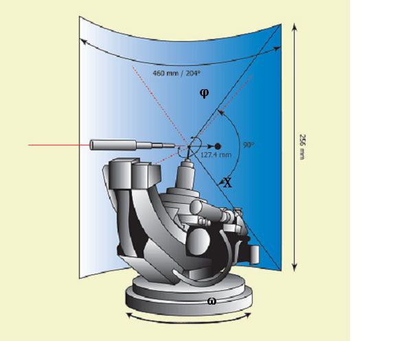
Figure 2 shows a schematic drawing of the R-AXIS RAPID II X-ray optics. The detector is 460
mm in the horizontal direction and 256 mm in the vertical direction with a radius of 127.4 mm.
This provides a 2θ range from -60° to +144° in the horizontal direction and a 2θ range of ±45° in
the vertical direction. The pixel size is 100 µm. A Rigaku ultraX rotating anode X-ray generator
with either a Cu (λ = 1.5418 Å) or Mo (0.7107 Å) target and a graphite monochromator was used in
the study. A 0.3 mm double pinhole collimator was used to spatially condition the X-ray beam. The
X-ray generator was operated at 50 kV and 90 mA. Both CuKα and MoKα radiation were used to
study the X-ray absorption characteristics of the NIST calcium hydroxyapatite reference sample,
but only MoKα radiation was used to study the bone samples. The exposure time for the NIST
apatite reference sample was 10 min. The bone samples were exposed for 1 min.
The samples were oriented on a 3-circle Eulerian goniometer with a manual XYZ stage. The
Eulerian angles were ω, χ, and φ. The φ-axis was closest to the sample and provided for complete
rotation of the sample around that axis. The χ-axis was next and used to tilt the sample as shown in
Figure 2. In general, the ω-axis is used as the scanning axis. However, in this study the sample was
fixed for each exposure. The XYZ stage was used to select the desired position on the bone sample
for study. The distribution of the X-ray absorption of the bone samples were measured using a
Copyright ©JCPDS-International Centre for Diffraction Data 2008 ISSN 1097-0002                                   157

        Rigaku X-ray scintillation counter operating in integrating mode. The bone sample was placed in
        the beam and the X-ray intensity of the transmitted beam was measured as function of the X
        direction. To measure along Anterior-Posterior direction, the φ-axis was rotated 90°.

        X-ray diffraction data were recorded using the Rigaku RAPID Auto software. The data were
        processed as follows. For each bone sample, 10 measurements were taken three times. First 10 mm
        × 45° segments in both the equatorial and meridional directions of the images (Figure 3) were
        extracted and converted to 2θ versus I profiles. Next the (002) and (310) reflections were identified
        and the integrated intensity was calculated.

                                                                            Figure 2. Schematic drawing of the
            Figure 1. Mouse femurs used for the study.                     Rigaku R-AXIS RAPID II goniometer.

                                 Figure 3. Schematic drawing of the experimental conditions.
Copyright ©JCPDS-International Centre for Diffraction Data 2008 ISSN 1097-0002 158
RESULTS AND DISCUSSION
X-ray micro CT
The results of the CT measurements are shown in Figure 4. The op/op bone sample shows a
spongier structure with thinner walls for the same diameter bone as compared to the control
(normal) indicating presence of disease in the op/op sample.
Absorption
X-ray absorption is described by the equation
I = I 0 e − µt (1)
where I is the intensity of the transmitted X-ray beam in counts per second, I0 is the intensity of the
incident X-ray beam in counts per second, µ is the absorption coefficient in cm-1, and t is the
thickness of the sample in cm. In general, X-ray absorption decreases with decreasing wavelength.
Assuming a nominal density of 3.19 g/cm2, µ = 159.09 cm-1 for CuKα and µ = 19.12 cm-1 for MoKα
calcium hydroxyapatite. Substituting these values into Eq. (1) with a thickness of 0.05 cm for the
bone sample indicates 99.96% of the Cu X-rays would be absorbed but only 38.4% for the Mo
X-rays. This suggests that MoKα radiation is a much better choice for this experiment.
An absorption profile of the two bones is shown in Figure 5. The absorption profile shows that the
peak transmission is about 40% to 20%, which is well within the range of reasonable values for
transmission diffractometry. The curves show higher bone density at the edges for the control
sample and higher density in the center for the op/op sample, consistent with the micro CT
described above.
Figure 4. Results of X-ray micro CT (left: op/op
mouse; right: control mouse).
Figure 5. Distribution of X-ray absorption of
MoKα radiation (upper: Lateral to Medial
direction; lower: Anterior to Posterior
direction).Copyright ©JCPDS-International Centre for Diffraction Data 2008 ISSN 1097-0002 159
Angular resolution and diffraction plane
The disadvantage of MoKα radiation is the reduced angular resolution of the reflections. Figure 6
shows a typical MoKα diffraction pattern of the NIST calcium hydroxyapatite. The present work
compares the ratio of the integrated intensities of the (002) and (310) reflections in BAp. Figure 6
also displays the region 10° ≤ 2θ ≤ 20° and shows these reflections which are well resolved with
MoKα radiation.
It is known that the crystallites in BAp orient preferentially and that the geometry that optimizes
the ratio of the (002) and (310) reflection requires a tilt of the bone axis of 6°, the 2θ angle of the
(002) reflection for MoKα radiation, but the preferred orientation of the c-axis to the longitudinal
axis of the bone is very broad. This suggests that careful orientation of the bone is not necessary to
get meaningful results. The longitudinal axis of the bone simply needs to be within a few degrees
of being aligned to the vertical axis.
Figure 6. X-ray diffraction pattern taken by MoKα X-ray radiation. The intensity in
equatorial 100 pixels (10 mm) on the equator were accumulated and converted to 2θ-I
data.
Measurement of distribution of bone quality
Finally, we measured a series of 10 diffraction patterns at equidistant points along the longitudinal
axis of the bone samples. The measurement points are shown in Figure 1. The observed images at
point numbers 1, 3, 5, 7, and 9 for both op/op and control bones are shown in Figure 7. Each
diffraction pattern was analyzed as described earlier and the resultant (002)/(310) ratios are plotted
in Figure 8 with standard deviations. Crystallites with the c-axis oriented horizontally contribute to
the equatorial sections of the diffraction pattern, and crystallites with the c-axis oriented vertically
contribute to the meridional sections of the diffraction pattern. The meridional regions of the
diffraction show a strong dependence to the distribution of BAp within the bone. Furthermore, the
data show a weak dependence of the equatorial regions with the distribution of BAp in the bone.
Both the control and op/op bone samples display a maximum (002)/(310) ratio near the center of
the bone. For the control sample, the ratio of (002)/(310) is 14.0, almost the same as observed by
Nakano et al. [5], at the maximum values, corresponding to the center of the bone. The op/op bone
sample displays a (002)/(310) ratio value of 5.9 which indicates less preferred orientation and
lower bone quality, consistent with the micro CT observations.Copyright ©JCPDS-International Centre for Diffraction Data 2008 ISSN 1097-0002 160
Figure 7. X-ray diffraction patterns for sample position numbers 1, 3, 5, 7, and 9.
Figure 8. Distribution of the (002)/(310) intensity ratio distribution
along the longitudinal direction.
CONCLUSION
We have shown that MoKα radiation is a better choice for data collection for the determination of
preferred orientation of BAp in bone samples because of reduced absorption. We have also shown
that the reduced angular resolution is inconsequential because the (002) and (310) reflections are
adequately resolved with MoKα radiation. An important aspect of this work is the demonstration
that it is not necessary to tilt the bone by the Bragg angle of the (002) reflection to correct for the
pole of BAp. Furthermore, simply orienting the bone vertically (or nearly so) is sufficient to collect
meaningful data. Finally, we have shown that there is a clear difference in the (002)/(310) ratio
depending on the quality of the bone being measured.Copyright ©JCPDS-International Centre for Diffraction Data 2008 ISSN 1097-0002 161
The method we propose has several advantages. First, it is not necessary to orient the bone to better
than a few degrees to get meaningful results. Second, it is possible to obtain c-axis distribution
along the longitudinal direction of the bone without sectioning the bone. Finally, this work has
shown it is possible to measure the c-axis distribution easily with little sample preparation. This
method can be used to study mechanism of bone growth through the c-axis distribution.
ACKNOWLEDGMENTS
The authors thank Yukinori Hara of Rigaku Corporation for the measurement of the micro CT, and
Youji Moritani of Rigaku Corporation for help with the X-ray diffraction measurements. The
authors are also grateful to Hisashi Homma and Koichiro Ito of Rigaku Corporation for their
advice and suggestions.
REFERENCES
[1] Shibata, A.; Sasaki, K.; Kinefuchi, T. Adv. X-ray Anal. 1992, 35, 407–413.
[2] Sasaki, K.; Hirose, Y.; Sasaki, T. Adv. X-ray Anal. 1994, 37, 483–490.
[3] Marcus, R.; Wong, M.; Heath III, H.; Stock, J. L. Endocrine Rev. 2002, 23, 16–37.
[4] Veenland, J. F.; Link, T. M.; Konermann, W.; Meier, N.; Grashuis, J. L.; Gelsema, E. S.
Calcified Tissue Int. 1997, 61, 474–479.
[5] Nakano, T.; Kaibara, K.; Tabata, Y.; Nagata, N.; Enomoto, S.; Marukawa, E.; Umakoshi, Y.
Bone 2002, 31, 479–487.
[6] Nakano, T.; Kaibara, Y.; Tabata, Y.; Nagata, N.; Enomoto, S.; Marukawa, E.; Umakoshi, Y. In
Tissue Engineering for Therapeutic Use 6; Ikada, Y.; Umakoshi, Y.; Hotta, T., Eds.; Elsevier:
Tokyo, 2002; pp. 95–104.
[7] Nakano, T.; Ishimoto, T.; Umakoshi, Y.; Tabata, Y. Mater. Sci. Forum 2007, 539–543,
675–680.
[8] Sasaki, N.; Matsushima, N.; Ikawa, T.; Yamamura, H.; Fukuda, A. J. Biomech. 1989, 22,
157–159.You can also read