The effect of collagen hydrogels on chondrocyte behaviors through restricting the contraction of cell/hydrogel constructs
←
→
Page content transcription
If your browser does not render page correctly, please read the page content below
Regenerative Biomaterials, 2021, 1–10
                                                                                                                       doi: 10.1093/rb/rbab030
                                                                                                                              Research Article

The effect of collagen hydrogels on chondrocyte
behaviors through restricting the contraction of
cell/hydrogel constructs
Longpeng Dong, Qingli Liu, Yongli Gao, Hengxing Jia, Wenling Dai,

                                                                                                                                                                          Downloaded from https://academic.oup.com/rb/article/8/4/rbab030/6312113 by guest on 19 September 2021
Likun Guo*, Hongsong Fan, Yujiang Fan and Xingdong Zhang
National Engineering Research Center for Biomaterials, Sichuan University, 29 Wangjiang Road, Chengdu, Sichuan
610064, PR China

*Correspondence address. National Engineering Research Center for Biomaterials, Sichuan University, 29 Wangjiang
Road, Chengdu, Sichuan 610064, PR China. E-mail: likunguo@scu.edu.cn
Received 7 March 2021; revised 12 May 2021; accepted on 21 May 2021

Abstract
Collagen is a promising material for tissue engineering, but the poor mechanical properties of col-
lagen hydrogels, which tend to cause contraction under the action of cellular activity, make its ap-
plication challengeable. In this study, the amino group of type I collagen (Col I) was modified with
methacrylic anhydride (MA) and the photo-crosslinkable methacrylate anhydride modified type I
collagen (CM) with three different degrees of substitution (DS) was prepared. The physical proper-
ties of CM and Col I hydrogels were tested, including micromorphology, mechanical properties
and degradation properties. The results showed that the storage modulus and degradation rate of
hydrogels could be adjusted by changing the DS of CM. In vitro, chondrocytes were seeded into
these four groups of hydrogels and subjected to fluorescein diacetate/propidium iodide (FDA/PI)
staining, cell counting kit-8 (CCK-8) test, histological staining and cartilage-related gene expression
analysis. In vivo, these hydrogels encapsulating chondrocytes were implanted subcutaneously into
nude mice, then histological staining and sulfated glycosaminoglycan (sGAG)/DNA assays were
performed. The results demonstrated that contraction of hydrogels affected behaviors of chondro-
cytes, and CM hydrogels with suitable DS could resist contraction of hydrogels and promote the se-
cretion of cartilage-specific matrix in vitro and in vivo.

Keywords: type I collagen; contraction; photo-crosslinkable hydrogel; chondrocyte

Introduction
Tissue engineering is using ideal scaffolds to encapsulate cells for the                mesenchymal stem cells in Col I hydrogels can be induced to differ-
purpose of new tissue regeneration, and it has the potential to                         entiate into chondrocytes in the absence of growth factors in vitro
achieve long-term satisfying results compared to traditional clinical                   [6], reflecting the excellent properties of Col I.
repair methods such as marrow stimulation in cartilage repair [1–3].                        However, since collagen hydrogels formed by non-covalent
Collagen is the primary component of chondrocyte extracellular ma-                      crosslinking, they will contract rapidly under the action of cellular
trix (ECM) and plays an important role in cartilage tissue engineer-                    activity [7]. Many studies have shown that the excessive contraction
ing because of its excellent biocompatibility and its ability to                        of hydrogels often leads to the dedifferentiation of chondrocytes, the
support cell adhesion and proliferation [4]. The limited source and                     limitation of cell proliferation, the massive staining of Col I matrix,
complicated extraction process of type II collagen have restricted its                  and the contraction is often related to the formation of fibrocartilage
application. In contrast, Col I is widely used due to its wide source,                  [8, 9]. Some scholars have pointed out that the degree of hydrogels
low immunogenicity, and its ability to provide a cartilage-like mi-                     contraction is determined by cell concentration and mechanical
croenvironment for cells [5]. The literature has reported that                          properties of the scaffolds [10, 11].

C The Author(s) 2021. Published by Oxford University Press.
V                                                                                                                                                                     1
This is an Open Access article distributed under the terms of the Creative Commons Attribution License (http://creativecommons.org/licenses/by/4.0/), which permits
unrestricted reuse, distribution, and reproduction in any medium, provided the original work is properly cited.
2                                                                                                                                    Dong et al.

    In order to resist cell-mediated contraction and prevent the dedif-    with NaOH. The final concentration of the samples was 10 mg/ml
ferentiation of chondrocytes, it is necessary to introduce chemical        and the final concentration of the photoinitiator was 0.5 mg/ml. The
crosslinking into collagen hydrogels to improve their mechanical           precursor solution was filled into a cylindrical mold with 6 mm wide
property. As a natural biological cross-linker, genipin has been used      and 2 mm high. The precursor solution was placed at 37 C for
to chemically crosslink cartilage-derived matrix, which prevents           15 min and then exposed to UV light (OminiCure S1500, EXPO,
scaffold contraction and promotes stem cell differentiation into           Canada) for 30 s to prepare hydrogels. The wavelength of UV light
chondrocyte [9]. However, chemical crosslinking agents usually             was 320–480 nm, and the power density was 8 W/cm2.
have potential biocompatibility issues due to their cytotoxic nature
[12].                                                                      Characterization of hydrogel
    Photo-crosslinking is a relatively non-cytotoxic method to cross-      Morphology of hydrogels
link collagen [13]. In this study, methacrylamide groups were intro-       The microscopic morphology of hydrogels was viewed by scanning
duced into collagen to endow it with photo-chemical reactivity and         electron microscopy (SEM, Hitachi S-4800, Japan). The hydrogels
photo-chemical crosslinking ability. Three kinds of CM hydrogels           were washed with deionized water for three times, then the hydro-
with different physical properties were prepared by adjusting the

                                                                                                                                                    Downloaded from https://academic.oup.com/rb/article/8/4/rbab030/6312113 by guest on 19 September 2021
                                                                           gels were frozen with liquid nitrogen and immediately put into the
degrees of substitution (DS) of CM, and their ability in maintaining       freeze dryer for freeze-drying. After being cut off, the cross-section
chondrocyte phenotype was further explored in vitro and in vivo.           of samples was covered with a gold/platinum layer and then viewed
                                                                           by SEM.

Materials and methods
                                                                           Mechanical analysis
Materials                                                                  The hydrogels were prepared and soaked in PBS to achieve swelling
Col I was obtained from the skin of neonatal calves according to the       equilibrium, then the mechanical properties of hydrogels were char-
methods reported by previous scholars [14]. Photoinitiator                 acterized with a dynamic mechanical analyzer with frequencies of 1,
Irgacure2959, Methacrylic anhydride (MA), fluorescein diacetate            2 and 5 Hz at room temperature, and the amplitude was 20 mm, pre-
(FDA) and propidium iodide (PI) were all acquired from Sigma-              stress was 1 mN. Three parallel samples in each group were
Aldrich (St. Louis, USA). Safranine O, hematoxylin and eosin               detected, and the results were averaged.
(H&E) were purchased from Solarbio (Beijing, China). Acetic acid
and sodium hydroxide were acquired from Kelong Chemical                    Degradability measurement
(Chengdu, China).                                                          The initial mass of hydrogels was weighed, which was recorded as
                                                                           W1. Then the hydrogels were immersed in 50 mg/ml collagenase I so-
Synthesis of methacrylate anhydride modified type I                        lution at 37 C. At fixed time point, the mass was weighed and
collagen                                                                   recorded as W2. Three parallel samples were detected to get the av-
The methacrylate anhydride modified collagen was prepared accord-          erage value. The degradation rate of hydrogels was determined by
ing to the methods reported by previous scholars [13, 15]. In brief,       the following equation:
the pH value of the 2 mg/ml collagen solution (0.5 M acetic acid)
                                                                                       Degradationrate ¼ ðW2  W1 Þ=W1  100%
was adjusted to 8.0 with 5 M sodium hydroxide. A certain amount
of methacrylate anhydride (MA) was poured into the solution and
stirred continuously at 4 C for 4 h. At the end of reactions, the solu-   In vitro culture of chondrocytes in hydrogels
tion was moved to a dialysis bag with a molecular weight of 8000–          Chondrocyte isolation and encapsulation
14 000 Dalton. The dialysis bag was sequentially dialyzed in mixed         Chondrocytes were obtained from the cartilage of neonatal rabbits
solution of 0.5 M acetic acid and ethanol with the concentrations of       using formerly reported methods [17]. The experimental animals
80%, 50% and 20% for 3 h, respectively. Then the products were             were acquired from ENSIWEIER Biotechnology. All animal experi-
dialyzed in 0.5 M acetic acid solution for 4 days. CM with 30%,            ments in this study were authorized by the Animal Care and Use
50% and 80% theoretical degree of substitutions was synthesized            Committee of Sichuan University (approve number: KS2020372).
by adding different amounts of MA, which was named as CM30,                The chondrocytes were incubated in a-MEM medium with 10% se-
CM50 and CM80, respectively.                                               rum, 100 mg/ml penicillin and streptomycin solution and 50 mg/ml
                                                                           vitamin C. The culture dishes were placed in an environment con-
Characterization of methacrylate anhydride modified                        taining 95% air and 5% CO2 at 37 C. In this study, the second-gen-
type I collagen                                                            eration chondrocytes were collected and mixed with neutral
Five milligram of Col I, CM30, CM50 and CM80 were dissolved in             precursor solution of hydrogels, and the final cell concentration was
0.6 ml of DMSO respectively, then the nuclear magnetic resonance           5  106 cells/ml. The cell/hydrogel constructs were prepared as de-
spectrometer (AV11-400MH, bruker) was used to detect the 1H                scribed above and cultured in 24-well plate.
NMR spectra of the samples. The DS was calculated according to
the proton integral area of double bond and benzene ring using the         Live/dead staining
previously reported method [16].                                           The survival of chondrocytes was detected by FDA/PI staining.
                                                                           Specifically, the cell/hydrogel constructs were rinsed using PBS and
Fabrication of hydrogels                                                   stained with FDA/PI dye (2 mg/ml) for 2 min. The live chondrocytes
One milliliter of 0.5 M acetic acid solution was added into the cen-       were stained green with FDA and the dead chondrocytes were
trifuge tube containing 20 mg of Col I or CM and placed at 4 C to         stained red with PI. The confocal laser scanning microscope (CLSM,
dissolve the sample completely. The photoinitiator solution was            Leica-TCS-SP5, Germany) was used for the observation of
added to the solution, then the solution was regulated to neutral          constructs.
The effect of collagen hydrogels on chondrocyte behaviors                                                                                     3

Cell proliferation                                                      study lived in the environment with a regular cycle (12 h of light and
CCK-8 test was performed for the detection of cell proliferation in     12 h of dark) and were given water and food which could be freely
hydrogels. Specifically, the cell/hydrogel constructs were submerged    accessed. The cell/hydrogel constructs were fabricated and
in 500 ml of fresh serum-free medium with 10% CCK-8, then placed        implanted subcutaneously into the back of nude mice. At scheduled
at 37 C and cultured for 4 h. The absorbance of the reacted solution   time points, nude mice were anesthetized with pentobarbital sodium
at 450 nm was measured by a Microplate Reader (Multiskan FC,            salt solution by intraperitoneal injection and sacrificed, and the cell/
USA). Three parallel samples in each group were detected, and the       hydrogel constructs were taken out from the back of nude mice for
results were averaged.                                                  histological evaluation and biochemical assay.

Cytoskeleton staining                                                   Biochemical assay
The cytoskeleton of chondrocytes was stained red by rhodamine-          The cell/hydrogel constructs were collected from the back of nude
phalloidin and the nucleus was stained blue by DAPI. Specifically,      mice after cultured for 1, 2 and 4 weeks, and the content of sGAG
the cell/hydrogel constructs were fixed in 4% paraformaldehyde so-      and DNA was determined as described above. Three parallel sam-

                                                                                                                                                   Downloaded from https://academic.oup.com/rb/article/8/4/rbab030/6312113 by guest on 19 September 2021
lution for 15 min. Subsequently, the 0.1% Triton X-100 solution         ples in each group were detected, and the results were averaged.
was used to rupture cell membrane for 5 min. Then the constructs
were stained with rhodamine-phalloidin dye for 1 h at room temper-      Histological evaluation
ature and stained with DAPI for 15 min at 37 C. All the staining       The collected cell/hydrogel constructs were embedded in paraffin
processes were performed at dark. Lastly, the cytoskeleton was ob-      and sectioned. The sections were stained with H&E, TB and SO to
served with CLSM.                                                       analyze the morphology of chondrocytes and the secretion of ECM
                                                                        in vivo.
Histological evaluation
After being embedded with paraffin, the cell/hydrogel constructs        Statistical analysis
were sectioned by a microtome (RM2235, Leica, Germany), then            The results were given as mean 6 standard deviation (SD). The sta-
stained histologically to analyze cell morphology and ECM secre-        tistical significance between groups was analyzed with one-way
tion. The morphology of chondrocytes was observed with H&E              analysis of variance and the significant difference levels were set at
staining. Cartilage-specific ECM can be stained purple with tolui-      P < 0.05 (*), P < 0.01 (**), P < 0.001 (***).
dine blue (TB) and orange with safranin O (SO).

Biochemical assay
                                                                        Results and discussion
The cell/hydrogel constructs were collected after 2 and 3 weeks of      Synthesis and characterization of methacrylate
culture, then they were digested in papain buffer solution at 60 C     anhydride modified type I collagen
for 12 h. After digestion, they were centrifuged at 4000 rpm/s for      CM was prepared with Col I and MA at pH 8.0 and 4 C. The DS of
3 min in a centrifuge, and the supernatant was collected for testing.   CM was regulated by changing the proportion of Col I and MA.
The content of sulfated glycosaminoglycan (sGAG) was quantified         The synthetic scheme of CM was illustrated in Fig. 1a, and their
by the Blyscan sGAG assay kit (Biocolor, Newtown abbey, UK),            chemical structures were confirmed by 1H NMR spectrum as shown
then the absorbance value of the solution at 656 nm was determined      in Fig. 1b. CM had absorption peaks at 5.3 and 5.6 ppm in 1H
by a Microplate Reader, and the content of sGAG in the samples          NMR spectrum, which were the proton peaks of the methacryla-
was calculated using a standard curve. The DNA content was quan-        mide double bond (–C¼CH2). The integral area ratio of absorption
tified according to the standard procedure of the DNA                   peaks of the methacrylamide double bond to benzene ring became
Quantification Kit (Pico Green, Invitrogen). The fluorescence inten-    larger as the DS of CM increased, indicating that the modification
sity of the sample solution was determined at an excitation wave-       of collagen was successful and the CM with three different DSs was
length of 480 nm by a Microplate Reader, and the DNA                    synthesized. The DS of CM was 27%, 48%, 76%, respectively, as
concentration was calculated using a DNA standard curve. Three          calculated from 1H NMR spectra. These results confirmed that the
parallel samples in each group were detected, and the results were      DS of CM could be regulated by adjusting the ratio of Col I and
averaged.                                                               MA.

Real-time quantitative polymerase chain reaction analysis               Fabrication and characterization of hydrogels
After being extracted from cells with the RNeasy Mini Kit (Qiagen,      Fabrication of hydrogels
Germany), the mRNA was reverse transcribed to cDNA using the            The Col I hydrogels formed by physical self-assemble through hy-
iScript cDNA Synthesis Kit (Bio-Rad, USA), then the cDNA was            drogen bonding, electrostatic and hydrophobic interactions at 37 C
quantified by quantitative polymerase chain reaction (qPCR) using       [18], while CM hydrogels formed by both physical self-assemble
the SsoFast EvaGreen Supermix (Bio-Rad, USA). In this study, the        and chemical reaction initiated by UV irradiation. In addition to
gene expression level of Col II, Aggrecan, Sox9, Col X and Col I        self-assembling, the CM solution could form covalence through
was measured and normalized to GAPDH. The primer sequences of           chemical reaction occurred by UV irradiation, so there was physical
the relevant genes involved in this study were shown in Table 1.        and chemical crosslinking occurrence based on self-assemble of CM
                                                                        and chemical reaction of methacrylamide groups in CM hydrogels.
In vivo culture of chondrocytes in hydrogels
Subcutaneous implantation in nude mice                                  Morphology observation
Nude mice (20 g) were used for histological evaluation and bio-        The internal morphology of hydrogels has a significant impact on
chemical assay in vivo. All nude mice that stayed one month in this     cell behaviors, nutrient delivery and metabolite excretion [19]. In
4                                                                                                                                      Dong et al.

Table 1. Primers for qPCR amplification

Target                                          Forward primer (50 -30 )                                            Reverse primer (50 -30 )

GAPDH                                    ATCACTGCCACCCAGAAGAC                                                GTGAGTTTCCCGTTCAGCTC
Col II                                   GCCACCGTGCCCAAGAAGAACT                                              ACAGCAGGCGCAGGAAGGTCAT
Agg                                      CCTACCAGGACAAGGTCTCG                                                ACACCTTTCACCACGACCTC
Sox9                                     GGAAGCTCTGGAGACTGCTG                                                CGTTCTTCACCGACTTCCTC
Col I                                    AGAGGACCACGTGGAGAAAG                                                CCATCAAACTGAGCAGCAAA
Col X                                    GGAAAACAAGGGGAGAGAGG                                                CCAGGAGCACCATATCCTGT

                                                                           storage modulus of hydrogels was measured at the frequencies of 1,
                                                                           2 and 5 Hz. The results (Fig. 2a) showed that the storage modulus of
                                                                           CM hydrogels increased with the increase of DS, and the storage

                                                                                                                                                     Downloaded from https://academic.oup.com/rb/article/8/4/rbab030/6312113 by guest on 19 September 2021
                                                                           modulus of CM50 and CM80 was 8.1 kPa and 8.7 kPa at the fre-
                                                                           quency of 1 Hz, respectively, while CM30 was only 4.5 kPa. The
                                                                           storage modulus of Col I was 7.1 kPa at the frequency of 1 Hz,
                                                                           which was significantly higher than CM30 but lower than CM50
                                                                           and CM80. It indicated that the mechanical property of hydrogels
                                                                           was effectively regulated by crosslinking manners, meanwhile the
                                                                           crosslinking density of hydrogels also had a great impact on their
                                                                           mechanical properties. In these CM hydrogels, the higher DS meant
                                                                           the higher possibility of chemical crosslinking between collagen
                                                                           fibers, which was the reason that the storage modulus of CM hydro-
                                                                           gels increased with increase of DS. Compared to Col I hydrogels, the
                                                                           presence of methacrylamide groups may partly hinder the physical
                                                                           self-assemble of CM30, meanwhile the density of chemical cross-
                                                                           linking was low owing to the lower DS of CM30, and this may be
                                                                           the reason that the mechanical property of CM30 was lower than
                                                                           that of Col I hydrogels. It can be speculated that the mechanical
                                                                           properties of hydrogels are closely related to crosslinking manners,
                                                                           and the formation of chemical bonds among molecules can usually
Figure 1. Synthetic scheme of CM (a) and 1H NMR spectra of Col I, CM30,
CM50 and CM80 (b).                                                         give the materials higher mechanical strength.

                                                                           In vitro degradation
                                                                           The degradation property of hydrogels is a critical factor to consider
this study, the microstructure of hydrogels was observed by SEM            in clinical applications [27]. The hydrogels can provide adhesion
and the images were shown in Fig. 2c. The Col I hydrogels had
                                                                           and proliferation sites for cells in tissue engineering, and their rate
continuous fibrous network structure and well interconnected
                                                                           of degradation should match the generation of new tissue [28].
pores, which could provide the encapsulated cells with a suitable
                                                                           Collagen is a stable protein and its controlled degradation is critical
microenvironment for cell proliferation and matrix secretion as
                                                                           during physiological development [29]. In this study, the hydrogels
reported [20]. The CM hydrogels had not only fibrous structure
                                                                           were soaked in collagenase I solution at 37 C and the weight loss
but also obvious lamellar structure, and they had more lamellar
                                                                           percentage was measured at fixed time points as shown in Fig. 2b. It
structure and became more compact as the DS of CM increased. It
                                                                           can be clearly seen that the degradation rate of hydrogels was closely
indicated that the morphology of hydrogels was closely related to
                                                                           associated with the DS of CM. Col I, in which hydrogels formed by
its crosslinking manners and crosslinking density. Some studies
have shown that chondrocytes grow faster and secrete more ECM              physical self-assemble, had the fastest degradation rate. CM hydro-
in larger pores, but in smaller pores the cells often show a dediffer-     gels formed by physical and chemical crosslinking, and the higher
entiated form [21]. Microstructure of the scaffold materials such as       DS of CM led to the slower rate of degradation. It has been reported
pore size and pore volume has an effect on matrix secretion and            that the higher density of chemical crosslinking of hydrogels leads to
growth of cells [22]. It has been reported that the proliferation abil-    lower degradation rate [30]. The degradation of collagen hydrogels
ity and biological activity of the chondrocytes encapsulated in chi-       was initiated by cleavage of the three chains between residues 775
tosan matrix will be improved as the interconnected pore size              and 776 by collagenase [29]. The structure of collagen hydrogels be-
increases [23]. In this study, these findings suggested that the dif-      came more compact by chemical crosslinking and this steric hin-
ferences in the microstructure of hydrogels may have a critical im-        drance effect prevented the diffusion of collagenase to the cleavage
pact on cellular activity.                                                 sites [31, 32]. Besides, previous reports have indicated that the pres-
                                                                           ence of hydrophobic groups in hydrogels makes it hard for collage-
Mechanical property of hydrogels                                           nase to reach the cleavage sites [33]. In this study, the introduction
Many studies have shown that the stiffness of hydrogels has a signif-      of hydrophobic methacrylamide groups can resist the attack of col-
icant effect on cell behaviors, such as cell proliferation, cell mor-      lagenase and limit its degradation ability. Based on above evidence,
phology, and the formation of new tissues [24–26]. In this study, the      it can be speculated that the chemical crosslinking method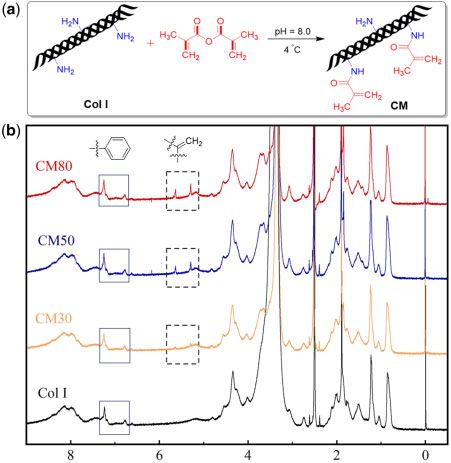
The effect of collagen hydrogels on chondrocyte behaviors                                                                                                     5

                                                                                                                                                                   Downloaded from https://academic.oup.com/rb/article/8/4/rbab030/6312113 by guest on 19 September 2021
Figure 2. The storage modulus of hydrogels at different frequencies (1, 2 and 5 Hz) (a), the degradation curves of hydrogels measured in PBS containing 50 mg/ml
collagenase I at 37 C (b) and SEM images of freeze-dried hydrogels (c) (*P < 0.05, **P < 0.01, ***P < 0.001).

contributes to the stable structure of hydrogels and slows down the               result of live/dead staining was shown in Fig. 3c, and it indicated
rate of degradation.                                                              that chondrocytes survived well in these hydrogels. The results sug-
                                                                                  gested that these hydrogels were all biocompatible and the CM
Chondrocytes culture in vitro                                                     hydrogels might be more suitable for cell survival and proliferation
Cell survival and proliferation                                                   compared to Col I hydrogels.
In order to evaluate the biocompatibility of hydrogels as tissue engi-
neering scaffolds, live/dead staining was performed for observation               Cytoskeleton staining
of cell survival in hydrogels and CCK-8 test was performed for                    Cell adhesion and spreading to the ECM is critical for tissue devel-
quantitative analysis of cell proliferation. The appearance of cell/hy-           opment and it directs cell survival, proliferation and the expression
drogel constructs after cultured for different intervals in vitro was             of differentiation phenotypes [38, 39]. The cytoskeleton staining re-
shown in Fig. 3a. It can be seen that the Col I hydrogels contracted              sult was shown in Fig. 3d and it demonstrated that all these hydro-
severely with the extension of culture time. However, the CM
                                                                                  gels could promote chondrocytes adhesion, spreading and
hydrogels did not contract obviously for up to 3 weeks of culture. It
                                                                                  proliferation with the extension of culture time. Peptide sequences
is reported that the movement of cells in the hydrogels may generate
                                                                                  on collagen, including RGD and GFOGER, can provide adhesion
a large tension field and exert traction forces to hydrogels in order
                                                                                  sites for cells [40, 41]. It can be speculated that the modification of
to push themselves outward, then causing significant contraction of
                                                                                  collagen in this study may not lead to a reduction of cell adhesion
the hydrogels [34]. It indicated that the chemical crosslinking helped
                                                                                  sites and has almost no bad effect on cell adhesion and spreading.
to resist cell-mediated contraction of hydrogels in this study. The re-
sult of CCK-8 test demonstrated that the proliferation of chondro-
cytes in CM hydrogels was better than in Col I hydrogels as shown                 Biochemical assay and chondrogenic gene expression
in Fig. 3b. Some studies have showed that cells ceased dividing when              The content of sGAG and DNA in cell/hydrogel constructs was mea-
gels contracted, which may be attributed to the fact that the contrac-            sured to detect ECM secretion of chondrocytes in different hydro-
tion of gels greatly reduced the space for cell growth and the cells              gels. The result showed that the value of sGAG/DNA in CM30
stopped growing due to contact inhibition [35, 36]. The contracted                hydrogels was statistically higher than other groups as shown in Fig.
hydrogels cannot provide an environment beneficial for the growth                 4g, which meant that chondrocytes in CM30 hydrogels had a
and proliferation of the encapsulated cells as reported [37]. The                 greater ability to secrete glycosaminoglycans in vitro. The CM30
6 Dong et al.
Downloaded from https://academic.oup.com/rb/article/8/4/rbab030/6312113 by guest on 19 September 2021
Figure 3. The appearance of cell/hydrogel constructs after being cultured for 1, 3 and 7 days in vitro (a), CCK-8 test for the proliferation of chondrocytes encapsu-
lated in hydrogels (b), the images of live/dead staining (c) and cytoskeleton staining (d) (**P < 0.01, ***P < 0.001).
hydrogels could resist cell-mediated contraction though they were Col I and CM80 hydrogels, while CM30 and CM50 hydrogels might
minimally modified, which allowed them to maintain a more porous be beneficial for hyaline cartilage formation in vitro.
microstructure that was beneficial for nutrient transport than CM50
and CM80. The severe contraction of Col I hydrogels may adversely Histological staining
affect the normal metabolic activity of chondrocytes as reported The histological staining was performed to observe chondrocyte
[35–37]. morphology and cartilage-specific ECM secretion after cell/hydrogel
The gene expression level associated with chondrogenesis [type II constructs were cultured for 2 or 3 weeks. The chondrocytes in all
collagen (Col II), aggrecan (AGG), and Sox9] and chondrocytes de- hydrogels showed round morphology by H&E staining as shown in
differentiation [type I collagen (Col I), type X collagen (Col X)] was Fig. 4a, but more cartilage lacunas were observed in CM30 and
analyzed by real-time PCR and the results were shown in Fig. 4b–f. CM50 hydrogels. Meanwhile, it can be found that lots of chondro-
The results showed that cells in CM30 and CM50 hydrogels had cytes aggregated at the edge of Col I hydrogels. The possible reason
higher gene expression level of key articular marker gene of Col II for the concentration of chondrocytes at the edge of Col I hydrogels
and AGG. Some studies have shown that cells synthesized little colla- was that the hydrogel contraction led to densification of the collagen
gen in contracted hydrogels [42], so the lowest Col II and AGG gene fibers, which limited the cell growth space and the supply of
expression level of cells in Col I hydrogels may be attributed to the nutrients to the inside of the gel [43]. Secreted ECM was detected by
severe contraction of hydrogels. The gene expression level of Sox 9 TB staining and SO staining as shown in Fig. 4a, and the results
had no significant difference between all groups. The cells in CM80 demonstrated that there was more ECM secreted in CM30 and
hydrogels had the highest gene expression level of Col I at 2 weeks, CM50 hydrogels. Similar to the results of H&E staining, the se-
and the expression level significantly decreased in all groups when creted matrix in Col I hydrogels was also detected to concentrate at
the culture duration was extended to 3 weeks. It indicated that the the edge of the hydrogel. The internal structure of hydrogels can in-
chondrocytes in CM80 hydrogels may be more prone to dedifferenti- fluence the matrix secretion of cells [19]. The CM80 hydrogels may
ation than in other groups due to the higher expression of Col I at be not convenient for nutrient transport due to the lamellar structure
the early culture duration. The gene expression level of Col X was resulted from the higher crosslinking density compared with others,
significantly higher in cells encapsulated in Col I and CM80 hydro- which caused the lower ECM synthesis. These results in vitro sug-
gels than that in CM30 and CM50 hydrogels at 3 weeks, and the gested that the CM30 and CM50 hydrogels were more suitable for
value of P between CM30 and CM80 hydrogels was 0.0051. These the maintenance of the chondrocyte phenotype and there was more
findings indicated that chondrocytes were prone to hypertrophy in ECM secretion compared to Col I and CM80 hydrogels.The effect of collagen hydrogels on chondrocyte behaviors 7
Downloaded from https://academic.oup.com/rb/article/8/4/rbab030/6312113 by guest on 19 September 2021
Figure 4. Histological staining (a), real-time qPCR quantification of gene expression associated with chondrogenesis (Col II (b), AGG (c) and Sox 9 (d)) or chondro-
cytes dedifferentiation (Col I (e), Col X (f)) , and quantitative determination of sGAG/DNA (g) of cell/hydrogel constructs cultured in vitro for 2 and 3 weeks
(*P < 0.05, **P < 0.01, ***P < 0.001).
Chondrocytes culture in vivo showed that Col I and CM30 hydrogels both contracted with the ex-
Biochemical assay tension of culture time and the former was more severe. The con-
Glycosaminoglycans are primary components of cartilage ECM traction of CM30 may be attributed to the mechanical force of cells
[44]. The highly sulfated glycosaminoglycans are covalently bound on the gels and the complex mechanical environment in vivo. Cells
to the core proteins of proteoglycans, which are crucial for cartilage would reorganize collagen fibers to form a non-covalent stable
mechanical property [45]. The appearance of hydrogels in Fig. 5a structure resulting in the contraction of gels and smaller gaps8 Dong et al.
Downloaded from https://academic.oup.com/rb/article/8/4/rbab030/6312113 by guest on 19 September 2021
Figure 5. The appearance of cell/hydrogel constructs after a certain culture time in vivo (a), quantitative determination of sGAG/DNA (b), H&E staining (c), TB
staining (d) and SO staining (e) of cell/hydrogel constructs after being cultured in vivo for 1, 2 and 4 weeks (*P < 0.05, **P < 0.01, ***P < 0.001).
between adjacent collagen fibers [46]. The contraction of hydrogels 5d and e) indicated that chondrocytes in CM hydrogels secreted a
may lead to enhanced mechanical interaction between the cells and large amount of ECM, and the ECM-specific staining was more ob-
the scaffold materials. vious than that in Col I hydrogels. However, a small amount of car-
The value of sGAG/DNA can reflect the cartilage-specific ECM tilage lacunas and matrix secretion were observed in the Col I
secretory ability of chondrocytes. In this study, the content of sGAG hydrogels. These results suggested that there was more ECM secre-
and DNA in hydrogels was measured separately after being cultured tion in CM hydrogels compared to that in Col I hydrogels, and it is
for 1, 2 and 4 weeks in vivo. The result of sGAG/DNA in Fig. 5b in- consistent with the result of sGAG/DNA. Previous studies have
dicated that the ECM secretion ability of chondrocytes in CM demonstrated that photo-crosslinked collagen hydrogels can pro-
hydrogels was statistically higher than that in Col I hydrogels, and mote mesenchymal stem cell differentiation toward chondrogenesis
the P value between Col I and CM50 hydrogels was 0.00096 at by inhibiting the expression of contraction-related signaling path-
4 weeks. The value of sGAG/DNA in CM50 and CM80 hydrogels ways in vivo, while the contraction of collagen hydrogels can cause
was significantly higher than that in CM30 hydrogels at 4 weeks, dedifferentiation by exerting mechanical stimulation on the cells
and the P value between CM30 and CM50 hydrogels was 0.0094. and activating these signaling pathways [47]. Mechanical effects of
These results demonstrated that the hydrogel contraction in vivo the environment on cells regulated cell adhesion, migration, gene
limited the maintenance of the chondrocyte phenotype and discour- expression and fate [48]. Chondrocytes respond to mechanical over-
aged chondrocytes from secreting-specific matrix. load by disrupting the composition and structure of the ECM, but
sustained mechanical overload can damage the normal function of
Histological staining chondrocytes [49]. In this study, the severe contraction of hydrogels
The results of HE, TB and SO staining in vivo experiments were may promote the hypertrophy trend of chondrocytes by reducing
shown in Fig. 5. The H&E staining (Fig. 5c) showed that there the ability to maintain their phenotype due to the excessive mechan-
were significant cartilage lacunas in CM hydrogels in the later ical stimulation exerted on the chondrocytes. It can be speculated
stages compared to that in Col I hydrogels when the implantation that a certain degree of modification of collagen to introduce chem-
time was extended to 4 weeks. It can be seen that there were a large ically crosslinkable groups was beneficial for the maintenance of
number of cell clusters in the CM hydrogels, which may be caused chondrocytes phenotype due to the limitation of hydrogel
by cell migration and proliferation. The TB and SO staining (Fig. contraction.The effect of collagen hydrogels on chondrocyte behaviors 9
Conclusions 16. Yang J, Li Y, Liu Y et al. Influence of hydrogel network microstructures
on mesenchymal stem cell chondrogenesis in vitro and in vivo. Acta
In this study, the modified collagen was prepared by controlling the
Biomater 2019;91:159–72.
ratio of MA to Col I. The CM hydrogels had stronger mechanical 17. Yuan L, Li B, Yang J et al. Effects of composition and mechanical property
properties and slower degradation rate as DS of CM increased. CM of injectable collagen i/ii composite hydrogels on chondrocyte behaviors.
hydrogels were able to resist cell-mediated contraction better than Tissue Eng A 2016;22:899–906.
Col I hydrogels both in vitro and in vivo. CM30 and CM50 hydro- 18. Fang M, Goldstein EL, Matich EK et al. Type I collagen self-assembly: the
gels promoted chondrocyte ECM secretion in vitro, while CM50 roles of substrate and concentration. Langmuir 2013;29:2330–8.
and CM80 had better ability to promote chondrocyte ECM secre- 19. Balakrishnan B, Banerjee R. Biopolymer-based hydrogels for cartilage tis-
sue engineering. Chem Rev 2011;111:4453–74.
tion in vivo. In summary, the MA-modified collagen showed some
20. Hwang CM, Sant S, Masaeli M et al. Fabrication of three-dimensional po-
excellent properties and could be an ideal candidate for tissue engi-
rous cell-laden hydrogel for tissue engineering. Biofabrication 2010;2:
neering scaffolds. 035003.
21. Lien SM, Ko LY, Huang TJ. Effect of pore size on ECM secretion and cell
growth in gelatin scaffold for articular cartilage tissue engineering. Acta
Downloaded from https://academic.oup.com/rb/article/8/4/rbab030/6312113 by guest on 19 September 2021
Funding Biomater 2009;5:670–9.
This work was sponsored by the National Key Research and Development 22. Ranucci CS, Kumar A, Batra SP et al. Control of hepatocyte func-
Program of China (2019YFA0110600), Science and Technology Support tion on collagen foams: sizing matrix pores toward selective induc-
Program of Sichuan Province (2019YJ0161) and Guangxi Key Research and tion of 2-D and 3-D cellular morphogenesis. Biomaterials 2000;21:
Development Plan (GuikeAB16450003). 783–93.
23. Griffon DJ, Sedighi MR, Schaeffer DV et al. Chitosan scaffolds: intercon-
Conflict of interest statement. None declared.
nective pore size and cartilage engineering. Acta Biomater 2006;2:
313–20.
24. Hadjipanayi E, Mudera V, Brown RA. Close dependence of fibroblast pro-
References liferation on collagen scaffold matrix stiffness. J Tissue Eng Regen Med
1. Athanasiou KA, Darling EM, Hu JC. Articular cartilage tissue engineer- 2009;3:77–84.
ing. E Biomed J Regen Med 2010;1:99–114. 25. Branco da Cunha C, Klumpers DD, Li WA et al. Influence of the stiffness
2. Temenoff JS, Mikos AG. Review: tissue engineering for regeneration of ar- of three-dimensional alginate/collagen-I interpenetrating networks on fi-
ticular cartilage. Biomaterials 2000;21:431–40. broblast biology. Biomaterials 2014;35:8927–36.
3. Huey DJ, Hu JC, Athanasiou KA. Unlike bone, cartilage regeneration 26. Karamichos D, Brown RA, Mudera V. Collagen stiffness regulates cellular
remains elusive. Science 2012;338:917–21. contraction and matrix remodeling gene expression. J Biomed Mater Res
4. Sadeghi-Avalshahr A, Nokhasteh S, Molavi AM et al. Synthesis and char- A 2007;83:887–94.
acterization of collagen/PLGA biodegradable skin scaffold fibers. Regen 27. Hong Y, Song H, Gong Y et al. Covalently crosslinked chitosan hydrogel:
Biomater 2017;4:309–14. properties of in vitro degradation and chondrocyte encapsulation. Acta
5. Revell CM, Athanasiou KA. Success rates and immunologic responses of Biomater 2007;3:23–31.
autogenic, allogenic, and xenogenic treatments to repair articular cartilage 28. Feng Q, Zhu M, Wei K et al. Cell-mediated degradation regulates human
defects. Tissue Eng B Rev 2009;15:1–15. mesenchymal stem cell chondrogenesis and hypertrophy in MMP-sensitive
6. Zhang L, Yuan T, Guo L et al. An in vitro study of collagen hydrogel to in- hyaluronic acid hydrogels. PLoS One 2014;9:e99587.
duce the chondrogenic differentiation of mesenchymal stem cells. J 29. Brodsky B, Ramshaw JAM. The collagen triple-helix structure. Matrix
Biomed Mater Res A 2012;100:2717–25. Biol 1997;15:545–54.
7. Cai G, Wang J, Qian K et al. Extremely stretchable strain sensors based on 30. Ni Y, Tang Z, Yang J et al. Collagen structure regulates MSCs behavior
conductive self-healing dynamic cross-links hydrogels for human-motion by MMPs involved cell-matrix interactions. J Mater Chem B 2018;6:
detection. Adv Sci (Weinh) 2017;4:1600190. 312–26.
8. Rowland CR, Colucci LA, Guilak F. Fabrication of anatomically-shaped 31. Tian Z, Liu W, Li G. The microstructure and stability of collagen hy-
cartilage constructs using decellularized cartilage-derived matrix scaf- drogel cross-linked by glutaraldehyde. Polym Degrad Stabil 2016;130:
folds. Biomaterials 2016;91:57–72. 264–70.
9. Cheng NC, Estes BT, Young TH et al. Genipin-crosslinked cartilage-de- 32. Saito H, Taguchi T, Kobayashi H et al. Physicochemical properties of gel-
rived matrix as a scaffold for human adipose-derived stem cell chondro- atin gels prepared using citric acid derivative. Mat Sci Eng C Mater 2004;
genesis. Tissue Eng A 2013;19:484–96. 24:781–5.
10. Corin KA, Gibson LJ. Cell contraction forces in scaffolds with varying 33. Yan M, Jiang X, Wang G et al. Preparation of self-assembled collagen fi-
pore size and cell density. Biomaterials 2010;31:4835–45. brillar gel from tilapia skin and its formation in presence of acidic polysac-
11. Lee CR, Grodzinsky AJ, Spector M. The effects of cross-linking of colla- charides. Carbohydr Polym 2020;233:115831.
gen-glycosaminoglycan scaffolds on compressive stiffness, chondrocyte- 34. Harris A, Wild P, Stopak D. Silicone rubber substrata: a new wrinkle in
mediated contraction, proliferation and biosynthesis. Biomaterials 2001; the study of cell locomotion. Science 1980;208:177–9.
22:3145–54. 35. Sarber R, Hull B, Merrill C et al. Regulation of proliferation of fibroblasts
12. Sung HW, Huang RN, Huang LLH et al. In vitro evaluation of cytotoxic- of low and high population doubling levels grown in collagen lattices.
ity of a naturally occurring cross-linking reagent for biological tissue fixa- Mech Ageing Dev 1981;17:107–17.
tion. J Biomater Sci Polym Ed 1999;10:63–78. 36. Weinberg CB, Bell E. Regulation of proliferation of bovine aortic endothe-
13. Brinkman WT, Nagapudi K, Thomas BS et al. Photo-cross-linking of type lial cells, smooth muscle cells, and adventitial fibroblasts in collagen latti-
I collagen gels in the presence of smooth muscle cells: mechanical proper- ces. J Cell Physiol 1985;122:410–4.
ties, cell viability, and function. Biomacromolecules 2003;4:890–5. 37. Yang K, Sun J, Wei D et al. Photo-crosslinked mono-component type
14. Lu J, Lin X, Jiang B et al. Preparation and characterization of collagen by II collagen hydrogel as matrix to induce chondrogenic diffrentiation of
hydrogel formation method. Key Eng Mater 2005;288–289:377–80. bone marrow mesenchymal stem cells. J Mater Chem B 2017;10:
15. Van DB, An I, Bogdanov B et al. Structural and rheological properties of C7TB02348K.
methacrylamide modified gelatin hydrogels. Biomacromolecules 2000;1: 38. Arcangelis AD, Georges-Labouesse E. Integrin and ECM functions: roles
31–8. in vertebrate development. Trends Genet 2000;16:389–95.10 Dong et al.
39. Danen EH, Sonnenberg A. Integrins in regulation of tissue development sensitive poly(ethylene glycol) hydrogels with localized transforming
and function. J Pathol 2003;200:471–80. growth factor beta3. Acta Biomater 2019;93:97–110.
40. Mineur P, Guignandon A, Lambert CA et al. RGDS and DGEA-induced 45. Kraan PMVD, Vitters EL, Vries BJD et al. High susceptibility of human
[Ca2þ]i signalling in human dermal fibroblasts. Biochim Biophys Acta articular cartilage glycosaminoglycan synthesis to changes in inorganic
2005;1746:28–37. sulfate availability. J Orthop Res 1990;8:565–571.
41. Knight CG, Morton LF, Peachey AR et al. The collagen-binding A- 46. Guidry C, Grinnell F. Contraction of hydrated collagen gels by fibroblasts:
domains of integrins alpha(1)beta(1) and alpha(2)beta(1) recognize the evidence for two mechanisms by which collagen fibrils are stabilized. Coll
same specific amino acid sequence, GFOGER, in native (triple-helical) col- Relat Res 1987;6:515–29.
lagens. J Biol Chem 2000;275:35–40. 47. Yang K, Sun J, Guo Z et al. Methacrylamide-modified collagen hydrogel
42. Nusgens B, Merrill C, Lapiere C et al. Collagen biosynthesis by cells in a with improved anti-actin-mediated matrix contraction behavior. J Mater
tissue equivalent matrix in vitro. Coll Relat Res 1984;4:351–63. Chem B 2018;6:7543–55.
43. Yao Y, Wang P, Li X et al. A di-self-crosslinking hyaluronan-based hydro- 48. Gallant ND, Michael KE, Garcia AJ. Cell adhesion strengthening: contri-
gel combined with type I collagen to construct a biomimetic injectable car- butions of adhesive area, integrin binding, and focal adhesion assembly.
tilage-filling scaffold. Acta Biomater 2020;111:197–207. Mol Biol Cell 2005;16:4329–40.
44. Schneider MC, Chu S, Randolph MA et al. An in vitro and in vivo compari- 49. Bader DL, Salter DM, Chowdhury TT. Biomechanical influence of carti-
Downloaded from https://academic.oup.com/rb/article/8/4/rbab030/6312113 by guest on 19 September 2021
son of cartilage growth in chondrocyte-laden matrix metalloproteinase- lage homeostasis in health and disease. Arthritis 2011;2011:979032.You can also read