Structured Waters Mediate Small Molecule Binding to G-Quadruplex Nucleic Acids
←
→
Page content transcription
If your browser does not render page correctly, please read the page content below

pharmaceuticals

Article
Structured Waters Mediate Small Molecule Binding to
G-Quadruplex Nucleic Acids
Stephen Neidle
                                          The School of Pharmacy, University College London, 29-39 Brunswick Square, London WC1N 1AX, UK;
                                          s.neidle@ucl.ac.uk

                                          Abstract: The role of G-quadruplexes in human cancers is increasingly well-defined. Accordingly,
                                          G-quadruplexes can be suitable drug targets and many small molecules have been identified to date
                                          as G-quadruplex binders, some using computer-based design methods and co-crystal structures. The
                                          role of bound water molecules in the crystal structures of G-quadruplex-small molecule complexes
                                          has been analyzed in this study, focusing on the water arrangements in several G-quadruplex
                                          ligand complexes. One is the complex between the tetrasubstituted naphthalene diimide compound
                                          MM41 and a human intramolecular telomeric DNA G-quadruplex, and the others are in substituted
                                          acridine bimolecular G-quadruplex complexes. Bridging water molecules form most of the hydrogen-
                                          bond contacts between ligands and DNA in the parallel G-quadruplex structures examined here.
                                          Clusters of structured water molecules play essential roles in mediating between ligand side chain
                                          groups/chromophore core and G-quadruplex. These clusters tend to be conserved between complex
                                          and native G-quadruplex structures, suggesting that they more generally serve as platforms for
                                          ligand binding, and should be taken into account in docking and in silico studies.

                                          Keywords: G-quadruplex; ligands; naphthalene diimides; acridines; crystal structures; water clusters;
                                          hydrogen bonding


Citation: Neidle, S. Structured
Waters Mediate Small Molecule
Binding to G-Quadruplex Nucleic           1. Introduction
Acids. Pharmaceuticals 2022, 15, 7.            G-quadruplexes (GQs) are higher-order four-stranded structures that can form in DNA
https://doi.org/10.3390/
                                          and RNA sequences by the folding of repetitive short guanine (G)-tracts [1–4]. These are
ph15010007
                                          typically interspersed with short runs of general sequence. The G-tracts self-associate by
Academic Editor:                          Hoogsteen hydrogen bonding to form guanine (G)-quartets, several of which stack on one
Alfredo Berzal-Herranz                    another to form a G-quadruplex (GQ). The quartets are held together by the phosphodiester
                                          backbones, and by the general sequences, which typically form extra-helical loops. GQ
Received: 29 November 2021
                                          folding and hence the nature of the loop can occur in several ways, such that the four
Accepted: 20 December 2021
                                          strands can be all-parallel, all-anti-parallel, or various combinations [2,5]. GQ prevalence
Published: 22 December 2021
                                          in the human genome is non-random [6,7]. In eukaryotic cells, they occur in telomeres [8,9]
Publisher’s Note: MDPI stays neutral      and are over-represented in genomic promoter [10–14] and untranslated sequences [15–17],
with regard to jurisdictional claims in   especially in genes and pathways involved in cancer initiation and progression [14,18–22].
published maps and institutional affil-   GQ-forming sequences have been found in the genomes of many organisms, ranging
iations.
                                          from viruses [23–27] to bacteria [28–31] and malaria [32–34]. GQs have been visualized in
                                          fixed [35–38] and in live cells [39–42], where their existence may be more than transient,
                                          with several roles in gene function [3,4,12]; for example, GQ folding is associated with sites
                                          of active transcription and precedes transcription itself [13,22].
Copyright:     © 2021 by the author.
Licensee MDPI, Basel, Switzerland.
                                               The presence of GQ sequences in the promoters of oncogenes, such as hTERT, MYC,
This article is an open access article
                                          KRAS, BCL2, and c-KIT as well as their potential for destabilizing telomere maintenance
distributed under the terms and
                                          in cancer cells (and their involvement in replication and genomic instability [12,43–45]),
conditions of the Creative Commons        has focused attention on GQs as promoter or telomeric therapeutic targets in viral and
Attribution (CC BY) license (https://     bacterial diseases as well as human cancers. The strategy of stabilizing them with appropriate
creativecommons.org/licenses/by/          small-molecule compounds has resulted in many chemically diverse chemotypes being
4.0/).

Pharmaceuticals 2022, 15, 7. https://doi.org/10.3390/ph15010007                                  https://www.mdpi.com/journal/pharmaceuticals
Pharmaceuticals 2022, 15, 7                                                                                                                                      2 of 18

                                      investigated, notably against oncogene GQ promoter targets [12,46,47]. Formation of high-
                                      affinity GQ-ligand complexes within promoters has been demonstrated to reduce or even
                                      abolish transcription of a target gene or genes, see for example refs. [48–58], which can result in
                                      cell growth inhibition and in vivo anti-cancer activity in animal models of human cancers [59].
                                                 Most of these GQ-binding compounds share common structural features of planar het-
                                      eroaromatic groups and side chains carrying cationic groups, albeit in a wide variety of
                                      chemotypes [60–66]. Structure-activity studies have optimized activity for numerous series of
                                      these compounds, for for example several libraries of acridine [67–76] and naphthalene di-
                                      imide [18,19,77–94] derivatives. The lead naphthalene diimide derivatives CM03 and SOP1812
                                      (Figure 1) show evidence of target engagement and in vivo anti-tumor activity [18,19]. Crys-
                                      tallographic and NMR studies have provided detailed information on GQ-ligand interactions
                                      in these and other GQ-ligand complexes [50,51,71,74–76,80,81,95–100], some of which have
                                      been used in several hit-to-lead optimization projects [18,19,50,81,90,97,99]. Quantitative and
                                      semi-quantitative computer modeling methods including docking procedures [101–111] have
                                      also been extensively employed to screen virtual libraries, with the aims of aiding optimization
                                      and identifying plausible new chemotypes and lead compounds for future
                              Pharmaceuticals 2022, 14, x FOR PEER REVIEW
                                                                                                                       drug development
                                                                                                                  3 of 20
                                      and eventual clinical trial.

                                                        Figure 1. Structures of the tri-substituted naphthalene diimide derivatives (CM03), two tetra-sub-
                                   Figure 1. Structures            of the
                                                    stituted derivatives (MM41 tri-substituted
                                                                                 and SOP1812), and thenaphthalene           diimide
                                                                                                        trisubstituted acridine compoundderivatives
                                                                                                                                         BRACO19.          (CM03), two
                                                    CM03: 2,7-bis(3-morpholinopropyl)-4-((2-(pyrrolidin-1-yl)ethyl)amino) benzo[lmn][3,8] phenan-
                                   tetra-substitutedthroline-1,3,6,8(2H,7H)-tetraone;
                                                        derivatives (MM41 and                 SOP1812), and the trisubstituted acridine compound
                                                                                        SOP1812: 2,7-bis(3-morpholinopropyl)- 4-((2-(pyrrolidin-1-
                                   BRACO19. CM03:        2,7-bis(3-morpholinopropyl)-4-((2-(pyrrolidin-1-yl)ethyl)amino)
                                                    yl)ethyl)amino)-9-(4-(pyrrolidin-1-ylmethyl)phenyl)        benzo[lmn][3,8]phenanthroline-1,3,6,8-    benzo[lmn][3,8]
                                                    (2H,7H)-tetraone); MM41: (4,9-bis((3-(4-methylpiperazin-1-yl)propyl)amino)-2,7-bis(3-morpho-
                                   phenanthroline-1,3,6,8(2H,7H)-tetraone;                SOP1812: 2,7-bis(3-morpholinopropyl)-
                                                    linopropyl) benzo[lmn][3,8] phenanthroline-1,3,6,8(2H,7H)-tetraone);  BRACO19: 3,6-bis(3-pyrroli- 4-((2-(pyrrolidin-1-
                                                    din-1-ylpropionamido)-9-(4-dimethylaminophenylamino) acridine.
                                   yl)ethyl)amino)-9-(4-(pyrrolidin-1-ylmethyl)phenyl)                     benzo[lmn][3,8] phenanthroline-1,3,6,8-(2H,7H)-
                                   tetraone); MM41: (4,9-bis((3-(4-methylpiperazin-1-yl)propyl)amino)-2,7-bis(3-morpholinopropyl)
                                   benzo[lmn][3,8] phenanthroline-1,3,6,8(2H,7H)-tetraone); BRACO19:                                           3,6-bis(3-pyrrolidin-1-
                                   ylpropionamido)-9-(4-dimethylaminophenylamino) acridine.
Pharmaceuticals 2022, 15, 7                                                                                         3 of 18

                                    Several high-resolution crystallographic studies of native GQs [112–115] and GQ-
                              ligand complexes [71,74,75,80,81,98,100,116,117] have reported that water molecules are
                              intimately associated with GQ sites and with bound ligand. It is well-established for
                              DNA-, RNA- [118], and protein-ligand complexes [119–123] that water molecules can play
                              critical target-ligand mediation roles. The present work analyzes water arrangements
                              in several GQ-ligand complexes (PDB id 3CE5, 3NZ7, and 3UYH) [74,81,100] that are of
                              sufficient resolution to give confidence in the significance of the arrangements, to better
                              understand the role and the consequences of discrete water molecules associated with
                              ligand binding. The naphthalene diimide complex [81] was also chosen since it involves the
                              tetra-substituted compound MM41 (Figure 1). This is the direct precursor of two other more
                              recently designed naphthalene diimide compounds, CM03 and SOP1812 [18,19] (Figure 1),
                              which are currently in pre-clinical development stages. Hydration features of this complex
                              and other complexes examined here, it is suggested, have wider implications for GQ-based
                              drug design and hit/lead selection.

                              2. Results
                                    Only the crystal structures containing acridine, berberine, and naphthalene diimide
                              (ND) derivatives fulfilled the acceptance criteria summarized in the preceding section. Two
                              co-crystal structures are available (Table 1) for the tetrasubstituted naphthalene diimide
                              compound MM41 (Figure 1) complexed to intramolecular human telomeric GQs, for which
                              it has high binding affinity. Structure PDB id 3UYH is at the higher resolution of the two
                              and consequently a greater number of ligand-associated water molecules were observed
                              in electron density maps and included in the final refined crystal structure [81]. Hence
                              it was chosen for further detailed analysis (Tables 1 and 2). Structure 3CDM [124] has
                              2 G-quadruplexes, 158 water molecules, and 4 substituted naphthalene diimides in the
                              asymmetric unit, i.e., 79 waters per G-quadruplex. However, few water molecules in
                              this structure are resolved in the vicinity of the two stacked naphthalene diimide ligands
                              compared to structure 3UYH and so this structure was not chosen for detailed analysis. In
                              addition, the nature of naphthalene diimide substituents in 3CDM is not directly relevant
                              to MM41 and hence not to CM03 or SOP1812, so this structure was not considered any
                              further in the present analysis.

                              2.1. MM41 Side Chain Contacts and Water Environment
                                   MM41 has two side chains terminating in N-methyl-piperazine groups and two with
                              terminal morpholino groups. Each of these groups can be assumed to be protonated at
                              physiological pH, with N-methyl-piperazine having a pK of 8.5 compared to the slightly less
                              basic morpholino group, with a pK of 9.2 [81]. Figure 2a shows a view of structure 3UYH
                              projected onto the planes of the G-quartets and the naphthalene diimide core, highlighting
                              the grooves of the GQ. Each MM41 side chain is positioned in or close to the mouth of
                              a GQ groove, although only three of the four end groups are actually situated within a
                              groove. The fourth, having a terminal morpholino ring, is oriented away from the quartet
                              plane and a detailed examination of the crystal structure has indicated that rotation of the
                              side chain to place the morpholino group into groove 4 is sterically hindered by the small
                              surface area of the naphthalene diimide core compared to that of the quartet [81].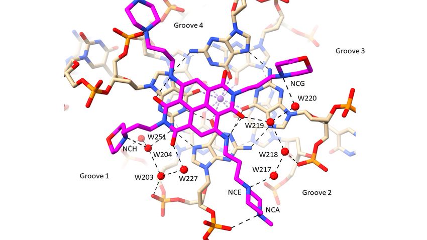
Pharmaceuticals 2022, 15, 7 4 of 18
Pharmaceuticals 2022, 14, x FOR PEER REVIEW 5 of 20
Table 1. G-quadruplex-small molecule crystal structures, taken from the Protein Data Bank, for which
at the minimum a first shell of water molecules around the DNA and ligand have been reported. The
number of water molecules associated with each complete G-quadruplex is quoted, as taken from the
11-mer biomolecular human
PDB entry. Tetrasubstituted
The structures naphthalene
discussed here (PDB ids 3CE5, 3NZ7
3CCO 2.20and 3UYH) are
28 highlighted124
in bold.
telomeric diimide
PDB Id G-Quadruplex Type TetrasubstitutedCompound naphthalene Resoln (Å) No. of Waters/AU Ref.
3CDM
3CE5
22-mer human telomeric
12-mer bimolecular human telomeric 3,6,9- trisubstituted
2.10 158 12474
diimide BRACO19
acridine 2.5 54
12-mer bimolecular Oxytricha nova
3NZ7
telomeric telomeric
Tetrasubstituted
3,6- naphthalene
disubstituted acridine, F substituents 1.10 187 100
4DA3 21-mer human 2.40 25 81
3NYP 12-mer bimolecular Oxytricha nova telomeric diimide MM41
3,6- disubstituted acridine, F substituents 1.18 176 100
3EM2 12-mer
12-mer bimolecular
bimolecular human
Oxytricha nova telomeric 3,6- disubstituted acridine 2.3 64 75
6S15 Pyridine derivative of berberine 1.70 23 98
3EQW telomeric
12-mer bimolecular Oxytricha nova telomeric 3,6- disubstituted acridine 2.2 66 75
3EUI 12-mer bimolecular Oxytricha nova telomeric 3,6- disubstituted acridine 2.2 159 75
3ERU 2.1.nova
12-mer bimolecular Oxytricha MM41 Side
telomeric Chain Contacts and Water
3,6- disubstituted acridineEnvironment 2.0 71 75
3ES0 12-mer bimolecular Oxytricha nova telomeric
MM41 has two 3,6-chains
side disubstituted acridine
terminating in 2.2
N-methyl-piperazine 56
groups 75
and two with
3ET8 12-mer bimolecular Oxytricha nova telomeric
terminal morpholino 3,6- disubstituted
groups. Each acridine
of these 2.45 51
groups can be assumed to be protonated at 75
3EUM 12-mer bimolecular Oxytricha nova telomeric pH, with3,6-
physiological disubstituted acridine
N-methyl-piperazine 1.78of 8.5 compared
having a pK 52 to the slightly
75
1L1H 12-mer bimolecular Oxytricha
lessnova telomeric
basic morpholino group,3,6- disubstituted
with acridine
a pK of 9.2 [81]. Figure1.75 2a shows a view
146 of structure
75
Tetrasubstituted naphthalene diimide
3UYH projected onto the planes of the G-quartets and the1.95 naphthalene diimide core, high-
3UYH 22-mer human telomeric 51 81
MM41
lighting the grooves of the GQ. Each MM41 side chain is positioned in or close to the
Tetrasubstituted naphthalene diimide
3T5E 22-mer human telomeric
mouth of a GQ groove, although BMSG-SH-4only three of the four 2.10 38
end groups are actually 80
situated
3CCO within
11-mer biomolecular human a groove. The
telomeric fourth, having
Tetrasubstituted naphthalenea terminal
diimide morpholino
2.20 ring, is oriented
28 away from
124
3CDM the quartet plane and
22-mer human telomeric a detailed
Tetrasubstituted examination
naphthalene diimide of the crystal
2.10 structure has158indicated 124that
4DA3 rotation of the side
21-mer human telomeric chain tonaphthalene
Tetrasubstituted place thediimide
morpholino
MM41 group2.40into groove 4 is
25 sterically 81
hin-
6S15 dered
12-mer bimolecular human by the small surface
telomeric area of of
Pyridine derivative the naphthalene diimide
berberine 1.70 core compared 23 to that of98the
quartet [81].
(a)
Figure 2. Cont.
Pharmaceuticals 2022, 14, x FOR PEER REVIEW 6 of 20
Pharmaceuticals 2022, 15, 7 5 of 18
(b)
(c)
Figure
Figure 2. Views
2. Views ofcrystal
of the the crystal structure
structure of MM41
of MM41 (with(with its carbon
its carbon atomsatoms colored
colored magenta)
magenta) boundbound
to to
a human intramolecular telomeric G-quadruplex, PDB id 3UYH. (a) The view is projected onto the
a human intramolecular telomeric G-quadruplex, PDB id 3UYH. (a) The view is projected onto the
G-quartet plane and shows the extent of overlap with the naphthalene diimide core chromophore.
G-quartet plane and shows the extent of overlap with the naphthalene diimide core chromophore.
The water molecules that are in direct or indirect contact with the MM41 molecule, are shown as
The water molecules
red spheres, withthat are in direct
hydrogen bondsorindicated
indirect contact withlines.
by dashed the MM41
This andmolecule, are shown
the subsequent as redwere
figures
spheres,
drawnwith hydrogen
using bonds indicated
the ChimeraX package by(https://www.cgl.ucsf.edu/chimerax/,
dashed lines. This and the subsequent last figures were drawn
accessed on 16 De-
using the ChimeraX
cember package
2021) [125]. (b) A(https://www.cgl.ucsf.edu/chimerax/,
view of the 3UYH complex looking into last accessed
groove on 16
1. The December
four water mole-
2021)cules are(b)shown
[125]. A viewthat
of are hydrogen
the 3UYH bonded
complex to theinto
looking morpholino
groove 1.group in this
The four watergroove and the
molecules areOAF
carbonyl
shown that areoxygen atombonded
hydrogen of the naphthalene diimidegroup
to the morpholino core. Ainnearby clusterand
this groove of water
the OAFmolecules
carbonyl is also
shown, embedded deep in the groove and adjacent to a TTA loop. (c) A view
oxygen atom of the naphthalene diimide core. A nearby cluster of water molecules is also shown, of the 3UYH complex
midway
embedded between
deep in the grooves
groove and2 and 3, highlighting
adjacent to a TTA the
loop.group
(c) Aof fourofwater
view molecules
the 3UYH hydrogen
complex midway bond-
ing to an N-methyl-piperazine and a morpholino group in these grooves.
between grooves 2 and 3, highlighting the group of four water molecules hydrogen bonding to an
N-methyl-piperazine and a morpholino group in these grooves.
All four basic end groups of MM41 have their protonated nitrogen atoms in hydro-
gen bond/electrostatic contact with atoms in the GQ grooves (Table 2). However, only two
Pharmaceuticals 2022, 15, 7                                                                                                     6 of 18

                              Table 2. Hydrogen bond interactions. (a) In structure 3UYH, involving the tetrasubstituted naph-
                              thalene diimide MM41, a human intramolecular telomeric G-quadruplex, and water molecules.
                              Hydrogen-bond distances are shown (d1-2 in Å), together with the reported crystallographic B factor
                              values (in Å2 ) for MM41-bound waters and associated MM41 and DNA atoms. MM41 atoms are
                              highlighted in bold red type. Waters in direct contact with MM41 atoms are highlighted in bold blue
                              type. Numbering is as in the PDB entry. (b) In structure PDB id 3CE5 [74], involving the trisubstituted
                              compound BRACO19, a human intermolecular bimolecular telomeric G-quadruplex, and water
                              molecules. Parameter definitions and color coding are as in Table 2a.

                                                                                (a)
                                                    Atom1             Atom2             d1-2        B Factor Atom1   B Factor Atom2
                                  Groove 1
                                                     NCH              W204              3.2               64               32
                                                     W204             W203              3.0               32               32
                                                     W203           OP2 dG10            2.9               32               32
                                                     W203             W227              3.4               32               45
                                                     W227             OAF               2.5               45               29
                                                     W251            OP2 dG9            2.9               27               30
                                                     W204            N2 dG4             2.9               32               20
                                                     W204             W251              2.8               32               27
                                  Groove 2
                                                     NCA            OP2 dT11            3.1               46               35
                                                     NCE              W217              2.7               40               46
                                                     W217             W218              2.6               46               48
                                                     W218             W219              3.0               48               43
                                                     W219             ODX               2.7               43               31
                                                     W218           OP2 dG16            3.4               48               45
                                  Groove 3
                                                     NCG              W220              2.9               48               50
                                                     W219             W220              2.9               43               50
                                  Groove 4
                                                     NCF             OP2 dG4            2.9               58               39
                                                                                (b)
                                                    Atom1             Atom2             d1-2        B Factor Atom1   B Factor Atom2
                                  Groove 1
                                                      N7              W52               2.7               11               21
                                                     W52             N3 dT24            2.9               21               12
                                                     W52             O6 dG5             3.0               21               18
                                                     W52              W53               3.1               21               33
                                                     W53             O2 dT24            3.3               33               18
                                                     W53               O52              3.1               33               14
                                  Groove 2
                                                     W56               N39              3.4               23               27
                                                      N17             W55               3.0               14               38
                                                     W55             O2 dT12            3.3               38               17
                                  Groove 4
                                                      N21            O4 dT24            3.0               13               17
                                                      N47             W41               3.0               21               28
                                                     W41              W44               2.9               28               29
                                                     W44            OP2 dG23            2.5               29               21
                                                     W41             N2 dG17            2.9               28               18

                                  All four basic end groups of MM41 have their protonated nitrogen atoms in hydrogen
                              bond/electrostatic contact with atoms in the GQ grooves (Table 2). However, only two of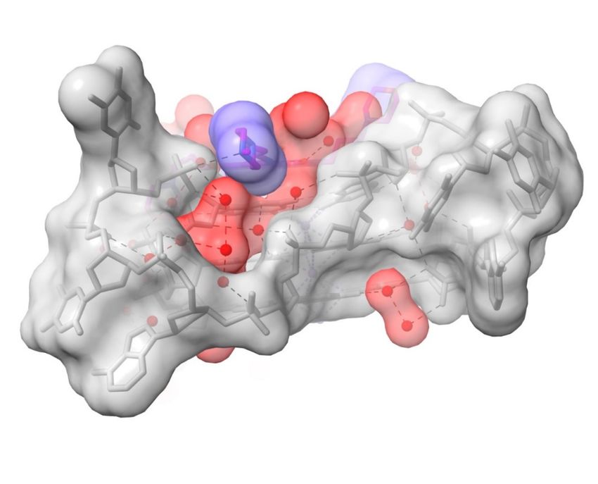
Pharmaceuticals 2022, 15, 7                                                                                                      7 of 18

                                      these contacts, each involving the terminal nitrogen atom of an N-methyl-piperazine group,
                                      has a direct nitrogen-phosphate group hydrogen bond interaction (N . . . OP distances
                                      of 2.9 and 3.1 į ). The three other end groups all have water contacts with ring nitrogen
                                      atoms (Figure 2b,c), which in the case of the morpholino groups, are presumed to be
                                      protonated. Groove 1 (Figure 2b) has the morpholino group positioned at the mouth of
                                      the groove. A small linear cluster of four water molecules extends from the morpholino
                                      basic nitrogen atom, with one water contacting two further waters, which contact with
                                      two neighboring phosphate oxygen atoms. One of these waters OP2 dG10 also contacts
                                      a fourth water molecule, which in turn contacts the adjacent oxygen substituent on the
                                      naphthalene diimide chromophore. The other water, contacting a phosphate oxygen atom
                                      (OP2 G9), also contacts and is thus the link to a water molecule in a second water network
                                      that fills the rest of this groove. The N-methyl-piperazine group in groove 2 (Figure 2c),
                                      which is situated at the mouth of the groove, has its terminal nitrogen atom (NCA) in
                                      close contact with a phosphate oxygen atom, OP2 dT11, suggesting that this nitrogen atom
                                      carries a proton. The inner piperazine ring nitrogen atom contacts another linear group of
                                      four water molecules which extends into groove 3 and terminates with a hydrogen bond
                                      contact with the second morpholino group. The second water in this array has a contact
                                      with a phosphate oxygen atom (OP2dG16), and the third is in hydrogen bond contact with
                                      carbonyl oxygen atom OAG of the ND core. The second N-methyl-piperazine ring, also
                                      situated at the mouth of groove 4, has a direct contact involving the outer ring nitrogen and
                                      a phosphate oxygen atom (Figure 2a), but does not have any associated water molecules.
                                            The above section describes water molecules involved in ligand contacts; other waters
                                      fill out the remaining space in the grooves (the non-completeness in some grooves is most
                                                                                                  ¯ with only a fraction of the potential
                                      likely due to limitations of the crystal structure at 1.95 Ä
                                      total number of water molecules located in electron density). Groove 2 is one of the more
                                      completely resolved grooves in terms of hydration (Figure 3), with waters embedded
Pharmaceuticals 2022, 14, x FOR PEER REVIEW
                                      deep into the groove. This complex array of 12 water molecules, of which the majority      9 of 20
                                                                                                        0         0
                                      are first-shell, hydrogen bond with phosphate oxygens, O4 and O5 atoms, and guanine
                                      base edges (which form the floor of the groove). The net effect is to maintain the relative
                                      positions of TTA loop and the groove.

                                    Figure 3. Surface representation of a view into groove 2 in the 3UYH complex. The N-methyl-
                                    piperazine substituent of MM41 is oriented end-on and is colored blue. Note that the groove space
                                    is filled out by water molecules, colored red. The semi-transparent surface of the G-quadruplex is
                                 Figure 3. Surface representation of a view into groove 2 in the 3UYH complex. The N-methyl-piper-
                                    colored grey.
                                 azine substituent of MM41 is oriented end-on and is colored blue. Note that the groove space is
                                 filled out by water molecules, colored red. The semi-transparent surface of the G-quadruplex is col-
                                 ored grey.

                                 2.2. MM41 and Water Mobility
                                      It is notable that there is remarkably little overlap between the ND four-ring core and
                                 the individual guanines in the top quartet, as seen in Figure 2a. The surface area of the
                                 ND core of MM41 is too small to allow simultaneous overlap with more than one guanine
Pharmaceuticals 2022, 15, 7                                                                                                 8 of 18

                              2.2. MM41 and Water Mobility
                                     It is notable that there is remarkably little overlap between the ND four-ring core and
                              the individual guanines in the top quartet, as seen in Figure 2a. The surface area of the
                              ND core of MM41 is too small to allow simultaneous overlap with more than one guanine
                              of the top G-quartet. Computational experiments were undertaken using two different
                              energy minimization protocols (in the ARGUSLAB and AVOGADRO packages) to assess
                              the effect of removing all the water molecules from the structure. Minimization in both
                              cases resulted in movement of the ND core by ca 1.5 Å to produce improved overlap with a
                              guanine base of the G-quartet. The cationic groups in the side chains tended to move closer
                              to the phosphate oxygen atoms. Attempts to dock the MM41 molecule onto the water-free
                              GQ using AutoDock Vina 1.1.2 as installed within the database G4LDB 2.2, resulted in a
                              series of almost equi-energetic poses in which three out of four side chains were positioned
                              away from the grooves. This was not pursued further.
                                     The side chain heterocyclic groups in MM41 have greater mobility than the ND core,
                              as revealed by their individual atomic temperature factors (see the PDB entry for 3UYH
                              and Table 2). The five nitrogen atoms in these terminal rings, which are hydrogen-bonded
                              to waters or phosphate groups, have a mean B value of 51 Ä      ¯ 2 , corresponding to a  of
                                   ¯
                              0.8 Ä. The water molecules in the mini cluster around and contacting the morpholino ring
                              in groove 1 have lower B values, with most in the range 27–32 Ä       ¯ 2 , corresponding to a
                                       ¯
                              of 0.6 Ä. The cluster of waters in groove 2/3 have slightly greater mobility.
                                     The extent to which water molecules located in the 3UYH crystal structure correspond
                              to those found in the native structure was examined by superimposing on 3UYH the
                              native crystal structure 1KF1 [126], overlaying the G-quartets (Figure 4a,b). Overlap of
                              the quartets was good, as expected. However, the loops in the MM41-bound structure
                              adopt distinct conformations compared to those in the native structure. Systematics of loop
                              conformations in GQ crystal structures have been previously reported [95] and will not be
                              further discussed here. Detailed comparison of water positions revealed that the cluster
                              of four waters at the mouth of groove 1 that mediate between ono the morpholino end
                              groups of MM41 and G-quadruplex is also present in the native structure (Figure 4a), with
                              distances between each pair of waters (i.e., a 3UYH water . . . .1KF1 water) 0.6–1.0 Ä     ¯ . Since
                                                                               ¯
                              these waters are mobile with a  of 0.6 Ä, they can be considered to likely occupy the
                              same space. Three conserved water molecules are also present at the mouth of groove 3
                              (Figure 4b), close to the second morpholino end group.

                              2.3. Water Mediation in Acridine-G-Quadruplex Structures
                                   The crystal structure (PDB id 3CE5) of the complex between the experimental drug
                              BRACO19 and a bimolecular human GQ [74], shows that, in common with the MM41
                              complex, the GQ has adopted a parallel topology. In both instances, the ligand is stacked
                              onto one end of the GQ, onto a terminal G-quartet. The BRACO19 molecule has three
                              cationic charges at physiological pH, one in each side chain pyrrolidino ring and one on the
                              central ring nitrogen atom in the acridine ring. None of these are directly hydrogen-bonded
                              to anionic phosphate groups. Instead (Table 2 and Figure 5a), they are hydrogen bonded to
                              water molecules. The waters hydrogen bonded to the pyrrolidino cationic nitrogen atoms
                              do eventually link indirectly to phosphate groups, via further water molecules. Water
                              molecule W52 hydrogen bonded to the acridine central ring nitrogen, appears to play
                              a crucial role, hydrogen bonding both to an O6 of a guanine from the adjacent stacked
                              G-quartet, and to N3 of a thymine in-plane with the acridine. W52 also hydrogen bonds to
                              W53, which in turn hydrogen bonds to the carbonyl oxygen atom of one of the amide side-
                              chains on BRACO19. The other amide group is trans to this and its amide nitrogen atom
                              hydrogen bonds to O4 of this thymine—this is the sole direct drug—GQ hydrogen bond,
                              with the remaining five being water-mediated. An overlay of the MM41 and BRACO19
                              complexes (Figure 5c) shows that several of the key water molecules are conserved between
                              the two structures, with distances between pairs of waters < 1 Å, using the argument
                              outlined in the previous section.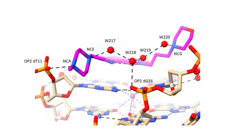
end groups of MM41 and G-quadruplex is also present in the native structure (Figure 4
                                           with distances between each pair of waters (i.e., a 3UYH water ….1KF1 water) 0.6–1.0
                                           Since these waters are mobile with a  of 0.6 Ǟ, they can be considered to likely occu
                                           the same space. Three conserved water molecules are also present at the mouth of groo
Pharmaceuticals 2022, 15, 7                3 (Figure 4b), close to the second morpholino end group.                  9 of 18

                                                                               (a)

                                                                               (b)
                                           Figure 4. Superposition
                              Figure 4. Superposition                   of the of
                                                           of the G-quartets   G-quartets
                                                                                   native and of MM41-complexed
                                                                                                 native and MM41-complexed
                                                                                                                       G-quadruplex G-quadruplex
                                                                                                                                       crystal       crys
                                           structures,
                              structures, 1KF1          1KF1 respectively,
                                                and 3UYH,      and 3UYH, respectively,
                                                                            viewed into viewed
                                                                                           groove 1.into
                                                                                                      Thegroove
                                                                                                            native1.structure
                                                                                                                     The native  structure
                                                                                                                              is colored    is colored lig
                                                                                                                                         light
                                           red, the ligand-bound
                              red, the ligand-bound    is cyan, and is
                                                                    thecyan,
                                                                         MM41andmolecule
                                                                                   the MM41    molecule
                                                                                            is shown      is shown
                                                                                                      magenta.       magenta.
                                                                                                                  Only          Only
                                                                                                                         the water    the water molecu
                                                                                                                                    molecules
                              in the groove are shown, colored as in their G-quadruplex structures. Those water molecules inmolecules
                                           in the groove    are shown,  colored   as in their G-quadruplex     structures.  Those  water   the        in t
                                           two structures that are < 1.0 Å to each other are enclosed in red circles. (a) Viewed into groove 1.
                              two structures that are
pears to play a crucial role, hydrogen bonding both to an O6 of a guanine from the adja-
cent stacked G-quartet, and to N3 of a thymine in-plane with the acridine. W52 also hy-
drogen bonds to W53, which in turn hydrogen bonds to the carbonyl oxygen atom of one
of the amide side-chains on BRACO19. The other amide group is trans to this and its am-
ide nitrogen atom hydrogen bonds to O4 of this thymine—this is the sole direct drug—
GQ hydrogen bond, with the remaining five being water-mediated. An overlay of the
Pharmaceuticals 2022, 15, 7 MM41 and BRACO19 complexes (Figure 5c) shows that several of the key water molecules 10 of 18
are conserved between the two structures, with distances between pairs of waters < 1 Å,
using the argument outlined in the previous section.
Pharmaceuticals 2022, 14, x FOR PEER REVIEW 12 of 20
(a)
(b)
(c)
Figure Figure 5. (a) View of the BRACO19 complex with a human telomeric bimolecular G-quadruplex, as
5. (a) View of the BRACO19 complex with a human telomeric bimolecular G-quadruplex, as
observed in the crystal structure PDB id 3CE5 [74], projected onto the acridine plane. The carbon
observed in the crystal
atoms of the ligand are structure PDB id
colored magenta 3CE5
and water[74], projected
molecules onto
are colored as the acridine
red spheres. plane. The carbon
Hydrogen
bonds are shown as dotted lines. Some water molecules not directly involved
atoms of the ligand are colored magenta and water molecules are colored as red spheres. Hydrogen in ligand interactions
have been omitted from this view to enhance clarity. (b) View of the complex involving a disubsti-
bonds are
tuted shown
acridineaswith
dotted lines.
a fluorine atomSome water
attached molecules
to each terminal not
side directly involved
chain pyrrolidino ring,inbound
ligandto interactions
have been omittednova
an Oxytricha from this viewG-quadruplex
bimolecular to enhance[100].clarity. (b)coding
Color Viewisofasthe complex
in other involving
figures, a disubstituted
with the flu-
acridineorine
withatoms colored yellow. (c) Overlay of structures 3UYH (G-quadruplex in cyan and MM41 in
a fluorine atom attached to each terminal side chain pyrrolidino ring, bound to an
magenta) and 3CE5 (G-quadruplex in red and BRACO19 in dark blue), superimposed on the G-
Oxytricha nova bimolecular
quartets, viewed G-quadruplex
into groove 1 of the 3UYH[100]. Color
structure. Watercoding is as
molecules in in
theother
groovefigures,
are coloredwith
as the fluorine
in their G-quadruplex structures. Those water molecules in the two structures
atoms colored yellow. (c) Overlay of structures 3UYH (G-quadruplex in cyan and MM41 in magenta) that are < 1.0 Å dis-
tance to each other are enclosed in red circles.
and 3CE5 (G-quadruplex in red and BRACO19 in dark blue), superimposed on the G-quartets,
viewed intoRemoval
groove of 1 ofthethe 3UYH
water structure.
molecules from Water molecules
this structure in the
followed bygroove
energy are colored as in their
minimiza-
tion, resulted
G-quadruplex in movement
structures. Thoseofwater
the acridine by ca 2.5
molecules in Å,
theenabling the acridine
two structures ring
that are nitrogenPharmaceuticals 2022, 15, 7 11 of 18
Removal of the water molecules from this structure followed by energy minimization,
resulted in movement of the acridine by ca 2.5 Å, enabling the acridine ring nitrogen
atom to directly contact the thymine ring substituents. Such an arrangement has been
observed in the series of co-crystal structures [75] involving disubstituted acridines with the
bimolecular anti-parallel GQ from Oxytricha nova (Table 2). These structures, exemplified by
the two high-resolution structures [100] with fluorine substituents in the pyrrolidino side
chains, have direct O2 thymine and N3 hydrogen-bond contacts with the acridine ring and
an amide carbonyl oxygen atom (Figure 5b). A water molecule is involved in mediating
between a side chain amide and a phosphate oxygen atom. However, the Oxytricha nova
complexes all have a distinct anti-parallel GQ topology, with the acridine constrained
within a tetranucleotide diagonal loop, with little room for any associated water molecules.
3. Discussion
The number of water molecules located in nucleic acid and protein crystal structures
is invariably less than the total present in the crystal lattice. Thus, crystal structures 3UYH
and 3CE5, whose reported water molecules are analyzed here, both have an estimated 56%
solvent, which corresponds to >400 water molecules. A small fraction of this, 51 and 54
water molecules, respectively, were observed in electron density maps [74,81]. These are
almost all first or second shell immobilized waters.
Three principal findings emerge from the current analysis:
1. The morpholino end groups of MM41, which are assumed to be basic in the buffering
conditions of the crystallization experiment and in biological solution, do not directly
contact the GQ. Hydrogen bonding/electrostatic interactions with negative backbone
phosphate groups were anticipated but were not observed. Instead, the basic ring
nitrogen in each morpholino group hydrogen bonds to one of a group of four water
molecules positioned in the mouth of the relevant grooves (1 and 3). The waters are
in hydrogen bond contact with backbone phosphates. Similarly, the basic pyrrolidino
side chain terminal groups of BRACO19 do not directly contact phosphate groups in
its GQ complex, with water mediation being observed in the crystal environment.
2. A nitrogen atom on both N-methyl-piperazine groups of MM41, by contrast, directly
hydrogen bonds to a backbone phosphate oxygen atom, implying greater basicity
than morpholino for this end group.
3. The water clusters associated with the two morpholino groups of MM41 are highly
conserved between the native and the MM41-bound GQ structures. There is also
conservation of a number of the ligand-associated waters between the MM41 and
BRACO19 structures, and by implication, between the native and BRACO19 struc-
tures.
We suggest that the conserved water clusters have relevance to the observed structure-
activity relationships for MM41 derivatives [81]. Thus, replacing the morpholino groups
with isosteric groups such as hexose or ether groups, which lack the morpholino hydrogen
bonding ability, results in an almost complete loss of GQ affinity and reduced biological
activity compared to MM41. It is also notable that none of the four side chains are deeply
embedded in the GQ grooves, and one might have therefore expected reduced GQ affinity
compared to analogues with longer side chains. This is not the case since, as observed
here, the short side chains are effectively captured by hydrogen bonded to the conserved
water clusters, which would have to be displaced by longer side chains. This would be at a
significant entropic cost. The strategy of replacing two of the four strongly basic end groups
characteristic of earlier ND compounds (see, for example refs. [55,57,79]), by less strongly
basic morpholino groups [18,19,81], has culminated in the design and evaluation of MM41,
and subsequently compounds CM03 and SOP1812 (Figure 1), which are currently being
assessed as pre-clinical candidates. The rationale for lowering the highly cationic nature of
these ND compounds is that this could improve cellular uptake and tumor distribution
while retaining GQ affinity. The present analysis has shown that this substitution hasPharmaceuticals 2022, 15, 7 12 of 18
preserved the water structure around the perimeter of the grooves in which the morpholino
groups bind, with no diminution of GQ affinity [90].
The mediated waters in the BRACO19 structure also have relevance to the observed
structure-activity relationships for BRACO-GQ interactions [73,74], and for BRACO19
biology. Although BRACO19 may not be further developed as an anti-cancer drug [127],
its activity against a variety of anti-viral GQ targets [128–130] suggests the likelihood of
further anti-viral analogue programs, for which the 3CE5 structure and its structured water
features are of direct relevance.
The present analysis, although limited to two crystal structures, demonstrates that
water molecules can play an active role in GQ-ligand recognition. This indicates that in
silico and docking studies of ligand-GQ binding need to take account of reliably located
explicit water molecules. It is concluded that their omission will lead to misleading
conclusions on low-energy ligand binding states and interactions. Many such studies
still tend to ignore the role of water and really require input from high-resolution crystal
structures or reliable and well-validated water modelling/simulations. This has been
recognized in several studies for example, refs. [118,131]. Prediction of water positions
and mobilities in ligand complexes can be made using molecular dynamics [131], although
this has only rarely been been used to date for GQ systems [132]. The prediction of water
positions having low mobility in nucleic acids by use of a specially generated water force
field together with statistical scoring has led to the development of an automated method,
termed “SPLASH’EM” (Solvation Potential Laid around Statistical Hydration on Entire
Macromolecules) [118], which has given results for duplex DNA and some RNA structures
in good agreement with experiment. It will be interesting to see this type of approach
used for those GQ ligand complexes for which there is high resolution structural data, as
well as therapeutically important GQ drug targets such as that from the KRAS promoter
sequence [114]. Conserved groove water molecules have been identified in the grooves of
this crystal structure [114], as well as in other high-resolution GQ native structures [115].
By analogy with structures 3UYH and 3CE5, these conserved and structured waters should
be retained in docking studies. The present analysis indicates that such water platforms for
ligand binding can form an essential part of the total low-energy GQ interaction complex.
4. Materials and Methods
Crystal structures were downloaded from the Protein Data Bank and visualized by the
ChimeraX (https://www.cgl.ucsf.edu/chimerax/, last accessed on 16 December 2021) [125]
and BIOVIA Discovery Studio (https://www.3ds.com/products-services/biovia/, last
accessed on 16 December 2021) programs.
Criteria for further consideration of individual structures were:
1. Resolution ≤ 2.5 Å,
2. Having at least one water molecule contacting a ligand,
3. Hydrogen bonds were accepted in a structure if
donor-acceptor distances ≤3.25 Å
donor-hydrogen . . . acceptor angles were ≤30◦ from ideality, and
4. Relevance to current drug discovery.
We excluded the daunomycin complexes with d(G4 ) and d(TG4 T) (PDB ids 3TVB and
1O0K) from consideration, even though they are of high resolution and have large numbers
of localized water molecules [116,117]. Their relationship to human GQ ligand complexes
is unclear, because of their characteristics of multiple bound and stacked daunomycin
molecules.
Root-mean-square displacement (RMSD) values, (=)1/2 , in Å, were calcu-
lated from the deposited experimental crystallographic isotropic temperature factors (B
factors in Å2 ), using the relationship
= (B/8π2 )1/2Pharmaceuticals 2022, 15, 7 13 of 18
B factors, obtained from the refinement of a crystal structure, indicate the relative
isotropic thermal motions of individual atoms in a crystal structure.
Molecular mechanics and docking studies were performed on structure PDB id 3UYH
using the ARGUSLAB (http://www.arguslab.com/arguslab.com/Publications.html. Last
accessed on 23 August 2021) and AVOGADRO (https://avogadro.cc/, last accessed on 23
August 2021) packages. The UFF Universal Force Field [133] was used in these calculations.
Docking studies were also undertaken within the G-quadruplex ligand database G4LDB
2.2 (https://www.g4ldb.com. Last accessed on 16 December 2021) [134], which utilizes the
docking modules in AutoDock Vina 1.1.2 [135].
5. Conclusions
This study has analysed data from earlier crystal structure analyses and has shown that
1st and 2nd shell water molecules play an important role in the binding of two experimental
small-molecule drugs, MM41 and BRACO19 to human telomeric G-quadruplexes. These
waters mediate between cationic side-chain functional groups and phosphate backbones.
They also directly bridge the chromophore core of the drugs and other G-quadruplex
groups. Altogether, waters serve to maintain the drug molecules in their low-energy
binding positions and their removal would result in incorrect drug positions. This has
implications for drug design and virtual library screening and docking.
Funding: This research received no external funding.
Institutional Review Board Statement: Not applicable.
Informed Consent Statement: Not applicable.
Data Availability Statement: Data is contained within the article.
Acknowledgments: I am grateful to the former members of my group, Gavin Collie and Nancy
Campbell, who determined and refined numerous G-quadruplex-small molecule crystal structures,
resulting in high-quality and well-validated structures. Work in my laboratory supporting the
development of ND ligands has been funded by grants from Cancer Research UK, the European
Union, an MRC Confidence in Concept Award, and the UCL Technology Fund. I am grateful to all
these funders for their generous support.
Conflicts of Interest: The author declares no conflict of interest.
References
1. Gellert, M.; Lipsett, M.N.; Davies, D.R. Helix formation by guanylic acid. Proc. Natl. Acad. Sci. USA 1962, 48, 2013–2018.
[CrossRef]
2. Burge, S.; Parkinson, G.N.; Hazel, P.; Todd, A.K.; Neidle, S. Quadruplex DNA: Sequence, topology and structure. Nucleic Acids
Res. 2006, 34, 5402–5415. [CrossRef]
3. Bochman, M.L.; Paeschke, K.; Zakian, V.A. DNA secondary structures: Stability and function of G-quadruplex structures. Nature
Rev. Genet. 2012, 13, 770–780. [CrossRef] [PubMed]
4. Spiegel, J.; Adhikari, S.; Balasubramanian, S. The structure and function of DNA G-quadruplexes. Trends Chem. 2019, 2, 123–136.
[CrossRef] [PubMed]
5. Winnerdy, F.R.; Phan, A.T. Quadruplex structure and diversity. Ann. Rep. Med. Chem. 2020, 54, 45–73.
6. Todd, A.K.; Johnston, M.; Neidle, S. Highly prevalent putative quadruplex sequence motifs in human DNA. Nucleic Acids Res.
2005, 33, 2901–2907. [CrossRef] [PubMed]
7. Huppert, J.L.; Balasubramanian, S. Prevalence of quadruplexes in the human genome. Nucleic Acids Res. 2005, 33, 2908–2916.
[CrossRef] [PubMed]
8. Henderson, E.; Hardin, C.C.; Walk, S.K.; Tinoco, I., Jr.; Blackburn, E.H. Telomeric DNA oligonucleotides form novel intramolecular
structures containing guanine-guanine base pairs. Biochemistry 1987, 51, 899–908.
9. Williamson, J.R. G-quartet structures in telomeric DNA. Ann. Rev. Biophys. 1994, 23, 703–730. [CrossRef] [PubMed]
10. Huppert, J.L.; Balasubramanian, S. G-quadruplexes in promoters throughout the human genome. Nucleic Acids Res. 2007, 35,
406–413. [CrossRef]
11. Siddiqui-Jain, A.; Grand, C.L.; Bearss, D.J.; Hurley, L.H. Direct evidence for a G-quadruplex in a promoter region and its targeting
with a small molecule to repress c-MYC transcription. Proc. Natl. Acad. Sci. USA 2002, 99, 11593–11598. [CrossRef]
12. Varshney, D.; Spiegel, J.; Zyner, K.; Tannahill, D.; Balasubramanian, S. The regulation and functions of DNA and RNA G-
quadruplexes. Nature Rev. Mol. Cell Biol. 2020, 21, 459–474. [CrossRef]Pharmaceuticals 2022, 15, 7 14 of 18
13. Spiegel, J.; Cuesta, S.M.; Adhikari, S.; Hänsel-Hertsch, R.; Tannahill, D.; Balasubramanian, S. G-quadruplexes are transcription
factor binding hubs in human chromatin. Genome Biol. 2021, 22, 117. [CrossRef] [PubMed]
14. Hansel-Hertsch, R.; Beraldi, D.; Lensing, S.V.; Marsico, G.; Zyner, K.; Parry, A.; Di Antonio, M.; Pike, J.; Kimura, H.; Narita, M.;
et al. G-quadruplex structures mark human regulatory chromatin. Nature Genet. 2016, 48, 1267–1272. [CrossRef]
15. Huppert, J.L.; Bugaut, A.; Kumari, S.; Balasubramanian, S. G-quadruplexes: The beginning and end of UTRs. Nucleic Acids Res.
2008, 36, 6260–6268. [CrossRef] [PubMed]
16. Bugaut, A.; Balasubramanian, S. 50 -UTR RNA G-quadruplexes: Translation regulation and targeting. Nucleic Acids Res. 2012, 40,
4727–4741. [CrossRef] [PubMed]
17. Lee, D.S.M.; Ghanem, L.R.; Barash, Y. Integrative analysis reveals RNA G-quadruplexes in UTRs are selectively constrained and
enriched for functional associations. Nat. Commun. 2020, 11, 527. [CrossRef]
18. Marchetti, C.; Zyner, K.G.; Ohnmacht, S.A.; Robson, M.; Haider, S.M.; Morton, J.P.; Marsico, G.; Vo, T.; Laughlin-Toth, S.; Ahmed,
A.A.; et al. Targeting multiple effector pathways in pancreatic ductal adenocarcinoma with a G-quadruplex-binding small
molecule. J. Med. Chem. 2018, 61, 2500–2517. [CrossRef] [PubMed]
19. Ahmed, A.A.; Angell, R.; Oxenford, S.; Worthington, J.; Williams, N.; Barton, N.; Fowler, T.G.; O’Flynn, D.E.; Sunose, M.;
McConville, M.; et al. Asymmetrically substituted quadruplex-binding naphthalene diimide showing potent activity in pancreatic
cancer models. ACS Med. Chem. Lett. 2020, 11, 1634–1644. [CrossRef]
20. Zyner, K.G.; Mulhearn, D.S.; Adhikari, S.; Martínez Cuesta, S.; Di Antonio, M.; Erard, N.; Hannon, G.J.; Tannahill, D.; Balasubra-
manian, S. Genetic interactions of G-quadruplexes in humans. eLife 2019, 8, e46793. [CrossRef] [PubMed]
21. Hänsel-Hertsch, R.; Simeone, A.; Shea, A.; Hui, W.W.I.; Zyner, K.G.; Marsico, G.; Rueda, O.M.; Bruna, A.; Martin, A.; Zhang, X.;
et al. Landscape of G-quadruplex DNA structural regions in breast cancer. Nature Genet. 2020, 52, 878–883. [CrossRef]
22. Shen, J.; Varshney, D.; Simeone, A.; Zhang, X.; Adhikari, S.; Tannahill, D.; Balasubramanian, S. Promoter G-quadruplex folding
precedes transcription and is controlled by chromatin. Genome Biol. 2021, 22, 143. [CrossRef]
23. Perrone, R.; Nadai, M.; Poe, J.A.; Frasson, I.; Palumbo, M.; Palù, G.; Smithgall, T.E.; Richter, S.N. Formation of a unique cluster of
G-quadruplex structures in the HIV-1 Nef coding region: Implications for antiviral activity. PLoS ONE 2013, 8, e73121.
24. Ruggiero, E.; Richter, S.N. Viral G-quadruplexes: New frontiers in virus pathogenesis and antiviral therapy. Ann. Rep. Med. Chem.
2020, 54, 101–131.
25. Abiri, A.; Lavigne, M.; Rezaei, M.; Nikzad, S.; Zare, P.; Mergny, J.-L.; Rahimi, H.R. Unlocking G-quadruplexes as antiviral targets.
Pharmacol. Rev. 2021, 73, 897–923. [CrossRef] [PubMed]
26. Zhao, C.; Qin, G.; Niu, J.; Wang, Z.; Wang, C.; Ren, J.; Qu, X. Targeting RNA G-quadruplex in SARS-CoV-2: A promising
therapeutic target for COVID-19? Angew. Chem. Int. Ed. Engl. 2021, 60, 432–438. [CrossRef] [PubMed]
27. Belmonte-Reche, E.; Serrano-Chacón, I.; Gonzalez, C.; Gallo, J.; Bañobre-López, M. Potential G-quadruplexes and i-Motifs in the
SARS-CoV-2. PLoS ONE 2021, 16, e0250654. [CrossRef] [PubMed]
28. Rawal, P.; Kummarasetti, V.B.; Ravindran, J.; Kumar, N.; Halder, K.; Sharma, R.; Mukerji, M.; Das, S.K.; Chowdhury, S. Genome-
wide prediction of G4 DNA as regulatory motifs: Role in Escherichia coli global regulation. Genome Res. 2006, 16, 644–655.
[CrossRef]
29. Holder, I.T.; Hartig, J.S. A matter of location: Influence of G-quadruplexes on Escherichia coli gene expression. Chem. Biol. 2014, 21,
1511–1521. [CrossRef]
30. Du, X.; Wojtowicz, D.; Bowers, A.A.; Levens, D.; Benham, C.J.; Przytycka, T.M. The genome-wide distribution of non-B DNA
motifs is shaped by operon structure and suggests the transcriptional importance of non-B DNA structures in Escherichia coli.
Nucleic Acids Res. 2014, 41, 5965–5977. [CrossRef] [PubMed]
31. Yadav, P.; Kim, N.; Kumari, M.; Verma, S.; Sharma, T.K.; Yadav, V.; Kumar, A. G-quadruplex structures in bacteria—Biological
relevance and potential as antimicrobial target. J. Bacteriol. 2021, 203, e0057720. [CrossRef] [PubMed]
32. Smargiasso, N.; Gabelica, V.; Damblon, C.; Rosu, F.; De Pauw, E.; Teulade-Fichou, M.-P.; Rowe, J.A.; Claessens, A. Putative
DNA G-quadruplex formation within the promoters of Plasmodium falciparum var genes. BMC Genom. 2009, 10, 362. [CrossRef]
[PubMed]
33. Harris, L.M.; Monsell, K.R.; Noulin, F.; Famodimu, M.T.; Smargiasso, N.; Damblon, C.; Horrocks, P.; Merrick, C.J. G-quadruplex
DNA motifs in the malaria parasite plasmodium falciparum and their potential as novel antimalarial drug targets. Antimicrob.
Agents Chemother. 2018, 62, e01828-17. [CrossRef] [PubMed]
34. Gazanion, E.; Lacroix, L.; Alberti, P.; Gurung, P.; Wein, S.; Cheng, M.; Mergny, J.-L.; Gomes, A.R.; Lopez-Rubio, J.J. Genome
wide distribution of G-quadruplexes and their impact on gene expression in malaria parasites. PLoS Genet. 2020, 16, e1008917.
[CrossRef] [PubMed]
35. Biffi, G.; Tannahill, D.; McCafferty, J.; Balasubramanian, S. Quantitative visualization of DNA G-quadruplex structures in human
cells. Nat. Chem. 2013, 5, 182–186. [CrossRef]
36. Henderson, A.; Wu, Y.; Huang, Y.C.; Chavez, E.A.; Platt, J.; Johnson, F.B.; Brosh, R.M., Jr.; Sen, D.; Lansdorp, P.M. Detection of
G-quadruplex DNA in mammalian cells. Nucleic Acids Res. 2013, 42, 860–869. [CrossRef]
37. Biffi, G.; Di Antonio, M.; Tannahill, D.; Balasubramanian, S. Visualization and selective chemical targeting of RNA G-quadruplex
structures in the cytoplasm of human cells. Nat. Chem. 2014, 6, 75–80. [CrossRef]
38. Tseng, T.-Y.; Chien, C.-H.; Chu, J.-F.; Huang, W.-C.; Lin, M.-Y.; Chang, C.-C.; Chang, T.-C. Fluorescent probe for visualizing
guanine-quadruplex DNA by fluorescence lifetime imaging microscopy. J. Biomed. Opt. 2013, 18, 101309. [CrossRef]Pharmaceuticals 2022, 15, 7 15 of 18
39. Laguerre, A.; Hukezalie, K.; Winckler, P.; Katranji, F.; Chanteloup, G.; Pirrotta, M.; Perrier-Cornet, J.-M.; Wong, J.M.Y.; Monchaud,
D. Visualization of RNA-quadruplexes in live cells. J. Am. Chem. Soc. 2015, 137, 8521–8525. [CrossRef]
40. Summers, P.A.; Lewis, B.W.; Gonzalez-Garcia, J.; Porreca, R.M.; Lim, A.H.M.; Cadinu, P.; Martin-Pintado, N.; Mann, D.J.; Edel,
J.B.; Vannier, J.B.; et al. Visualising G-quadruplex DNA dynamics in live cells by fluorescence lifetime imaging microscopy. Nat.
Commun. 2021, 12, 162. [CrossRef]
41. Di Antonio, M.; Ponjavic, A.; Radzevičius, A.; Ranasinghe, R.T.; Catalano, M.; Zhang, X.; Shen, J.; Needham, L.M.; Lee, S.F.;
Klenerman, D.; et al. Single-molecule visualization of DNA G-quadruplex formation in live cells. Nat. Chem. 2020, 12, 832–837.
[CrossRef]
42. Long, W.; Zheng, B.-X.; Huang, X.-H.; She, M.-T.; Liu, A.-L.; Zhang, K.; Wong, W.-L.; Lu, Y.-J. Molecular recognition and imaging
of human telomeric G-quadruplex DNA in live cells: A systematic advancement of thiazole orange scaffold to enhance binding
specificity and inhibition of gene expression. J. Med. Chem. 2021, 64, 2125–2138. [CrossRef] [PubMed]
43. Wang, Y.; Yang, J.; Wild, A.T.; Wu, W.H.; Shah, R.; Danussi, C.; Riggins, G.J.; Kannan, K.; Sulman, E.P.; Chan, T.A.; et al.
G-quadruplex DNA drives genomic instability and represents a targetable molecular abnormality in ATRX-deficient malignant
glioma. Nat. Commun. 2019, 10, 943. [CrossRef]
44. Lerner, L.K.; Sale, J.E. Replication of G quadruplex DNA. Genes 2019, 10, 95. [CrossRef] [PubMed]
45. Amato, J.; Miglietta, G.; Morigi, R.; Iaccarino, N.; Locatelli, A.; Leoni, A.; Novellino, E.; Pagano, B.; Capranico, G.; Randazzo, A.
Monohydrazone based G-quadruplex selective ligands induce DNA damage and genome instability in human cancer cells. J.
Med. Chem. 2020, 63, 3090–3103. [CrossRef] [PubMed]
46. Balasubramanian, S.; Hurley, L.H.; Neidle, S. Targeting G-quadruplexes in gene promoters: A novel anticancer strategy? Nat. Rev.
Drug. Discov. 2011, 10, 261–275. [CrossRef] [PubMed]
47. Rigo, R.; Palumbo, M.; Sissi, C. G-quadruplexes in human promoters: A challenge for therapeutic applications. Biochim. Biophys.
Acta 2017, 1861, 1399–1413. [CrossRef]
48. Song, J.H.; Kang, H.-J.; Luevano, L.A.; Gokhale, V.; Wu, K.; Pandey, R.; Sherry Chow, H.-H.; Hurley, L.H.; Kraft, A.S. Small-
molecule-targeting hairpin loop of hTERT promoter G-quadruplex induces cancer cell death. Cell Chem. Biol. 2019, 26, 1110–1121.
[CrossRef]
49. Boddupally, P.V.; Hahn, S.; Beman, C.; De, B.; Brooks, T.A.; Gokhale, V.; Hurley, L.H. Anticancer activity and cellular repression of
c-MYC by the G-quadruplex-stabilizing 11-piperazinylquindoline is not dependent on direct targeting of the G-quadruplex in the
c-MYC promoter. J. Med. Chem. 2012, 55, 6076–6086. [CrossRef] [PubMed]
50. Calabrese, D.R.; Chen, X.; Leon, E.C.; Gaikwad, S.M.; Phyo, Z.; Hewitt, W.M.; Alden, S.; Hilimire, T.A.; He, F.; Michalowski, A.M.;
et al. Chemical and structural studies provide a mechanistic basis for recognition of the MYC G-quadruplex. Nat. Commun. 2018,
9, 4229. [CrossRef] [PubMed]
51. Wang, K.B.; Elsayed, M.S.A.; Wu, G.; Deng, N.; Cushman, M.; Yang, D. Indenoisoquinoline topoisomerase inhibitors strongly bind
and stabilize the MYC promoter G-quadruplex and downregulate MYC. J. Am. Chem. Soc. 2019, 141, 11059–11070. [CrossRef]
[PubMed]
52. Lavrado, J.; Brito, H.; Borralho, P.M.; Ohnmacht, S.A.; Kim, N.S.; Leitão, C.; Pisco, S.; Gunaratnam, M.; Rodrigues, C.M.; Moreira,
R.; et al. KRAS oncogene repression in colon cancer cell lines by G-quadruplex binding indolo[3,2-c]quinolines. Sci. Rep. 2015, 5,
9696. [CrossRef]
53. Brito, H.; Martins, A.C.; Lavrado, J.; Mendes, E.; Francisco, A.P.; Santos, S.A.; Ohnmacht, S.A.; Kim, N.S.; Rodrigues, C.M.;
Moreira, R.; et al. KRAS oncogene in colon cancer cells with 7-carboxylate indolo[3,2-b]quinoline tri-alkylamine derivatives. PLoS
ONE 2015, 10, e0126891. [CrossRef]
54. Wang, X.-D.; Ou, T.-M.; Lu, Y.-J.; Li, Z.; Xu, Z.; Xi, C.; Tan, J.-H.; Huang, S.-L.; An, L.-K.; Li, D.; et al. Turning off transcription
of the bcl-2 gene by stabilizing the bcl-2 promoter quadruplex with quindoline derivatives. J. Med. Chem. 2010, 53, 4390–4398.
[CrossRef] [PubMed]
55. Gunaratnam, M.; Collie, G.W.; Reszka, A.P.; Todd, A.K.; Parkinson, G.N.; Neidle, S. A naphthalene diimide G-quadruplex ligand
inhibits cell growth and down-regulates BCL-2 expression in an imatinib-resistant gastrointestinal cancer cell line. Bioorg. Med.
Chem. 2018, 26, 2958–2964. [CrossRef] [PubMed]
56. Pelliccia, S.; Amato, J.; Capasso, D.; Di Gaetano, S.; Massarotti, A.; Piccolo, M.; Irace, C.; Tron, G.C.; Pagano, B.; Randazzo, A.;
et al. Bio-inspired dual-selective BCL-2/c-MYC G-quadruplex binders: Design, synthesis, and anticancer activity of drug-like
imidazo[2,1-i]purine derivatives. J. Med. Chem. 2020, 63, 2035–2050. [CrossRef] [PubMed]
57. Gunaratnam, M.; Swank, S.; Haider, S.M.; Galesa, K.; Reszka, A.P.; Beltran, M.; Cuenca, F.; Fletcher, J.A.; Neidle, S. Targeting
human gastrointestinal stromal tumor cells with a quadruplex-binding small molecule. J. Med. Chem. 2009, 52, 3774–3783.
[CrossRef] [PubMed]
58. McLuckie, K.I.; Waller, Z.A.; Sanders, D.A.; Alves, D.; Rodriguez, R.; Dash, J.; McKenzie, G.J.; Venkitaraman, A.R.; Balasubrama-
nian, S. G-quadruplex-binding benzo[a]phenoxazines down-regulate c-KIT expression in human gastric carcinoma cells. J. Am.
Chem. Soc. 2011, 133, 2658–2663. [CrossRef] [PubMed]
59. Kosiol, N.; Juranek, S.; Brossart, P.; Heine, A.; Paeschke, K. G-quadruplexes: A promising target for cancer therapy. Mol. Cancer
2021, 20, 40. [CrossRef] [PubMed]
60. Monchaud, D.; Teulade-Fichou, M.-P. A hitchhiker’s guide to G-quadruplex ligands. Org. Biomol. Chem. 2008, 6, 627–636.
[CrossRef]Pharmaceuticals 2022, 15, 7 16 of 18
61. Müller, S.; Rodriguez, R. G-quadruplex interacting small molecules and drugs: From bench towards bedside. Expert. Rev. Clin.
Pharmacol. 2014, 7, 663–679. [CrossRef]
62. Islam, M.K.; Jackson, P.J.; Rahman, K.M.; Thurston, D.E. Recent advances in targeting the telomeric G-quadruplex DNA sequence
with small molecules as a strategy for anticancer therapies. Future Med. Chem. 2016, 8, 1259–1290. [CrossRef]
63. Neidle, S. Quadruplex nucleic acids as novel therapeutic targets. J. Med. Chem. 2016, 59, 5987–6011. [CrossRef]
64. Duarte, A.R.; Cadoni, E.; Ressurreição, A.S.; Moreira, R.; Paulo, A. Design of modular G-quadruplex ligands. ChemMedChem 2018,
13, 869–893. [CrossRef]
65. Asamitsu, S.; Bando, T.; Sugiyama, H. Ligand design to acquire specificity to intended G-quadruplex structures. Chemistry 2019,
25, 417–430. [CrossRef] [PubMed]
66. Savva, L.; Georgiades, S.N. Recent developments in small-molecule ligands of medicinal relevance for harnessing the anticancer
potential of G-quadruplexes. Molecules 2021, 26, 841. [CrossRef]
67. Harrison, R.J.; Gowan, S.M.; Kelland, L.R.; Neidle, S. Human telomerase inhibition by substituted acridine derivatives. Bioorg.
Med. Chem. Lett. 1999, 9, 2463–2468. [CrossRef]
68. Read, M.A.; Wood, A.A.; Harrison, J.R.; Gowan, S.M.; Kelland, L.R.; Dosanjh, H.S.; Neidle, S. Molecular modelling studies on
G-quadruplex complexes of telomerase inhibitors: Structure-activity relationships. J. Med. Chem. 1999, 42, 4538–4546. [CrossRef]
69. Read, M.A.; Harrison, R.J.; Romagnoli, B.; Tanious, F.A.; Gowan, S.H.; Reszka, A.P.; Wilson, W.D.; Kelland, L.R.; Neidle, S.
Structure-based design of selective and potent G quadruplex-mediated telomerase inhibitors. Proc. Natl. Acad. Sci. USA 2001, 98,
4844–4849. [CrossRef] [PubMed]
70. Heald, R.A.; Modi, C.; Cookson, J.C.; Hutchinson, I.; Laughton, C.A.; Gowan, S.M.; Kelland, L.R.; Stevens, M.F.G. Antitumor
polycyclic acridines. 8. Synthesis and telomerase-inhibitory activity of methylated pentacyclic acridinium salts. J. Med. Chem.
2002, 45, 590–597. [CrossRef] [PubMed]
71. Haider, S.M.; Parkinson, G.N.; Neidle, S. Structure of a G-quadruplex–ligand complex. J. Mol. Biol. 2003, 326, 117–125. [CrossRef]
72. Burger, A.M.; Dai, F.; Schultes, C.M.; Reszka, A.P.; Moore, M.J.; Double, J.A.; Neidle, S. The G-quadruplex-interactive molecule
BRACO-19 inhibits tumor growth, consistent with telomere targeting and interference with telomerase function. Cancer Res. 2005,
65, 1489–1496. [CrossRef] [PubMed]
73. Moore, M.J.; Schultes, C.M.; Cuesta, J.; Cuenca, F.; Gunaratnam, M.; Tanious, F.A.; Wilson, W.D.; Neidle, S. Trisubstituted acridines
as G-quadruplex telomere targeting agents. Effects of extensions of the 3,6- and 9-side chains on quadruplex binding, telomerase
activity, and cell proliferation. J. Med. Chem. 2006, 49, 582–599. [CrossRef] [PubMed]
74. Campbell, N.H.; Parkinson, G.N.; Reszka, A.P.; Neidle, S. Structural basis of DNA quadruplex recognition by an acridine drug. J.
Am. Chem. Soc. 2008, 130, 6722–6724. [CrossRef] [PubMed]
75. Campbell, N.H.; Patel, M.; Tofa, A.B.; Ghosh, R.; Parkinson, G.N.; Neidle, S. Selectivity in ligand recognition of G-quadruplex
loops. Biochemistry 2009, 48, 1675–1680. [CrossRef]
76. Collie, G.W.; Sparapani, S.; Parkinson, G.N.; Neidle, S. Structural basis of telomeric RNA quadruplex-acridine ligand recognition.
J. Am. Chem. Soc. 2011, 133, 2721–2728. [CrossRef]
77. Cuenca, F.; Greciano, O.; Gunaratnam, M.; Haider, S.; Munnur, D.; Nanjunda, R.; Wilson, W.D.; Neidle, S. Tri- and tetra-substituted
naphthalene diimides as potent G-quadruplex ligands. Bioorg. Med. Chem. Lett. 2008, 18, 1668–1673. [CrossRef]
78. Hampel, S.M.; Sidibe, A.; Gunaratnam, M.; Riou, J.-F.; Neidle, S. Tetrasubstituted naphthalene diimide ligands with selectivity for
telomeric G-quadruplexes and cancer cells. Bioorg. Med. Chem. Lett. 2010, 20, 6459–6463. [CrossRef]
79. Gunaratnam, M.; de la Fuente, M.; Hampel, S.M.; Todd, A.K.; Reszka, A.P.; Schatzlein, A.; Neidle, S. Targeting pancreatic cancer
with a G-quadruplex ligand. Bioorg. Med. Chem. 2011, 19, 7151–7157. [CrossRef] [PubMed]
80. Collie, G.W.; Promontorio, R.; Hampel, S.M.; Micco, M.; Neidle, S.; Parkinson, G.N. Structural basis for telomeric G-quadruplex
naphthalene diimide ligand targeting. J. Am. Chem. Soc. 2012, 134, 2723–2731. [CrossRef]
81. Micco, M.; Collie, G.W.; Dale, A.G.; Ohnmacht, S.A.; Pazitna, I.; Gunaratnam, M.; Reszka, A.P.; Neidle, S. Structure-based design
and evaluation of naphthalene diimide G-quadruplex ligands as telomere targeting agents in pancreatic cancer cells. J. Med.
Chem. 2013, 56, 2959–2974. [CrossRef]
82. Nadai, M.; Cimino-Reale, G.; Sattin, G.; Doria, F.; Butovskaya, E.; Zaffaroni, N.; Freccero, M.; Palumbo, M.; Richter, S.N.; Folini, M.
Assessment of gene promoter Gquadruplex binding and modulation by a naphthalene diimide derivative in tumor cells. Int. J.
Oncol. 2015, 46, 369–380. [CrossRef]
83. Ohnmacht, S.A.; Marchetti, C.; Gunaratnam, M.; Besser, R.J.; Haider, S.M.; Di Vita, G.; Lowe, H.L.; Mellinas-Gomez, M.; Diocou,
S.; Robson, M.; et al. A G-quadruplex-binding compound showing anti-tumour activity in an in vivo model for pancreatic cancer.
Sci. Rep. 2015, 5, 11385. [CrossRef]
84. Spinello, A.; Barone, G.; Grunenberg, J. Molecular recognition of naphthalene diimide ligands by telomeric quadruplex-DNA:
The importance of the protonation state and mediated hydrogen bonds. Phys. Chem. Chem. Phys. 2016, 18, 2871–2877. [CrossRef]
85. Lopergolo, A.; Perrone, R.; Tortoreto, M.; Doria, F.; Beretta, G.L.; Zuco, V.; Freccero, M.; Borrello, M.G.; Lanzi, C.; Richter, S.N.;
et al. Targeting of RET oncogene by naphthalene diimide-mediated gene promoter G-quadruplex stabilization exerts anti-tumor
activity in oncogene-addicted human medullary thyroid cancer. Oncotarget 2016, 7, 49649–49663. [CrossRef]
86. Răsădean, D.M.; Sheng, B.; Dash, J.; Pantoş, G.D. Amino-acid-derived naphthalenediimides as versatile G-quadruplex binders.
Chemistry 2017, 23, 8491–8499. [CrossRef] [PubMed]You can also read