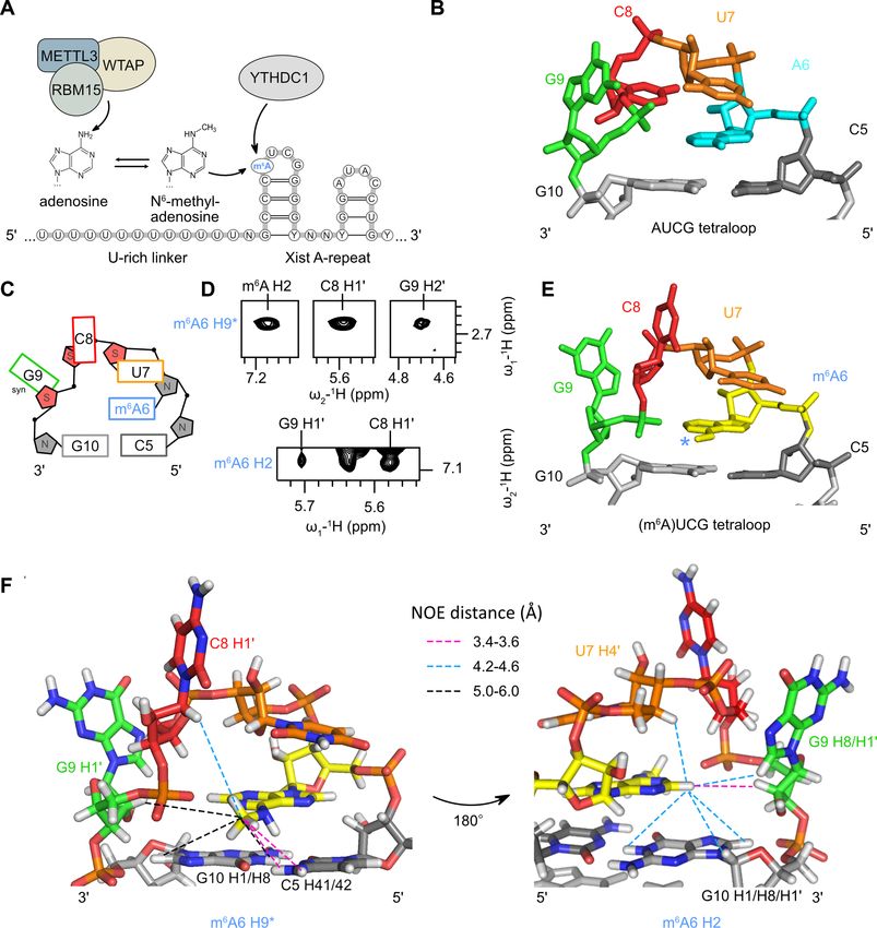
Structural effects of m6A modification of the Xist A-repeat AUCG tetraloop and its recognition by - YTHDC1
←
→
Page content transcription
If your browser does not render page correctly, please read the page content below
2350–2362 Nucleic Acids Research, 2022, Vol. 50, No. 4                                                                    Published online 15 February 2022
https://doi.org/10.1093/nar/gkac080

Structural effects of m6A modification of the Xist
A-repeat AUCG tetraloop and its recognition by
YTHDC1
Alisha N. Jones1,2,† , Ekaterina Tikhaia1,2,† , André Mourão1,2 and Michael Sattler                                                        1,2,*

1
 Institute of Structural Biology, Helmholtz Zentrum München, Ingolstädter Landstr. 1, 85764, Neuherberg, Germany
and 2 Bavarian NMR Center, Department of Chemistry, Technical University of Munich, Lichtenbergstr. 4,
85747, Garching, Germany

                                                                                                                                                                  Downloaded from https://academic.oup.com/nar/article/50/4/2350/6528901 by guest on 20 June 2022
Received September 13, 2021; Revised December 28, 2021; Editorial Decision January 19, 2022; Accepted February 11, 2022

ABSTRACT                                                                                 GRAPHICAL ABSTRACT
The A-repeat region of the lncRNA Xist is crit-
ical for X inactivation and harbors several N6 -
methyladenosine (m6 A) modifications. How the m6 A
modification affects the conformation of the con-
served AUCG tetraloop hairpin of the A-repeats and
how it can be recognized by the YTHDC1 reader pro-
tein is unknown. Here, we report the NMR solution
structure of the (m6 A)UCG hairpin, which reveals
that the m6 A base extends 5 stacking of the A-form
helical stem, resembling the unmethylated AUCG
tetraloop. A crystal structure of YTHDC1 bound to
the (m6 A)UCG tetraloop shows that the (m6 A)UC                                          INTRODUCTION
nucleotides are recognized by the YTH domain of                                          N6 -Methyladenosine (m6 A) is the most abundant post-
YTHDC1 in a single-stranded conformation. The m6 A                                       transcriptional base modification found in eukaryotic RNA
base inserts into the aromatic cage and the U and                                        (1). This modification, which is carried out by the ‘writer’
C bases interact with a flanking charged surface re-                                     methylation machinery (i.e. the METTL3-METTL14 com-
gion, resembling the recognition of single-stranded                                      plex), serves as a regulatory factor in the gene expression
m6 A RNA ligands. Notably, NMR and fluorescence                                          of over 7000 human genes (2–4). The N6 methyl group
quenching experiments show that the binding re-                                          in m6 A can adopt syn and anti conformations (5,6), and
quires local unfolding of the upper stem region of                                       may alter RNA secondary and tertiary structure: the pres-
the (m6 A)UCG hairpin. Our data show that m6 A can                                       ence of an N6 -methyl group may destabilize base pairing
be readily accommodated in hairpin loop regions,                                         in helical stem regions or may enhance stability when the
                                                                                         m6 A nucleotide is flanking the stem (7). The mechanism
but recognition by YTH readers requires local un-
                                                                                         by which m6 A regulates gene expression can involve the di-
folding of flanking stem regions. This suggests how                                      rect modulation of RNA recognition by RNA binding pro-
m6 A modifications may regulate lncRNA function by                                       teins, such as splicing factors (4), but in general depends on
modulating RNA structure.                                                                ‘reader’ proteins, which specifically recognize the m6 A-base
                                                                                         and flanking nucleotides. The recognition of m6 A by reader
                                                                                         proteins has been proposed to modulate RNA structure and
                                                                                         accessibility and thereby regulate interactions with RNA
                                                                                         binding proteins (7–10), although the underlying structural
                                                                                         mechanisms are poorly characterized. The most prevalent
                                                                                         reader proteins are the YT521-B homology (YTH) do-
                                                                                         main proteins (consisting of YTHDF1-3, YTHDC1 and
                                                                                         YTHDC2), that function to regulate processes that include

* To   whom correspondence should be addressed. Tel: +49 89 289 52600; Email: sattler@helmholtz-muenchen.de
†
    The authors wish it to be known that, in their opinion, the first two authors should be regarded as Joint First Authors.

C The Author(s) 2022. Published by Oxford University Press on behalf of Nucleic Acids Research.
This is an Open Access article distributed under the terms of the Creative Commons Attribution-NonCommercial License
(http://creativecommons.org/licenses/by-nc/4.0/), which permits non-commercial re-use, distribution, and reproduction in any medium, provided the original work
is properly cited. For commercial re-use, please contact journals.permissions@oup.com
Nucleic Acids Research, 2022, Vol. 50, No. 4 2351

alternative splicing, mRNA translation and mRNA decay             structure and retains base pairing in the helical stem. While
by specifically recognizing the m6 A base (10–14).                the m6 A base in the loop is stacked between C5 and U7,
   While the regulation of mRNAs by YTH domain pro-               the remaining two nucleotides of the apical loop, C8 and
teins is well-studied, other classes of RNAs, such as m6 A-       G9 are solvent-exposed, and oriented perpendicular to the
modified long non-coding RNAs (lncRNAs), are also ‘read’          m6 A base. This may contribute to the mechanism of how
by YTH proteins to influence m6 A-linked biological func-         the YTH domain can access the m6 A nucleotide. Our crys-
tions (15–17). One of these lncRNAs is the X-inactive spe-        tal structure of the YTH domain of YTHDC1 with the
cific transcript (Xist), a 17kb RNA that is responsible for X-    (m6 A)UCG-tetraloop reveals that the m6 A, U and C bases
chromosome inactivation (XCI) in female placental mam-            of the tetraloop are recognized in a single-stranded con-
mals (18,19). Through six interspersed conserved repeat do-       formation. Strikingly, NMR and fluorescence experiments
mains (A–F), the lncRNA Xist coats and transcriptionally          show that YTH binding leads to partial destabilization of
silences the inactive X-chromosome. This involves interac-        the upper stem, while base-pairing in the lower region of
tions of numerous RBPs to the Xist lncRNA (20), includ-           the stem remains intact. The required local unfolding of
ing SHARP/SPEN which is required for Xist-mediated si-            the closing base pair is reflected in a slightly reduced affin-

                                                                                                                                    Downloaded from https://academic.oup.com/nar/article/50/4/2350/6528901 by guest on 20 June 2022
lencing (20–25). Critically, the A-repeat domain, which is        ity compared to an interaction with a single-stranded m6 A
located at the 5 end of the transcript, is responsible for       RNA ligand. Our data provide insight into the structural
transcriptional silencing; in its absence, coating, but not si-   mechanism of m6 A recognition in the context of a hairpin
lencing, occurs. This region is comprised of a 24-nucleotide      structure to promote Xist-mediated X chromosome silenc-
conserved element that is repeated 8.5 times in human (26).       ing.
The first 14 nucleotides of this conserved element fold into
a thermodynamically stable AUCG tetraloop hairpin, while          MATERIALS AND METHODS
the remaining nucleotides promote duplex formation with
other A-repeat elements (27–29).                                  RNA transcription and purification
   Xist was recently found to harbor several hundred              The single-stranded hexamer RNAs, 5 -CCAUCG-3 and
N6 -methyladenosines along the length of the transcript,          5 -CC(m6 A)UCG-3 , and the m6 A-modified Xist A-repeat
including the adenosine located within the conserved              UG-mismatch RNA, 5 -GGCGU(m6 A)UCGGCGCC-
AUCG tetraloops of the A-repeats. (Figure 1A) (15).               3 , were purchased from Dharmacon as HPLC-
The m6 A modification of Xist is facilitated by the RBPs          or PAGE-purified and desalted RNA oligonu-
RBM15/RBM15B. RBM15, which directly binds the A-                  cleotides. The unlabeled and 13 C–15 N Xist A-repeat
repeats, associates and guides the ‘writer’ protein complex       tetraloop RNAs, 5 -GGCGCAUCGGCGCC-3 and 5 -
formed between WTAP (Wilms tumor-associated protein)              GGCGC(m6 A)UCGGCGCC-3 , were produced by in vitro
and METTL3 to the lncRNA Xist, thereby facilitating               transcription using in-house purified T7 polymerase and
the m6 A-modification (Figure 1A) (15,22,30). Through co-         purified as described previously (27). DNA template (5 -
immunoprecipitation assays, the m6 A-modified nucleotides         GGCGCCGATGCGCCTATAGTGAGTCGTATTA-3 )
were found to be bound by the YTH domain of the reader            containing the T7 promoter sequence (underlined) was
protein YTHDC1 (15). While the degree to which m6 A               purchased from Eurofins Genomics as a standard desalted
modification of Xist regulates XCI is disputed (15,24,30), it     DNA oligo. 13 C-15 N-labeled rCTP, rGTP and rUTP were
is widely accepted that recognition of m6 A-modified bases        purchased from Silantes, and N6 -methyl-ATP, for the
of Xist by YTHDC1 promotes Xist-mediated transcrip-               transcription of the m6 A modified RNA, was purchased
tional silencing (15).                                            from Jena Bioscience. RNAs were concentrated to 1 mM
   The unmodified AUCG tetraloop of the Xist A-repeats            in 25 mM NaCl and 25 mM sodium phosphate, pH 6.5.
adopts a unique fold, with the adenosine largely solvent in-
accessible. Extended 5 base stacking from C5 to U7 results
                                                                  Protein expression and purification
in the first two nucleotides of the tetraloop (AU) adopting
an A-form helical conformation (Figure 1B) (27). It is un-        The pETM28 vector-containing residues 345–509 of H.
clear how the addition of an m6 A methyl group may affect         sapiens YTHDC1 was purchased from Addgene (Addgene
the tetraloop conformation. Previous studies investigating        plasmid #64652). Nucleotides encoding an N-terminal
the effect of m6 A modification on RNA structure have re-         GGGG-linker were cloned between the YTH domain and
vealed both stabilizing and destabilizing effects. In RNA du-     TEV cleavage site to improve cleavage efficiency. Escherichia
plexes, m6 A modifications occurring at the termini of an A-      coli BL21 (DE3) cells transformed with the vector were
form helix result in stabilization of base stacking, whereas      grown either in Lysogeny broth (LB) medium for unlabeled
N6 methylation of base-paired adenosines results in destabi-      expression, or in M9 minimal medium supplemented with
lization of duplex structure (7). While structures have been      0.5 g/l 15 NH4 Cl and 4.0 g/l unlabeled glucose or 2.0 g/l
reported for the binding of YTH reader domains to m6 A            13
                                                                     C-labeled glucose (for 15 N- and 13 C–15 N-labeling, respec-
in single-stranded RNA regions (31–34), it is unknown if          tively). Expression and purification for all constructs was
and how YTH domains can recognize the N6 -methylated              carried out as previously described (33). During the final
adenosine in the context of a hairpin structure.                  purification by size-exclusion chromatography, the protein
   Here, we combine NMR, crystallography and biochem-             was eluted in NMR buffer (25 mM sodium phosphate pH
ical studies to study the effect of N6 -methylation of the        6.5, 150 mM NaCl and 5 mM dithiothreitol), and fractions
adenosine in the (m6 A)UCG-tetraloop of Xist. We show             containing the YTH domain were concentrated and stored
that the N6 methylation is compatible with the tetraloop          at 4◦ C.
2352 Nucleic Acids Research, 2022, Vol. 50, No. 4
Downloaded from https://academic.oup.com/nar/article/50/4/2350/6528901 by guest on 20 June 2022
Figure 1. Structure of the (m6 A)UCG tetraloop in Xist A-repeats. (A) Schematic overview of the reader (RBM15) and writer proteins (METTL3 and
WTAP) that facilitate m6 A modification of adenosines located within the tetraloop of the Xist AUCG tetraloop hairpin. YTHDC1 recognizes and binds
m6 A-modified Xist RNA. (B) Overview of the non-methylated AUCG tetraloop structure (PDB ID: 2Y95). (C) Schematic of base orientation and sugar
puckering of the (m6 A)UCG tetraloop. (D) NOEs between the m6 A H2 and methyl (H9*) protons, and inter-nucleotide NOEs between m6 A H2 and methyl
(H9*) protons to ribose protons of C8 and G9, as observed in the 1 H–1 H NOESY spectra at 150 ms mixing time. (E) Zoomed view of the (m6 A)UCG
tetraloop nucleotides in the lowest energy structure of the NMR ensemble of the (m6 A)UCG tetraloop hairpin. The methyl group of m6 A is marked with
a blue asterisk. (F) Zoomed view of the (m6 A)UCG tetraloop residues highlighting inter-residue NOEs between the m6 A H2 and methyl (H9*) protons.
NMR spectroscopy RNA assignments for the (m6 A)UCG tetraloop hair-
All NMR experiments were carried out on Bruker 500, 600, pin were partially facilitated using our previously re-
900 and 950 MHz Avance III spectrometers equipped with ported chemical shifts for the AUCG Xist tetraloop hair-
TCI or QCI cryoprobes. Backbone assignments for 60% of pin (27,38). Due to chemical shift differences induced by
the YTH domain of YTHDC1 were initially taken from the presence of the m6 A base, further NMR experiments
Theler et. al (32) (PDB 2MTV); completion of backbone were collected and analyzed to unambiguously assign reso-
1
H, 15 N, 13 C’, 13 C␣ and 13 C assignments for the YTH do- nances corresponding to the m6 A-modified RNA. All aro-
main were obtained from standard triple resonance experi- matic base (H2, H6, and H8) and anomeric ribose (H1 , H2
ments (CBCACONH, HNCACB, HNCO and HNCACO) and H3 ) protons were assigned to completion. 60% of res-
at 298 K (35). Data were processed using NMRPipe (36) onances corresponding to H4 , H5 and H5 were unam-
and analyzed using CCPN Analysis V2.5 (37). biguously assigned. The exchangeable 1 H resonances of the
Nucleic Acids Research, 2022, Vol. 50, No. 4 2353
(m6 A)UCG tetraloop hairpin of Xist were assigned using Isothermal titration calorimetry
homonuclear 1 H–1 H NOESY experiments at 280 K with All ITC measurements were performed on a MicroCal
graduated mixing times (50, 150 and 300 ms) in 90% H2 O PEAQ-ITC (Malvern Pananalytical Ltd., UK) at 25◦ C.
and 10% D2 O. The nonexchangeable 1 H resonances were The YTH domain and the unmodified and m6 A-modified
assigned using homonuclear 1 H-1 H NOESY (50, 150, and tetraloop hairpin RNA constructs were concentrated and
300 ms mixing times), homonuclear TOCSY (80 ms mix- dialyzed against NMR buffer (25 mM sodium phosphate,
ing time), and natural abundance 1 H–13 C HMQC experi- pH 6.5, 150 mM NaCl and 5 mM dithiothreitol) at 4◦ C
ments at 298 and 280 K on a sample dissolved in 99.99% overnight; the purchased single-stranded RNA oligos were
D2 O. Spectral overlap was resolved using 3D 13 C-edited diluted with dialysis buffer (25 mM sodium phosphate
1
H–1 H HMQC–NOESY–HMQC experiments implement- pH 6.5, 150 mM NaCl and 5 mM dithiothreitol or 1mM
ing ultrashort broadband cooperative pulses (39). Hydro- TCEP). Prior to measurement, all hairpin RNA samples
gen bond restraints of RNA base pairs were identified using were snap-cooled by heating at 96◦ C for 5 min, followed by
HNN COSY experiments (40,41). incubation on ice for 10 min. For hexamer and tetraloop
1
H–15 N SOFAST HMQC (42) titration experiments were hairpin RNA series, 9.5–10.5-fold (or 290–315 M) con-
Downloaded from https://academic.oup.com/nar/article/50/4/2350/6528901 by guest on 20 June 2022
recorded on a uniformly 15 N-labeled YTH domain that was centrated YTH domain was titrated into 30 M RNA
concentrated to 100 M. Prior to measurement, all hairpin in the cell, both in dialysis buffer. The experimental set-
RNA constructs were snap-cooled by heating at 96◦ C for tings include one 0.4 l injection followed by 19 injec-
5 min, followed by incubation on ice for 10 min. NMR ex- tions of 2 l with 120 s spacing for equilibration. Col-
periments were carried out at 298 K in NMR buffer sup- lected data were analyzed with MicroCal PEAQ-ITC Anal-
plemented with 10% D2 O. NMR spectra were processed in ysis Software using a one-site binding model; KD and ther-
TopSpin 3.5 (Bruker) and analyzed in CCPN Analysis V2.5. modynamic signatures of binding were determined based
The chemical shift perturbation plots were generated based on three technical replicates. Note, that the distinct en-
on chemical shifts obtained from titration experiments us- thalpy and entropy contribution seen for the binding of
ing the equation: the less stable RNA tetraloop with a closing UG base
pair, might reflect unfolding of the hairpin upon complex
2 2
CSP = (δ 1 H) + δ 15 N × α formation.
The scaling factor ␣ is calculated according to the range of
chemical shifts observed in bound states of each titration CD-monitored thermal denaturation
series and varies from 0.114 to 0.117 (43). Line broadening CD measurements were performed on Jasco J-715 Spec-
was evaluated as the change in peak intensity of the protein– tropolarimeter using a quartz cuvette with 10 mm path
RNA complex relative to the free protein. length. 50 M of RNA in NMR buffer (25 mM sodium
1
H 1D and homonuclear 1 H–1 H NOESY experiments phosphate buffer pH 6.4, 25 mM NaCl) was refolded by
with WATERGATE water flip-back were used for titra- heating at 96◦ C for 5 min followed by incubating on ice for
tion experiments. The NMR spectra of 200 M unmodified 10 min. The changes of CD as a function of wavelength for
and 160 M m6 A-modified Xist tetraloop hairpin RNA both unmodified and m6 A-modified RNAs were recorded
were acquired at 298 K in NMR buffer supplemented with at 25◦ C and averaged based on five replicates. The spe-
10% D2 O. Before measurements, RNA samples were snap- cific wavelength values were derived for both unmodified
cooled as described above. Protein to RNA molar ratios are (265 nm) and m6 A-modified (265.5 nm) RNAs and further
indicated in the figure legends. NMR spectra were processed used to measure temperature-dependent changes in elliptic-
in TopSpin 3.5 and analyzed in CCPN Analysis V2.5. ity from 10 to 100◦ C, with a 1◦ C/min slope and 5 min de-
lay. Tm values were calculated as described previously (27)
using the minima of first derivatives of the melting and re-
Fluorescence quenching assays folding curves.
The (m6 A)UCG Xist tetraloop hairpin RNA construct with
6-FAM conjugated at the 5 -end and BhQ-1 at the 3 -end
NMR structure calculations
were purchased from Dharmacon and Eurofins Genomics,
respectively, as PAGE-purified and desalted oligos. Prior to The structure of the Xist (m6 A)UCG tetraloop hairpin was
measurement, all RNA samples were diluted to 400 nM in calculated using restrained molecular dynamics followed
NMR buffer (25 mM sodium phosphate pH 6.5, and 25 mM by energy minimization in NIH-XPLOR using the RNA-
NaCl) and refolded by heating at 96◦ C for 5 min followed by ff1 force field (44). Briefly, an extended structure was gen-
incubation on ice for 10 min. A denaturing control was pre- erated and folded using NOE-derived distance restraints
pared by supplementing 400 nM RNA with 6 M urea and obtained from homonuclear 1 H–1 H NOESY experiments
heating the RNA to 95◦ C prior to measurement. YTH pro- and unambiguous hydrogen bond and planarity restraints
tein was added at concentrations as indicated in the main for base-paired nucleotides to generate 200 structures us-
figure. FAM fluorescence was excited at 495 nm with a slit ing the fold.py script. Based on qualitative assessment of
of 2 nm. Emission was recorded at 517 nm with a slit of 3 homonuclear TOCSY and NOESY spectra, U7, C8 and
nm for 0.5 s (integration time). The acquired data were nor- G9 riboses were restrained to adopt C2 -endo sugar puck-
malized against buffer control and plotted. Three biological ering (all other nucleotides adopt C3 -endo pucker confor-
replicates were performed. mation), and the G9 base was restrained to syn orienta-
2354 Nucleic Acids Research, 2022, Vol. 50, No. 4
Table 1. NMR structural statistics for the ensemble of 10 lowest energy RESULTS
structures
Structure of the m6 A modified Xist A-repeat hairpin
NMR restraints
# restraints 560 We previously reported the NMR-resolved structure of the
NOEs 202 thermodynamically stable AUCG tetraloop hairpin of Xist
Intra-residue 112 (27). Here, the adenosine (A6) of the AUCG loops adopts
Inter-residue 90
Torsional anglesa 126 an A-form helical conformation, and contributes to ex-
Planarity 15 tended 5 base stacking from C5 to U7. After U7, the phos-
H-bonds 15 phate backbone twists, causing the bases of C8 and G9
NMR ensemble to be flipped out in solution (Figure 1B). We first moni-
R.M.S.D. NOE restraints (Å) 0.037 ± 0.001
NOE violations (>0.5 Å) 0
tored thermal denaturation with circular dichroism exper-
Torsion violations (>5◦ ) 1.2 iments to investigate potential effects of the N6 methyl
R.M.S.D. from the mean coordinates (Å)b 0.34 ± 0.02 group of m6 A on the thermodynamic stability of the AUCG
R.M.S.D. from ideal geometry tetraloop hairpin. Notably, the (m6 A)UCG tetraloop hair-
Downloaded from https://academic.oup.com/nar/article/50/4/2350/6528901 by guest on 20 June 2022
Bond lengths (Å) 0.002 ± 0.000 pin is thermodynamically stable, with a melting tempera-
Bond angles (◦ ) 0.631 ± 0.061
Average XPLOR energy (kcal/mol) 1127.6 ± 33.46
ture of 85.25 ± 0.25◦ C (Supplementary Figure S1A), which
is comparable to the melting temperature of the unmodi-
a A-form duplex backbone torsion angle restraints derived from high- fied AUCG tetraloop hairpin (85.75 ± 0.25◦ C) (Supplemen-
resolution crystal structures were used for the helical stem: ␣ (300◦ ± 20◦ ), tary Figure S1B), and consistent with measurements for a
 (180 ± 10◦ ), ϒ (50◦ ± 10◦ ), ␦ (80◦ ± 30◦ ), ε (210◦ ± 10◦ ), (290◦ ±
◦
related tetraloop RNA (82.1 ± 0.4◦ C) (27).
20◦ ).
b all heavy atoms. Inspection of the imino proton NMR resonances of the
(m6 A)UCG tetraloop hairpin in 1 H–1 H NOESY spectra
recorded in H2 O revealed a complete ‘imino-walk’ (i.e.,
tion. Refinement of the lowest energy structures was carried correlations based on NOEs between the imino protons
out as described (44) and included the aforementioned di- of the guanosines involved in base-pairs) along the heli-
hedral angle restraints, with final convergence being estab- cal stem (G2-G12-G4-G10) (Supplementary Figure S2A).
lished when no NOE violations >0.5 Å or dihedral angle As expected, imino resonances corresponding to the first
violations >5◦ occurred in the majority of calculated struc- G–C base pair are not visible due to end-fraying. Strong
tures. Structural statistics are given in Table 1. Torsional vi- imino-amino inter-residue NOEs between each guanosine
olations listed in Table 1 arise from the sugar pucker of U7 H1 imino proton and its respective base-paired cytosine
in the apical loop. amino proton resonances are observed, confirming G–C
base pair formation (Supplementary Figure S2A). These re-
sults show that the addition of the N6 -methyl in m6 A is com-
patible with the hairpin structure and does not alter stem-
X-ray crystallography formation.
The 4:1 ratio of (m6 A)UCG Xist tetraloop hairpin:YTH Assignment and analysis of aromatic H6/H8 and
complex obtained from ITC was concentrated to 5.0 mg/ml anomeric protons using homonuclear NOESY and TOCSY
and screening was carried out with the Qiagen Classics I experiments (50) revealed that the N6 -methyl of A6 is in
Suite X-ray crystallography screening kit. A 400 nl drop vol- the syn conformation and stacked between residues C5 and
ume, comprised of 200 nl of complex and 200 nl of crystal- U7, following an A-form helix conformation. The intra-
lization solution, was used. Optimal crystals were formed and inter-residual aromatic H6/H8 to anomeric H1 proton
using the sitting drop, vapor diffusion method after one NOE walk shows sequential connectivities from the 5 end
week in 0.2 M lithium sulfate, 0.1M Tris–HCl pH 8.5, 30% of the RNA through the H6 proton of U7 (Supplementary
(w/v) Peg4000. Crystals were supplemented with 30% ethy- Figure S2B). The H8 proton of m6 A is shifted downfield
lene glycol for cryogenic preservation prior to flash cool- (7.68 ppm), which is characteristic of adenosines adopting
ing. Diffraction datasets were collected on the beamlines an A-form conformation (27,50). NOEs are observed be-
at the Swiss Light Source (SLS, Villigen, Switzerland) at tween the H5 and amino protons of C5 to the methyl pro-
beamlines PXIII. Diffraction data were collected at cryo- tons of m6 A (Supplementary Figure S2C). The H5/H6 pro-
genic temperatures (100 K) at wavelengths of 1.0 Å. The tons of U7 also show NOEs to the H8 proton of the m6 A
data were indexed with the XDS package (45) before scal- base, supporting an orientation where the m6 A base is sand-
ing with Aimless as part of the CCP4 package (46). The wiched between the nucleobases of C5 and U7 (Supplemen-
structure was solved by molecular replacement using two tary Figure S2B). Further supporting this stacking arrange-
crystal structures from the Protein Data Bank (PDB), codes ment, we find a weak-intensity NOE between the H1 proton
4R3I and 4RCJ, as search models in Phaser (47); the missing of G10 and the N6-methyl group protons of m6 A in the 1 H–
1
residues and RNA were built into the visible electron den- H NOESY spectra of the RNA recorded in H2 O (Supple-
sity using Coot modeling building software (48). The mod- mentary Figure S2A). A qualitative analysis of the 1 H–1 H
els were refined using the Phenix suite (49). Figures were TOCSY spectra reveals strong H1 –H2 and H1 –H3 cor-
made using Pymol (Schrodinger, LLC. The PyMOL Molec- relations for U7, C8 and G9, supporting C2 -endo sugar
ular Graphics System, Version 2.4, 2020). Structural statis- puckering for these nucleotides in the (m6 A)UCG tetraloop
tics are given in Table 3. (Figure 1C, Supplementary Figure S2D). All other ribosesNucleic Acids Research, 2022, Vol. 50, No. 4 2355
in the RNA stem region adopt a C3 -endo sugar pucker. may be necessary for binding to the YTH domain. To inves-
The base of G9 adopts a syn conformation, as indicated tigate this, we thought to destabilize the upper region of the
by the strong NOE observed between its H8 base and H1 helical stem by modifying the closing C5-G10 base pair to a
ribose protons (Figure 1C, Supplementary Figure S2B). less stable U5–G10 base pair (UG base pairs are supported
While these results are in good agreement with what was by two hydrogen bonds, whereas CG base pairs are sup-
observed for the AUCG tetraloop hairpin (Supplementary ported by three). Indeed, the binding affinity of YTHDC1
Figure S2E), further inspection of the 1 H–1 H NOESY spec- is improved to 2.05 ± 0.1 M for the UG Xist hairpin RNA
tra revealed several medium- to weak-intensity NOEs be- relative to the native CG closing base pair RNA hairpin
tween the H2 and methyl group protons of m6 A to the C8 (Figure 2C, Table 2). As expected, the binding affinity is still
and G9 residues of the (m6 A)UCG tetraloop that suggest somewhat reduced compared to what is observed with the
a differential arrangement of these nucleobases relative to single stranded m6 A-modified hexamer. These results sup-
m6 A. In particular, NOEs are observed between the C8 H1 port the notion that the energetic penalty of breaking or
proton and the m6 A methyl protons, and between the m6 A destabilizing the upper stem base pair decreases the affinity
H2 proton and the H2 and H8 protons of G9 (Figure 1D). to the hairpin versus the single-stranded RNA.
Downloaded from https://academic.oup.com/nar/article/50/4/2350/6528901 by guest on 20 June 2022
To provide high-resolution structural insight, we deter- Altogether, these data demonstrate that the N6 -methyl is
mined the NMR solution structure of the (m6 A)UCG required, as expected, for recognition by the YTH domain.
tetraloop hairpin using molecular dynamics/simulated an- The 3-fold reduced affinity of the recognition of m6 A in the
nealing in XPLOR-NIH (51,52). The structure is based on hairpin fold compared to a single-stranded RNA sequence,
202 NOE-derived distance restraints, and additional tor- and the increased binding to a weakened hairpin compared
sional angle and base planarity restraints (see Methods and to a more stable one, suggest that binding is accompanied
Table 1). The ensemble of the 10 lowest energy structures is with some energetic penalty due to required conformational
well converged (Supplementary Figure S2F, G) and shows rearrangements.
that the stem region adopts an A-form helical conforma- NMR spectroscopy was used to assess which regions
tion, with stacking extended at the 5 strand from C8 base of the YTH domain interact with the Xist A-repeat
is solvent-exposed (Figure 1E, F). The H1 proton of C8 is (m6 A)UCG tetraloop hairpin and the single-stranded 5 -
localized in close proximity to the methyl group of m6 A. CC(m6 A)UCG-3 sequence comprising the tetraloop nu-
The base of G9 folds back into the minor groove with the cleotides (Figure 2D, E, Supplementary Figure S4A–F).
H8 pointing toward the H2 of m6 A (Figure 1F). Following In 1 H–15 N HSQC NMR spectra of 15 N-labeled YTH,
G9, G10 is base paired with C5. Overall, the (m6 A)UCG large chemical shift perturbations (CSPs) are observed for
tetraloop hairpin forms a stable structure, with the UCG amide resonances upon titration of the single-stranded
bases of the apical loop exposed to the solvent. Thus, the 5 -CC(m6 A)UCG-3 RNA, while severe line-broadening
N6 -methyl modification of A6 does not disrupt the stem is induced in the presence of the structured (m6 A)UCG
formed by the RNA, but results in a rearrangement of nu- tetraloop hairpin (Figure 2D and E, Supplementary Fig-
cleotides in the loop region. ure S4A–F). The line-broadening observed in the NMR
spectra likely reflects a combination of increased relaxation
rates of the RNA–protein complex (due to the significantly
The YTH domain recognizes m6 A in the Xist A-repeat
increased molecular weight) and distinct binding kinetics
(m6 A)UCG tetraloop
of the complex with the hairpin RNA, which, in contrast
Previous studies have shown that the YTH domain of to the single-stranded RNA interaction, require conforma-
YTHDC1 binds single-stranded m6 A-modified RNAs with tion changes in the hairpin. Consistent with the binding
nanomolar to micromolar binding affinity, while unmodi- affinities determined by ITC, the single-stranded m6 A RNA
fied oligonucleotides of the same sequence are bound with shows slow binding kinetics on the NMR time scale, while
considerably weaker binding affinity (i.e. 100 nM and 5 the tetraloop exhibits fast-to-intermediate binding kinet-
M for 5’-UG(m6 A)CAC-3’ and 5’-UGACAC-3’ RNAs, ics associated with line-broadening (Supplementary Fig-
respectively) (32,33,53). We used isothermal calorimetry ure S4A, D). Importantly, the YTH amide resonances ex-
(ITC) to investigate the interactions of the YTH domain hibiting CSPs and line-broadening comprise residues re-
with unmodified and m6 A-modified Xist AUCG tetraloop ported to mediate m6 A-nucleotide recognition (31–33) (Fig-
RNA, both as a single-stranded oligonucleotide and in ure 2D, E), including W377, W428 and N367, which are a
the context of the structured hairpin (Figure 2A, B; Ta- part of the aromatic cage (see also Figure 3). We find very
ble 2). The YTH domain binds the single-stranded 5 - minor spectral changes upon addition of the unmodified
CC(m6 A)UCG-3 sequence with low micromolar affinity Xist A-repeat AUCG tetraloop or the corresponding single-
(dissociation constant KD = 1.27 ± 0.24 M), while no stranded RNA, consistent with the specificity of YTH bind-
binding is observed with the unmodified 5 -CCAUCG-3 ing to m6 A-modified RNAs (Supplementary Figure S3C–
RNA (Supplementary Figure S3A). Notably, the YTH do- F).
main binds the (m6 A)UCG tetraloop hairpin structure with Altogether, the ITC and NMR data show the specificity
reduced binding affinity (KD = 3.03 M ± 0.53), while no of YTHDC1 to m6 A-modified Xist sequences over unmod-
binding is observable with the unmodified AUCG tetraloop ified Xist RNA, and demonstrate the YTH domain is able
hairpin in by ITC (Supplementary Figure S3B). The re- to recognize and bind m6 A, despite its stacking onto the
duced binding affinity of the structured m6 A-modified A-form helical stem in the tetraloop hairpin. However, the
RNA relative to single-stranded m6 A-modified RNA sug- reduced binding affinity to the hairpin RNA compared to
gests that opening of base pairs in the upper stem region single-stranded RNA, suggests that an energetic penalty is2356 Nucleic Acids Research, 2022, Vol. 50, No. 4
Downloaded from https://academic.oup.com/nar/article/50/4/2350/6528901 by guest on 20 June 2022
Figure 2. Binding of m6 A modified RNAs by the YTH domain. Isothermal titration calorimetry experiments for binding of the YTH domain (A) to
5 -CC(m6 A)UCG-3 RNA, (B) to the m6 A-modified Xist A-repeat tetraloop hairpin and (C) to a variant of the m6 A tetraloop with a destabilized closing
base pair (CG to UG). (D, E) Zoomed-in regions of 1 H–15 N HSQC spectra overlays of the YTH domain free (black), (D) in complex with m6 A-modified
5 -CC(m6 A)UCG-3’ hexameric oligo (red: 1:1), and (E) when bound to the m6 A modified Xist A-repeat tetraloop hairpin (blue: 1:1).
Table 2. Isothermal titration calorimetry
RNA KD N G (kcal/mol) H (kcal/mol) –TS (kcal/mol)
CC(m6 A)UCG single-stranded 1.27 ± 0.24 M 0.88 ± 0.02 − 33.77 ± 0.52 − 41.37 ± 2.02 7.64 ± 2.39
CCAUCG single-stranded No binding N/A N/A N/A N/A
GGCGC(m6 A)UCGGCGCC 3.03 ± 0.53 M 0.827 ± 0.09 − 31.57 ± 0.42 − 40.07 ± 4.45 8.47 ± 4.89
hairpin
GGCGCAUCGGCGCC No binding N/A N/A N/A N/A
hairpin
GGCGU(m6 A)UCGGCGCC 2.05 ± 0.1 M 0.93 ± 0.07 − 32.5 ± 0.1 − 13 ± 1.7 − 19.5 ± 1.7
hairpin
associated with the recognition of the m6 A in the hairpin tained crystals of the complex that diffracted to 1.7 Å in
RNA, presumably due to conformational changes that are space group of P21 21 21 (Table 3). Nucleotides of the stem
required. flanking each end of the (m6 A)UCG tetraloop (G1-C5, and
G10-C14) are missing in the electron density map, despite
there being sufficient space in the crystal lattice for these
Crystal structure of the complex of YTH with the (m6 A)UCG
nucleotides. This is likely due to local destabilization of the
tetraloop
upper stem of the RNA upon binding by YTHDC1, which
To gain insight into the structural basis that defines YTH results in increased flexibility. However, the (m6 A)UCG
recognition of the Xist (m6 A)UCG tetraloop, we crys- tetranucleotides are clearly visible and structurally well-
tallized the YTH domain of YTHDC1 with the 14-mer defined (Supplementary Figure S5A). Consistent with pre-
(m6 A)UCG tetraloop hairpin of the Xist A-repeats. We ob- viously determined structures of the YTH domains withNucleic Acids Research, 2022, Vol. 50, No. 4 2357
Downloaded from https://academic.oup.com/nar/article/50/4/2350/6528901 by guest on 20 June 2022
Figure 3. Structure of YTH/(m6 A)UCG tetraloop complex. (A) Crystal structure of YTH bound to the (m6 A)UCG tetraloop of a Xist A-repeat. (B) The
aromatic cage that recognizes the m6 A is formed by W428, W377 and L439 side chains. (C) Hydrogen bonds (dashed lines) formed between S378, N367
and N363 mediate specific contacts to the m6 A base. (D) A positively charged surface area interacts with the backbone of (m6 A)UCG tetraloop and (E)
mediates electrostatic interactions with the RNA (dashed lines). (F) /cation stacking interaction between R475 and the U7 base.
RNA (31–33), the protein adopts an open ␣/ fold, con- Table 3. Crystallographic data collection and refinement statistics
sisting of five helices (␣0-␣4), six strands (1-6) and 310 Wavelength (Å) 1.00
helices (1-3) (Figure 3A). The (m6 A)UCG tetraloop of Resolution range (Å) 44.31–1.77 (1.833–1.77)
the Xist A-repeat hairpin RNA is bound by an arc-like sur- Space group P 21 21 21
face of the YTH domain (Figure 3A; Supplementary Figure Unit cell 78.438, 85.119, 88.617, 90,
90, 90
S5B). The N6 -methyl-adenosine adopts a syn conformation Unique reflections 57 778 (5063)
and is buried in a pocket that is formed by residues of ␣1, Completeness (%) 98.70 (87.53)
1 and 2 (amino acids 355 – 378), and the loop region con- Wilson B-factor (Å2 ) 29.26
necting 4 and 5 (amino acids 418-440). Three conserved Reflections used in refinement 57 723 (5027)
aromatic/hydrophobic residues, W377, W428 and L439, Reflections used for R-free 2883 (251)
R-work 0.1991 (0.4571)
form the aromatic cage that recognizes the m6 A methyl R-free 0.2313 (0.4734)
group by stacking of the m6 A base with the aromatic rings Number of non-hydrogen atoms 4387
of the two tryptophan residues, W377 and W428 (Figure macromolecules 4131
3B). The specificity for N6 -methyl adenine is achieved by ligands 106
base-specific hydrogen bonds of the nitrogen atoms of the solvent 234
Protein residues 495
m6 A base with the main chain amide of N363, the side chain RMS (bonds) (Å) 0.011
NH2 of N367, and the carbonyl oxygen of S378 (Figure 3C). RMS (angles) (◦ ) 1.22
The bases of the other three nucleotides of the (m6 A)UCG Ramachandran favored (%) 98.16
tetraloop, U7, C8 and G9, are stacked with each other while Ramachandran allowed (%) 1.84
Ramachandran outliers (%) 0
the phosphodiester backbone is bound by a charged sur- Rotamer outliers (%) 0
face pocket formed by K361, R404, K469, K472 and R475 Clashscore 3.14
in the YTH domain (Figure 3D,E). The 3 phosphate of the Average B-factor (Å2 ) 64.87
m6 A residue interacts with the R404 side chain (Figure 3E), Macromolecules 63.91
while the U7 base forms a -cation interaction with the Ligands 84.67
Solvent 65.02
guanidino group of R475 (Figure 3F). This binding inter-
face is in excellent agreement with the residues that show Statistics for the highest-resolution shell are shown in parentheses.
significant chemical shift changes and line-broadening in
our NMR titration experiments (Figure 2D, Supplementary
Figure S4B, E). Taken together these interactions provide single-stranded RNA ligands. A structural alignment of the
specific recognition of the loop region of the (m6 A)UCG YTH/(m6 A)UCG complex and the YTH/(m6 A)CU RNA
tetraloop. in PDB 6RT4 (34) shows that the two YTH domain struc-
Overall, the structure of the YTH/(m6 A)UCG com- tures are nearly identical, with a root mean square deviation
plex tetraloop structure resembles the recognition of m6 A (RMSD) of 0.146 Å for heavy atoms (Supplementary Fig-
seen in other reported structures of YTH domains with ure S5C). This indicates a conserved mode of m6 A recog-2358 Nucleic Acids Research, 2022, Vol. 50, No. 4
nition in a single-stranded RNA context and implies that intensity of the fluorescence emission of the RNA. Relative
the YTH binding requires at least a local conformational to an RNA-only control, which shows significant quench-
change of the (m6 A)UCG tetraloop, which avoids steric ing of fluorescence emission due to the spatial proximity of
clashes of the remaining helical stem region with the YTH fluorescent dye and quencher attached to the two arms of
domain (Supplementary Figure S5D). The helical stem is the helical stem, addition of YTH protein does not notably
thus oriented away from the YTH domain but with a dy- increase the fluorescence emission, even at a 4:1 protein to
namic orientation as indicated by the lack of observable RNA ratio (Figure 4C). As a positive control, denaturing
electron density in our crystal structure. of the RNA leads to more than a 5-fold increase in flu-
orescence emission (Supplementary Figure S6). Taken to-
gether the NMR and fluorescence quenching data indicate
Binding of YTHDC1 to the Xist (m6 A)UCG tetraloop leads
that binding of the YTH domain to the (m6 A)UCG hair-
local unfolding of the RNA stem
pin leads to local opening of the closing base pair, while the
In our NMR-derived structure of the (m6 A)UCG tetraloop lower part of the helical stem remains base paired.
hairpin RNA, the m6 A base is stacked with C5 and U7 and
Downloaded from https://academic.oup.com/nar/article/50/4/2350/6528901 by guest on 20 June 2022
is not fully solvent-exposed. The recognition of m6 A seen in
DISCUSSION
our crystal structure shows that the m6 A base is no longer
stacked, and the remaining residues of the tetraloop (U7, To date, >150 different RNA modifications have been iden-
C8 and G9, Figure 3A) are stacked with each other orien- tified across the RNA transcriptome (55). These modifica-
tated away from the protein surface. Notably, no electron tions are not limited to protein-encoding mRNA, but are
density is visible for the remaining residues of the RNA stem also prevalent in noncoding RNA transcripts (2,3,56). As
region. Thus, the conformation of the tetraloop residue has been demonstrated in several studies, RNA modifica-
within the (m6 A)UCG tetraloop hairpin and when bound tions increase the diversity of RNA function, influencing
to the YTH domain is drastically different and involves the regulation of numerous biological processes that include
substantial conformational rearrangements that likely af- pre-mRNA splicing, mRNA translation, and RNA export
fect destabilization of the helical stem region to enable m6 A and stability (1,4,10,11,13,57,58). The involvement of RNA
recognition. We therefore sought to determine whether the modifications in each of these cellular processes thus high-
YTH domain fully or only partially unwinds the stem-loop lights their integral role in the regulation of gene expression.
upon binding. However, the effects of the m6 A modification to RNA struc-
To investigate the conformation of the (m6 A)UCG ture and molecular interactions are still poorly understood.
tetraloop hairpin and potential changes induced by YTH Here, we have explored how the m6 A modification af-
binding we used NMR and fluorescence quenching exper- fects the structure of the conserved AUCG tetraloop hair-
iments. First, we followed intensity changes of imino res- pin structure located within the A-repeats of the lncRNA
onances of the (m6 A)UCG tetraloop stem by in 1D 1 H Xist, and the structural mechanisms that enable m6 A recog-
NMR and 1 H-1 H NOESY experiments upon addition of nition by the YTH domain of YTHDC1 reader protein. The
YTH to the RNA. While the guanine imino proton reso- ensemble of structures of the unmodified AUCG tetraloop
nances of the lower stem experience modest line-broadening hairpin structure shows that G9 in the tetraloop is solvent
upon addition of YTH protein, the G10 imino signal of exposed and that C8, G9 and G10, are conformationally dy-
the closing base pair becomes severely broadened already namic, while the A6 base is stabilized by extended stacking
at a 0.25:1 ratio of YTH to RNA (Figure 4A). Further- at the 5 arm of the RNA duplex (27). The final ensemble of
more, inspection of the 1 H-1 H NOESY spectra reveals that structures of the (m6 A)UCG tetraloop presented here re-
at this substoichiometric ratio, NOEs observed between the veals that the presence of the methylamino group on A6
amino protons of C5 (H41 and H42) and the imino pro- results in a minor but notable conformational rearrange-
ton of G10 (H1) are no longer present, while amino-imino ment of nucleotides of the tetraloop. The C8 nucleobase is
NOE correlations for other G-C base pairs are still ob- more solvent exposed and in closer proximity to U7, with
served. At a 1:1 ratio, imino proton resonances in both spec- its O2 group positioned down over the inside of the ma-
tra are broadened (due to increased relaxation associated jor groove and H5 and H6 protons exposed to solution.
with complex formation and conformational dynamics). On the other hand, our structure reveals that the N6 -methyl
However, while the diagonal peaks representing the imino group the m6 A base remains stacked with the 5 arm of the
protons of the base pairs in the lower stem region remain RNA stem, demonstrating that the N6 -methyl group does
visible, the G10 imino is no longer observed (Figure 4A). not destabilize the Xist AUCG tetraloop structure. This is
These results indicate that the closing C5-G10 base pair in distinct from the reported destabilization of RNA duplex
the upper stem of the (m6 A)UCG tetraloop hairpin is dis- regions upon introducing an m6 A base within an RNA he-
rupted upon YTH binding, whereas the lower stem remains lical stem (5,9). This is attributed to the requirement of an
folded. anti conformation of the N6 -methyl to sustain base pairing
To further support this conclusion, we performed flu- within an RNA helical region, while the energetically more
orescence quenching experiments using an (m6 A)UCG favorable syn conformation of the N6 -methyl in m6 A is fa-
tetraloop hairpin with a 6-FAM fluorophore and a Black vored in the single-stranded regions in the absence of base
Hole Quencher 1 (54) conjugated to the 5 and 3 ends of pairing. Indeed, our structure of the (m6 A)UCG tetraloop
the RNA, respectively (Figure 4B). Formation of the heli- shows that the m6 A base is not base-paired, but stacked on
cal stem in the absence and presence of increasing amounts top of the A-form helical stem. This structural arrangement
of the YTH domain of YTHDC1 is assessed based on the of the m6 A is compatible with the thermodynamically moreNucleic Acids Research, 2022, Vol. 50, No. 4 2359
Downloaded from https://academic.oup.com/nar/article/50/4/2350/6528901 by guest on 20 June 2022
Figure 4. YTH domain binding requires opening of the closing base pair of the (m6 A)UCG tetraloop stem. (A) 1 H 1D and 1 H–1 H NOESY spectra
showing broadening imino proton resonances upon addition of YTH domain protein (black: apo RNA, blue: 0.25:1, green: 0.5:1 and red: 1:1). The G10
imino proton resonance (as indicated by the dashed line) disappears even in the presence of 0.25 molar ratio of YTH protein. (B) Schematic representation
of the (m6 A)UCG tetraloop hairpin with a fluorophore conjugated to the 5 end and a quencher to the 3 end. When the two probes are separated, there is
emission of fluorescence intensity. (C) Bar plot showing the effect the YTH domain has on unwinding of the lower stem of the (m6 A)UCG tetraloop hairpin
at increasing concentration of protein. The RNA concentration was held constant at 400 nM. (D) Model illustrating the effects of m6 A modification and
YTHDC1 binding on the upper region of the Xist (m6 A)UCG stem–loop. The conformational changes induced in the RNA upon YTH binding might
modulate interactions with RNA-binding proteins or tertiary contacts involving the hairpin RNA.
favorable syn conformation of the N6 -methyl and does not RNA sequences (31–34). However, the fact that the m6 A
affect the stability of the Xist AUCG hairpin. base is not readily accessible in the (m6 A)UCG tetraloop
Our crystal structure of the YTH domain bound to the structure raises the question of how the YTH domain can
(m6 A)UCG tetraloop shows that the m6 A base is specifi- induce conformational changes in the hairpin RNA to en-
cally recognized by the aromatic cage of the YTH domain. able recognition of the m6 A. It has been suggested that the
The U7, C8 and G9 bases are stacked with each other, and interaction formed between the two nucleotides immedi-
their sugar-phosphate backbone interacts with a charged ately 3 of m6 A nucleotides (in our case U7 and C8) with
surface area (Figure 3D). These interactions are highly sim- the charged pocket may facilitate the recognition of m6 A
ilar to the recognition of single-stranded m6 A containing by the YTH domain through a two-step mechanism (34,59).2360 Nucleic Acids Research, 2022, Vol. 50, No. 4
Following a zipper-like mechanism of binding, the charged ing proteins (i.e. SHARP, which has been shown to bind
pocket on the surface of the YTH domain may first interact to the Xist A-repeat region) (23,25) and thereby modulate
with the sugar-phosphate backbone of the two nucleotides the A-repeat mediated silencing activity. (ii) Local unfold-
3 of the m6 A base, thus guiding the m6 A base into the ing of the RNA hairpin, i.e. by opening of the closing base
aromatic pocket. Our NMR structure of the m6 A modified pair, may expose single-stranded RNA sequence motifs that
AUCG hairpin indeed shows that the U7, C8 and G9 nu- can then be recognized by an RNA binding protein to fur-
cleotides are solvent-exposed in the apo-RNA, which could ther modulate the fate of the RNA (9). (iii) YTH binding
enable an initial interaction by the charged surface in the can attract additional RNA binding proteins (i.e. SRSF3)
YTH domain (Figure 3D). The stacking of R475 with U7 via protein-protein interactions to modulate biological ac-
and charged interactions with the phosphate-ribose back- tivity, such as alternative splicing (10). (iv) Finally, confor-
bone may then alter the backbone conformation of the mational changes of the RNA structure may modulate ter-
RNA and thereby trigger a reorientation of the m6 A base tiary contacts, which, in the case of the A-repeat regions are
into a position that enables its recognition by the aromatic expected to be required for its biological function (29). In
cage of the YTH domain. This binding mechanism may ra- general, the specific effects of m6 A will depend on whether
Downloaded from https://academic.oup.com/nar/article/50/4/2350/6528901 by guest on 20 June 2022
tionalize the conformational rearrangement of the hairpin the modification occurs in a single-stranded RNA region,
loop that is required for YTH binding. Indeed, the orienta- in the middle of an RNA duplex, or flanking a helical stem
tion of the RNA in the crystal structure does not align with as seen with the Xist AUCG tetraloop hairpin.
the tetraloop residues of our NMR-resolved RNA struc- In conclusion, our data demonstrate that m6 A modifi-
ture (Supplementary Figure S5D). Furthermore, this con- cation of lncRNAs are compatible with retaining overall
formational change may be reflected in energetic penalty structural features but may increase the potential and com-
that leads to the reduced binding affinity of the (m6 A)UCG plexity of fine-tuning and regulating their functional activ-
RNA tetraloop compared to the binding to single-stranded ity by modulating local structure and accessibility, for ex-
m6 A RNA, and an improved binding to the m6 A-modified ample, for RNA binding proteins.
tetraloop when the closing base pair is destabilized while
the rest of the stem remains base-paired (Figure 2, Table
2). Our ITC data indicate that the differences in binding DATA AVAILABILITY
affinity result from distinct enthalpic and entropic contribu- Atomic coordinates and structural restraints for the NMR
tions. These likely reflect the more complex binding mech- structure of the (m6 A)UCG tetraloop RNA and the crys-
anisms and conformational changes that are coupled with tal structure of the complex with the YTHDC1 YTH do-
the recognition of m6 A in a hairpin RNA by the YTH do- main have been deposited with the Protein Data Bank under
main. As expected the binding enthalpy with the hairpin accession numbers 7POF and 7PO6, respectively. Assign-
RNA is slightly reduced compared to the single-stranded ments and chemical shifts for the reported NMR structure
m6 A RNA due to the local unfolding of the closing base have been deposited in the BMRB with accession number
pair. Interestingly, recognition of m6 A in the context of both 51079.
the single-stranded or the hairpin RNA ligand by the YTH
domain shows slightly unfavorable binding entropy (consis-
tent with previous reports), although the release of solvent SUPPLEMENTARY DATA
molecules from the pocket of the protein that accommo-
Supplementary Data are available at NAR Online.
dates the N6 methyl is expected to show favorable entropic
contributions for binding (60,61).
The crystal structure of the YTH complex with the ACKNOWLEDGEMENTS
(m6 A)UCG RNA and our NMR data indicate that the clos-
ing C5-G10 base pair must be opened to enable recognition The authors are thankful to Arie Geerlof for prepara-
of the m6 A sequence by YTH. Consistent with this, a re- tion of T7 polymerase, Sam Asami and Gerd Gemmecker
cent analysis of m6 A/YTH domain interactions suggests for assistance with NMR experiments, Grzegorz Popow-
that residues located upstream, i.e. 5 , of the m6 A residue icz for advice with crystallography, Charles Schwieters for
do not form stable interactions with the protein and re- help with m6 A parameter and topology file generation, and
main flexible (34). Strikingly, our NMR and fluorescence Winfried Meining for assistance with the computational
data demonstrate that the lower region of the stem remains setup. The authors thank Giovanni Bussi, Hyun-Seo Kang,
base paired, and thus, the RNA hairpin is not completely and Santiago Martinez-Lumbreras for comments and dis-
unfolded. Considering that the stem is comprised only of cussions. E.T. is grateful for support by the International
stable G-C base pairs, it is reasonable that the fold of the he- Max-Planck Research School for Molecular Life Sciences
lical stem is partially maintained even in the complex with (IMPRS-LS) and Ludwig-Maximilians-Universität Grad-
the YTH domain. uate School Life Science Munich.
What are the consequences of the m6 A of a loop residue
in the Xist tetraloop hairpin? The binding of the YTH do-
FUNDING
main and the conformational changes induced in the hair-
pin structure may modulate molecular interactions of the Deutsche Forschungsgemeinschaft (German Research
RNA as a consequence of m6 A modification in multiple Foundation) [SFB 1309 – 325871075 to M.S.]. Funding for
ways. (i) The binding of YTH domain to the (m6 A)UCG open access charge: internal budget.
tetraloop may sterically block interactions with RNA bind- Conflict of interest statement. None declared.Nucleic Acids Research, 2022, Vol. 50, No. 4 2361
REFERENCES 22. Moindrot,B., Cerase,A., Coker,H., Masui,O., Grijzenhout,A.,
Pintacuda,G., Schermelleh,L., Nesterova,T.B. and Brockdorff,N.
1. Zhao,B.S., Roundtree,I.A. and He,C. (2017) Post-transcriptional
(2015) A pooled shRNA screen identifies rbm15, spen, and wtap as
gene regulation by mRNA modifications. Nat. Rev. Mol. Cell Biol.,
factors required for Xist RNA-Mediated silencing. Cell Rep., 12,
18, 31–42.
562–572.
2. Dominissini,D., Moshitch-Moshkovitz,S., Schwartz,S.,
23. Dossin,F., Pinheiro,I., Zylicz,J.J., Roensch,J., Collombet,S., Le
Salmon-Divon,M., Ungar,L., Osenberg,S., Cesarkas,K.,
Saux,A., Chelmicki,T., Attia,M., Kapoor,V., Zhan,Y. et al. (2020)
Jacob-Hirsch,J., Amariglio,N., Kupiec,M. et al. (2012) Topology of
SPEN integrates transcriptional and epigenetic control of
the human and mouse m6A RNA methylomes revealed by m6A-seq.
X-inactivation. Nature, 578, 455–460.
Nature, 485, 201–206.
24. Nesterova,T.B., Wei,G., Coker,H., Pintacuda,G., Bowness,J.S.,
3. Meyer,K.D., Saletore,Y., Zumbo,P., Elemento,O., Mason,C.E. and
Zhang,T., Almeida,M., Bloechl,B., Moindrot,B., Carter,E.J. et al.
Jaffrey,S.R. (2012) Comprehensive analysis of mRNA methylation
(2019) Systematic allelic analysis defines the interplay of key
reveals enrichment in 3 UTRs and near stop codons. Cell, 149,
pathways in X chromosome inactivation. Nat. Commun., 10, 3129.
1635–1646. 25. McHugh,C.A., Chen,C.K., Chow,A., Surka,C.F., Tran,C.,
4. Mendel,M., Delaney,K., Pandey,R.R., Chen,K.M., Wenda,J.M.,
McDonel,P., Pandya-Jones,A., Blanco,M., Burghard,C.,
Vagbo,C.B., Steiner,F.A., Homolka,D. and Pillai,R.S. (2021) Splice Moradian,A. et al. (2015) The Xist lncRNA interacts directly with
site m(6)A methylation prevents binding of U2AF35 to inhibit RNA SHARP to silence transcription through HDAC3. Nature, 521,
Downloaded from https://academic.oup.com/nar/article/50/4/2350/6528901 by guest on 20 June 2022
splicing. Cell, 184, 3125–3142. 232–236.
5. Roost,C., Lynch,S.R., Batista,P.J., Qu,K., Chang,H.Y. and Kool,E.T.
26. Wutz,A., Rasmussen,T.P. and Jaenisch,R. (2002) Chromosomal
(2015) Structure and thermodynamics of N6-methyladenosine in silencing and localization are mediated by different domains of Xist
RNA: a spring-loaded base modification. J. Am. Chem. Soc., 137, RNA. Nat. Genet., 30, 167.
2107–2115.
27. Duszczyk,M.M., Wutz,A., Rybin,V. and Sattler,M. (2011) The Xist
6. Shi,H., Liu,B., Nussbaumer,F., Rangadurai,A., Kreutz,C. and
RNA A-repeat comprises a novel AUCG tetraloop fold and a
Al-Hashimi,H.M. (2019) NMR chemical exchange measurements platform for multimerization. RNA, 17, 1973–1982.
reveal that N(6)-Methyladenosine slows RNA annealing. J. Am. 28. Maenner,S., Blaud,M., Fouillen,L., Savoye,A., Marchand,V.,
Chem. Soc., 141, 19988–19993.
Dubois,A., Sanglier-Cianferani,S., Van Dorsselaer,A., Clerc,P.,
7. Zhou,K.I., Parisien,M., Dai,Q., Liu,N., Diatchenko,L.,
Avner,P. et al. (2010) 2-D structure of the A region of Xist RNA and
Sachleben,J.R. and Pan,T. (2016) N(6)-Methyladenosine modification its implication for PRC2 association. PLoS Biol., 8, e1000276.
in a long noncoding RNA hairpin predisposes its conformation to 29. Jones,A.N. and Sattler,M. (2019) Challenges and perspectives for
protein binding. J. Mol. Biol., 428, 822–833.
structural biology of lncRNAs-the example of the Xist lncRNA
8. Zaccara,S., Ries,R.J. and Jaffrey,S.R. (2019) Reading, writing and A-repeats. J. Mol. Cell Biol., 11, 845–859.
erasing mRNA methylation. Nat. Rev. Mol. Cell Biol., 20, 608–624.
30. Coker,H., Wei,G., Moindrot,B., Mohammed,S., Nesterova,T. and
9. Liu,N., Dai,Q., Zheng,G., He,C., Parisien,M. and Pan,T. (2015)
Brockdorff,N. (2020) The role of the Xist 5 m6A region and RBM15
N(6)-methyladenosine-dependent RNA structural switches regulate
in X chromosome inactivation. Wellcome Open Res, 5, 31.
RNA-protein interactions. Nature, 518, 560–564.
31. Luo,S. and Tong,L. (2014) Molecular basis for the recognition of
10. Xiao,W., Adhikari,S., Dahal,U., Chen,Y.S., Hao,Y.J., Sun,B.F.,
methylated adenines in RNA by the eukaryotic YTH domain. Proc.
Sun,H.Y., Li,A., Ping,X.L., Lai,W.Y. et al. (2016) Nuclear m(6)A
Natl. Acad. Sci. U.S.A., 111, 13834–13839.
reader YTHDC1 regulates mRNA splicing. Mol. Cell, 61, 507–519.
32. Theler,D., Dominguez,C., Blatter,M., Boudet,J. and Allain,F.H.
11. Li,A., Chen,Y.S., Ping,X.L., Yang,X., Xiao,W., Yang,Y., Sun,H.Y.,
(2014) Solution structure of the YTH domain in complex with
Zhu,Q., Baidya,P., Wang,X. et al. (2017) Cytoplasmic m(6)A reader
N6-methyladenosine RNA: a reader of methylated RNA. Nucleic
YTHDF3 promotes mRNA translation. Cell Res., 27, 444–447.
Acids Res., 42, 13911–13919.
12. Roundtree,I.A. and He,C. (2016) Nuclear m(6)A reader YTHDC1
33. Xu,C., Wang,X., Liu,K., Roundtree,I.A., Tempel,W., Li,Y., Lu,Z.,
regulates mRNA splicing. Trends Genet., 32, 320–321.
He,C. and Min,J. (2014) Structural basis for selective binding of m6A
13. Roundtree,I.A., Luo,G.Z., Zhang,Z., Wang,X., Zhou,T., Cui,Y.,
RNA by the YTHDC1 YTH domain. Nat. Chem. Biol., 10, 927–929.
Sha,J., Huang,X., Guerrero,L., Xie,P. et al. (2017) YTHDC1
34. Li,Y., Bedi,R.K., Wiedmer,L., Huang,D., Sledz,P. and Caflisch,A.
mediates nuclear export of N(6)-methyladenosine methylated
(2019) Flexible binding of m(6)A reader protein YTHDC1 to its
mRNAs. Elife, 6, e31311.
preferred RNA motif. J. Chem. Theory Comput., 15, 7004–7014.
14. Shi,H., Wang,X., Lu,Z., Zhao,B.S., Ma,H., Hsu,P.J., Liu,C. and
35. Sattler,M., Schleucher,J. and Griesinger,C. (1999) Heteronuclear
He,C. (2017) YTHDF3 facilitates translation and decay of
multidimensional NMR experiments for the structure determination
N(6)-methyladenosine-modified RNA. Cell Res., 27, 315–328.
of proteins in solution employing pulsed field gradients. Prog. Nucl.
15. Patil,D.P., Chen,C.K., Pickering,B.F., Chow,A., Jackson,C.,
Magn. Reson. Spectrosc., 34, 93–158.
Guttman,M. and Jaffrey,S.R. (2016) m(6)A RNA methylation
36. Delaglio,F., Grzesiek,S., Vuister,G.W., Zhu,G., Pfeifer,J. and Bax,A.
promotes XIST-mediated transcriptional repression. Nature, 537,
(1995) NMRpipe - a multidimensional spectral processing system
369–373.
based on Unix pipes. J. Biomol. NMR, 6, 277–293.
16. Wang,X., Liu,C., Zhang,S., Yan,H., Zhang,L., Jiang,A., Liu,Y.,
37. Vranken,W.F., Boucher,W., Stevens,T.J., Fogh,R.H., Pajon,A.,
Feng,Y., Li,D., Guo,Y. et al. (2021) N(6)-methyladenosine
Llinas,M., Ulrich,E.L., Markley,J.L., Ionides,J. and Laue,E.D. (2005)
modification of MALAT1 promotes metastasis via reshaping nuclear
The CCPN data model for NMR spectroscopy: development of a
speckles. Dev. Cell, 56, 702–715. software pipeline. Proteins, 59, 687–696.
17. Liu,H., Xu,Y., Yao,B., Sui,T., Lai,L. and Li,Z. (2020) A novel 38. Duszczyk,M.M. and Sattler,M. (2012) (1)H, (1)(3)C, (1)(5)N and
N6-methyladenosine (m6A)-dependent fate decision for the lncRNA
(3)(1)P chemical shift assignments of a human Xist RNA A-repeat
THOR. Cell Death. Dis., 11, 613.
tetraloop hairpin essential for X-chromosome inactivation . Biomol.
18. Avner,P. and Heard,E. (2001) X-chromosome inactivation: counting, NMR Assign., 6, 75–77.
choice and initiation. Nat. Rev. Genet., 2, 59. 39. Asami,S., Kallies,W., Gunther,J.C., Stavropoulou,M., Glaser,S.J. and
19. Brockdorff,N., Bowness,J.S. and Wei,G. (2020) Progress toward
Sattler,M. (2018) Ultrashort broadband cooperative pulses for
understanding chromosome silencing by Xist RNA. Genes Dev., 34, multidimensional biomolecular NMR experiments. Angew. Chem.
733–744. Int. Ed. Engl., 57, 14498–14502.
20. Chu,C., Zhang,Q.C., da Rocha,S.T., Flynn,R.A., Bharadwaj,M., 40. Dallmann,A., Simon,B., Duszczyk,M.M., Kooshapur,H., Pardi,A.,
Calabrese,J.M., Magnuson,T., Heard,E. and Chang,H.Y. (2015)
Bermel,W. and Sattler,M. (2013) Efficient detection of hydrogen
Systematic discovery of xist RNA binding proteins. Cell, 161, bonds in dynamic regions of RNA by sensitivity-optimized NMR
404–416. pulse sequences. Angew. Chem. Int. Ed. Engl., 52, 10487–10490.
21. Monfort,A., Di Minin,G., Postlmayr,A., Freimann,R., Arieti,F., 41. Dingley,A.J. and Grzesiek,S. (1998) Direct observation of hydrogen
Thore,S. and Wutz,A. (2015) Identification of spen as a crucial factor
bonds in nucleic acid base pairs by internucleotide 2JNN couplings.
for Xist function through forward genetic screening in haploid J. Am. Chem. Soc., 120, 8293–8297.
embryonic stem cells. Cell Rep., 12, 554–561.You can also read


