Spinal transection switches the effect of metabotropic glutamate receptor subtype 7 from the facilitation to inhibition of ejaculation
←
→
Page content transcription
If your browser does not render page correctly, please read the page content below
Spinal transection switches the effect of
metabotropic glutamate receptor subtype 7 from
the facilitation to inhibition of ejaculation
Miwako Masugi-Tokita ( masugi@belle.shiga-med.ac.jp )
Shiga University of Medical Science: Shiga Ika Daigaku https://orcid.org/0000-0002-1846-5901
Shigehisa Kubota
Shiga University of Medical Science: Shiga Ika Daigaku
Kenichi Kobayashi
Shiga University of Medical Science: Shiga Ika Daigaku
Tetsuya Yoshida
Shiga University of Medical Science: Shiga Ika Daigaku
Susumu Kageyama
Shiga University of Medical Science: Shiga Ika Daigaku
Hirotaka Sakamoto
Okayama University Graduate School of Natural Science and Technology: Okayama Daigaku Daigakuin
Shizen Kagaku Kenkyuka
Akihiro Kawauchi
Shiga University of Medical Science: Shiga Ika Daigaku
Research Article
Keywords: mGluR7, ejaculation, lumbar spinothalamic (LSt) cells, ejaculation generator, lumbosacral
spinal cord, spinalization
Posted Date: March 11th, 2022
DOI: https://doi.org/10.21203/rs.3.rs-1415285/v1
License: This work is licensed under a Creative Commons Attribution 4.0 International License.
Read Full License
Page 1/20
Abstract
Metabotropic glutamate receptor subtype 7 (mGluR7) is a member of the group III mGluRs, which localize
to presynaptic active zones of the central nervous system. We previously reported that mGluR7 knockout
(KO) mice exhibit ejaculatory disorders, although they have normal sexual motivation. We hypothesized
that mGluR7 regulates ejaculation by potentiating the excitability of the neural circuit in the lumbosacral
spinal cord, because administration of the mGluR7-selective antagonist into that region inhibits drug-
induced ejaculation. In the present study, to elucidate the mechanism of impaired ejaculation in mGluR7
KO mice, we eliminated the influence of the brain by spinal transection (spinalization). Unexpectedly,
sexual responses of mGluR7 KO mice were stronger than those of wild-type mice after spinalization.
Histological examination indicated that mGluR7 controls sympathetic neurons as well as
parasympathetic neurons. In view of the complexity of its synaptic regulation, mGluR7 might control
ejaculation by multi-level and multi-modal mechanisms. Our study provides insight into the mechanism
of ejaculation as well as a strategy for future therapies to treat ejaculatory disorders in humans.

Introduction
Sexual dysfunction includes erectile dysfunction (ED) and ejaculatory disorders [1,2]. With the advent of
effective pharmaceutical treatments (e.g., phosphodiesterase type 5 inhibitors such as sildenafil), our
understanding of ED has improved, although ejaculatory disorders remain perhaps the least studied and
least understood of all male sexual dysfunctions [3,4]. Lack of knowledge of the physiology of
ejaculation has prevented the development of treatments.

Metabotropic glutamate receptors (mGluRs) constitute a superfamily of seven transmembrane domain
receptors that are linked via G proteins to intracellular signaling cascades. Eight different family members
are subdivided into three groups on the bases of sequence homology, pharmacological properties, and
second messenger coupling [5–7]. mGluR7 belongs to group III mGluRs, which localize presynaptically,
close to neurotransmitter release sites [8]. mGluR7 shows target cell-specific distribution that is
differentially concentrated at particular axon terminals, depending on the nature of the postsynaptic
target neuron [9]. Interestingly, mGluR7 undergoes agonist-induced internalization, which underlies long
term depression and a rapid switch to long term potentiation [10,11]. Although these unique properties
suggest the importance of mGluR7 in neural circuits, the physiological relevance of this receptor remains
to be fully elucidated.

Previously, we reported that mGluR7 knockout (KO) mice exhibited ejaculatory disorders [12], and greatly
reduced intermale aggression [13]. mGluR7 KO mice do not investigate the anogenital region of male
intruders, which is necessary for social recognition, although they can discriminate odors. We have also
reported that c-fos induction by urine odors is reduced in the bed nucleus of the stria terminalis of
mGluR7 KO mice; consistent with this, aggressive behavior is impaired by microinjection of a mGluR7
antagonist into the bed nucleus of the stria terminalis [13]. These results strongly suggest that mGluR7
KO mice fail to recognize other male mice as intruders to be attacked because of a deficit in processing

                                                  Page 2/20
pheromonal information, and hence exhibit reduced aggression. Because both aggressive and sexual
behavior depend on olfactory information, we had expected that mGluR7 KO mice would also show a
reduced interest in female mice. Unexpectedly, however, mGluR7 KO mice did sniff the anogenital region
of female mice and mounted them to the same extent as wild-type littermates, indicating that they have
normal sexual motivation [14]. The mechanism responsible for the mGluR7 KO mouse ejaculatory
disorder thus appears to be completely different from that of reduced aggression, although these two
phenotypes manifest at the same time because both are highly dependent on olfaction.

The existence of a spinal ejaculation generator in the lumbosacral spinal cord had long been
hypothesized because animals, including humans, with a complete transection of the spinal cord above
the thorax, can still ejaculate on vibratory or electrical stimulation [15,16]. The exact anatomical location
was relatively recently identified using the targeted neurotoxin SSP-SAP ([Sar9,Met(O2)11]-substance P
conjugated to saporin) for specific ablation of lumbar spinothalamic (LSt) cells [17]. LSt cells have
axonal projections to sympathetic and parasympathetic preganglionic neurons, to pudendal motor
neurons, as well as to the paracellular submuscular nucleus of the thalamus [18]. Recently, a human
spinal ejaculation generator has been identified [19], but the accompanying neural circuits that modulate
the main ejaculatory pathway from LSt cells are still not well-understood.

The spinal network controlling sexual function is tonically inhibited by input from the brainstem, whereby
the serotonergic (5-HT) system is a major source of descending inhibitory fibers [20–22]. We previously
found that mGluR7 in the lumbosacral spinal cord regulates ejaculation, because administration of a
mGluR7-selective antagonist into that region inhibited chemically-induced ejaculation [12]. However, we
need to be aware of the potential lack of specificity of intrathecal administration of mGluR7 antagonist.
In the present study, we definitively shut off any effects of the brain by spinalization, allowing us to
confirm the location of the mechanism responsible for ejaculatory disorders in mGluR7 KO mice.

Materials And Methods
Animals

Sexually inexperienced, gonadally-intact male mice (10–19 weeks) were employed in this study. mGluR7
KO and wild-type littermates were from heterozygous mating couples, produced by back-crossing the
mGluR7 KO line lacking the first coding exon and neighboring intron of the mGluR7 gene [23,24] onto the
C57BL/6N background (RRID:MGI:5705075) for at least 11 generations. All animal protocols were
compliant with the National Institutes of Health Guide for the Care and Use of Laboratory Animals and
were approved by the Committee for Animal Research, Shiga University of Medical Science. Animals were
euthanized by cervical dislocation or CO2 inhalation following approved protocols.

All mice tested in behavioral studies were maintained on a 12/12 h light/dark cycle (light off at 8 PM).
Food and water were provided ad libitum. Animals were identified by arbitrarily assigned numbers, such

                                                   Page 3/20
that the investigators performing the experiments were blinded to genotype. Additionally, the order of the
experiments was changed each day.
Spinal transection (spinalization)
Spinalization was accomplished as in previous reports [25,26]. Under anesthesia with isoflurane, the
spinal cord was completely transected intervertebrally using microscissors inserted between the 9th and
10th thoracic vertebrae. After filling the space of the removed spinal cord with Gelfoam (Pfizer,
Washington, DC, USA), the incised site was sutured. As spinalization caused loss of control of urination,
the Credé maneuver [27] was applied 3 times a day (early morning, early afternoon, and late afternoon) to
assist voiding, i.e. gentle pressure was applied to the bulging bladder to cause urination. Bladders were
emptied manually until a spontaneous return of micturition. Complete transection was confirmed by
flaccid hindlimbs and post-mortem examination of the lesioned area stained with neutral red.
Penile responses during manual urination
During manually-assisted urination in the morning (when the bladder was full), penile responses (n = 9 for
each genotype) were observed for 3 consecutive days. Before voiding, the bladder was palpated and
urinary retention was estimated, and only those mice with full bladders were included. Counting started
when urine began to flow and was observed for 90 sec. We defined penile responses according to 2
distinct categories as follows [28,29]:
1. flips: dorsiflexions of the penis due to a straightening of the penile body.
2. cups: intense erections with a flaring of the engorged glans penis.
The number of flips and cups was recorded. Additionally, the presence or absence of seminal emissions
(materials) during manual urination was noted.
Penile reflex test
On day 1 (6 hrs after spinalization) and day 2, penile reflexes (n = 9 for each genotype) were assessed in
a manner similar to a previous report [29]. Briefly, the mice were placed on their backs, restrained by strips
of adhesive tape across the midsection and at the base of the hind legs, and the glans penis was
extracted as far as possible. Adhesive tape was placed at the base of the sheath to maintain exposure of
the glans. Flips and cups were recorded. Testing ended 10 min after retraction of the penile sheath. Prior
to thoracic transection, mice were restrained using a 50 ml conical tube and adhesive tape, and the glans
penis was extracted. No flip or cup was observed in spinally-intact mGluR7 KO or wild-type mice.
Clonidine-induced ejaculation
All mice were tested on the 5th day post-transection according to a previous report [26]. The effects of
administration of 2.5 mg/kg, i.p. clonidine (Sigma, St-Louis, MO, USA) were examined. Three hrs prior to
drug administration, the bladder was manually voided and the penis was examined immediately prior to
Page 4/20testing to confirm the absence of seminal fluids or plugs. Black paper was placed in the cage to facilitate
the observation of seminal emissions. The ejaculatory response was observed every 10 min for 30 min
after clonidine administration by checking the coagulated seminal materials retrieved from the paper or
from the shaft of the penis. Seminal emissions (materials) were collected, allowed to dry on a filter paper
and weighed.
Immunohistochemistry
Wild-type and mGluR7 KO mice were anesthetized with medetomidine (0.3 mg/kg), midazolam (4 mg/kg)
and butorphanol (5 mg/kg), and then perfused through the left ventricle with formaldehyde (4%; freshly
depolymerized from paraformaldehyde) in 0.1 M sodium phosphate buffer (pH 7.4), after which the
spinal cords were removed. These were then saturated with 30% sucrose/0.1 M sodium phosphate buffer
at 4ºC, and cut into 35-µm coronal sections using a cryostat (CM3050 S; Leica Microsystems); these
sections were treated as free-floating. Because we found that immunostaining with anti-mGluR7 antibody
(07-239, Millipore; RRID:AB_310459) yielded a high background, we preabsorbed the antibody solution
with excess mGluR7 KO brain sections to prevent non-specific immunoreactivities, as previously reported
[12].
For triple-immunofluorescence histochemistry, some sections were incubated with a mixture of rabbit
anti-mGluR7 antibody (1:6,000 final dilution in Can Get Signal immunostain Solution A), rat anti-5-HT
antibody (clone YC5/45, MAB352, Sigma-Aldrich; 1:75; BRID:AB_11213564), and mouse anti-neuronal
nitric oxide synthase (nNOS; A-11, sc-5302, Santa Cruz Biotechnology; 1:8,000 dilution; RRID:AB_626757)
antibody. Sections were then treated with a mixture of Alexa Fluor 488-labelled goat anti-rabbit IgG (Life
Technologies), Alexa Fluor 594-labelled goat anti-guinea pig IgG (Life Technologies), and Alexa Fluor 647-
labelled goat anti-mouse IgG (Life Technologies) secondary antisera, as previously described [30]. Some
sections were incubated with a mixture of rabbit anti-mGluR7 antibody, guinea pig anti-galanin antibody
(Peninsula Laboratories, Inc; 1:16,000 dilution; RRID:AB_518351), and mouse anti-nNOS antibody. The
sections were then treated with a mixture of Alexa Fluor 488-labelled goat anti-rabbit IgG (Life
Technologies), goat anti-guinea pig Alexa Fluor 594-labelled goat anti-guinea pig IgG (Life Technologies),
and Alexa Fluor 647-labelled goat anti-mouse IgG (Life Technologies) secondary antibodies, and lastly,
examined under a confocal laser scanning microscope (LSM780, Zeiss) using appropriate filters.
Immuno-histochemical analysis was performed as described previously [30]. Briefly, the sections were
incubated sequentially with (1) rabbit anti-mGluR7 antibody (1:12,000 final dilution in Can Get Signal
immunostain Solution A), (2) 10 μg/ml goat biotinylated anti-rabbit IgG antibody (Vector Labs.,
Burlingame, CA), and (3) avidin-biotinylated peroxidase complex (ABC Standard, Vector Labs.,
Burlingame, CA; 1:100 dilution); all incubation media were prepared using 25 mM phosphate-buffered
saline (PBS) containing 0.1% Triton X-100 (PBS-Tx). Bound peroxidase was visualized by incubation with
0.02% diaminobenzidine tetrahydrochloride (DAB), and 0.003% H2O2 in 50 mM Tris-HCl (pH 7.6).
Statistics
Page 5/20Behavioral data were analyzed by two-way analysis of variance (ANOVA) for repeated measurements, or
unpaired t test for independent samples. Some data (in which variances were not homogeneous between
genotype groups) were analyzed by nonparametric tests (Mann-Whitney U test).
Results
Spinalized mGluR7 KO mice show enhanced penile
responses
Previously, we suggested that mGluR7 in the lumbosacral spinal cord regulates ejaculation [12], but we
had not completely excluded the possibility of an effect of the brain. To confirm the site responsible for
the regulation of ejaculation by mGluR7, we shut off any inputs from the brain by spinalization at the
thoracic level, and compared the sexual function of mGluR7 KO mice with their wild-type littermates.
Spinalized mice were unable to urinate by themselves and needed manual assistance to urinate.
Unexpectedly, we noticed that the penile responses were clearly enhanced during this procedure in the
mGluR7 KO mice, in contrast to the responses of wild-type mice, which were feeble.
To examine this enhanced response precisely, we conducted 3 daily tests of penile responses of
spinalized mGluR7 KO male mice during manually-assisted urination (measuring flips defined as
dorsiflexions of the penis, and cups defined as erections with flaring of the distal end of the glans)
[31,29]. On day 2–3, bladders of all mice were full when we performed manually-assisted urination in the
morning, but by day 4, a few mice had recovered urinary control. These mice were excluded from the
analysis, as were all data from day 5 when more mice had recovered urinary control. We found that the
mean numbers of penile flips (main effect of genotype, F(1, 16) = 7.78, p = 0.013; main effect of trial, F(2, 16)
= 7.84, p < 0.001; genotype × trial interaction, F(2, 16) = 0.37, p = 0.69; Fig. 1a) and of penile cups (main
effect of genotype, F(1, 16) = 6.63, p = 0.020; main effect of trial, F(2, 16) = 0.76, p = 0.48; genotype × trial
interaction, F(2, 16) = 0.81, p = 0.45; Fig. 1b) of the mGluR7 KO mice during manually-assisted urination
were significantly greater than those of their wild-type littermates, as assessed by repeated measures
ANOVA. There were no significant differences in pressure applied or time required for manually-assisted
urination between mGluR7 KO and wild-type mice.
We also observed that 4 of 9 mGluR7 KO mice, but only 1 of 9 wild-type mice emitted seminal material
during manual urination; in one mGluR7 KO mouse this even occurred three times over the experimental
course. mGluR7 KO mice already showed a tendency towards enhanced penile responses only 3 hrs after
spinalization (data not shown). At that time, responses during manual urination were weak in both
genotypes, probably because their bladders were not sufficiently full.
Having incidentally found intense penile responses of mGluR KO mice, we then employed the penile reflex
test [28] in order to confirm this finding using more traditional methods. Penile reflexes are elicited by the
retraction of the penile sheath, and stereotyped reflexes including flips and cups occur [31,29]. We found
that the mean number of penile flips (main effect of genotype, F(1, 16) = 5.72, p = 0.030; main effect of trial,
Page 6/20F(2, 16) = 4.88, p = 0.042; genotype × trial interaction, F(2, 16) = 1.14, p = 0.30; Fig. 1c) of the spinalized
mGluR7 KO mice during penile reflex tests was significantly greater than in their wild-type littermates, as
assessed by repeated measures ANOVA on day 1–2. However, there was no significant difference in the
number of penile cups (main effect of genotype, F(1, 16) = 0.55, p = 0.47; main effect of trial, F(2, 16) = 18.74,
p < 0.01; genotype × trial interaction, F(2, 16) = 1.34, p = 0.26; Fig. 1d) between the spinalized mGluR7 KO
mice and wild-type mice during penile reflex tests, as assessed by repeated measures ANOVA. These
results show that the degree of enhancement of penile reflexes of mGluR7 KO mice after spinalization
was more intense than in wild-type controls, implying a shift from facilitation of sexual responses by
mGluR7 to their inhibition on spinalization.
Spinalized mGluR7 KO mice show enhanced ejaculatory responses
As the penile reflex test is a simple observation of penile movement, to test the ejaculatory function of
mGluR7 KO more directly, we next used a clonidine-induced ejaculation model. Clonidine is an adrenergic
agonist, and elicits seminal emissions in spinalized mice [26]. We found that the weight of seminal
materials emitted following clonidine administration was greater from mGluR7 KO than wild-type
littermates (z = − 1.94, p = 0.047, Mann-Whitney U test; Fig. 2). In line with the results of the penile reflex
test, these data show that the degree of enhancement of drug-induced ejaculation by mGluR7 KO mice
caused by spinalization was greater than in wild-type controls.
mGluR7 is expressed in axon terminals forming synapses not only with parasympathetic but also
sympathetic preganglionic neurons
The spinal cord receives strong descending 5-HT innervation from the brain, and 5-HT released via this
pathway exerts inhibitory effects on sexual responses [20–22]. Therefore, eliminating inhibition from the
brain by spinalization is known to lower the threshold for sexual responses. We then focused on the
regions regulating ejaculation which have innervation of 5-HT. We found 5-HT-like immunoreactivity (LI)
around the intermediolateral cell column (IML) in spinal segments (T13-L2). At high magnification, sites
of apposition between large mGluR7-LI puncta and soma and dendrites of nNOS-LI-positive putative
sympathetic preganglionic neurons were revealed by triple labeling of mGluR7, 5-HT, and nNOS
(arrowheads; Fig. 3). These resembled what we had found for parasympathetic neurons [12]. Varicose
fibers immunoreactive for 5-TH making contact with the dendrites of putative preganglionic neurons were
also observed (arrows; Fig. 3), consistent with a previous report [32], although these appositions were not
co-localized.
Previously, we reported that, observed at low magnification, mGluR7-LI was distributed very similarly to
the parasympathetic branch of the ejaculatory pathway which is recognized by galanin-LI, a marker for
LSt cells, but was not present in the sympathetic nor in the pudendal branches. These results indicate
that by modulating the parasympathetic branch of the ejaculatory pathway, mGluR7 might be acting to
potentiate the activity of preganglionic neurons, which stimulate emission [12]. In the present study, we
found mGluR7-LI-positive puncta on sympathetic preganglionic neurons similar to parasympathetic ones,
although there was less mGluR7-LI than galanin-LI in the sympathetic branch of the ejaculatory pathway.
Page 7/20We further investigated whether mGluR7 is expressed at some other location in the parasympathetic area,
which would form staining patterns similar to galanin-LI. We found that mGluR7-LI was intense in the
region of the sacral parasympathetic nucleus (SPN; double arrowhead; Fig. 4a), the dorsal gray
commissure (DGC), and around the putative dendrites extending medially from the SPN to the DGC
(arrows; Fig. 4a and 4b) at the lumbosacral spinal cord (L6 − S1), as we already reported [12]. A magnified
view shows the beaded appearance of neuronal profiles with granular staining for mGluR7-LI (Fig. 4b), as
previously reported (Kinoshita, 1998). Contours of somatic (open star; Fig. 4c) and dendritic profiles
(arrowheads; Fig. 4c) decorated with granular staining for mGluR7-LI were found in the vicinity of the
SPN.
Discussion
We previously reported that mGluR7 KO mice exhibit ejaculatory disorders despite having normal sexual
motivation, and that intrathecal administration of an mGluR7 antagonist inhibits chemically-induced
ejaculation, strongly suggesting that mGluR7 controls ejaculation at the lumbosacral level [12]. It is
generally thought that the descending 5-HT innervation from the brain to the lumbosacral spinal cord
exerts a strong inhibitory control over sexual reflexes [20–22]. Thus, in the present study, we had
anticipated that if the sexual responses of mGluR7 KO mice remained less than those of wild-type
littermates after shutting off inputs from the brain by spinalization, this would clearly demonstrate that
mGluR7 regulates ejaculation at the spinal level. However, contrary to our expectations, the sexual
responses of spinalized mGluR7 KO mice were much higher than those of the wild-type controls.
What is the reason for switch of the effect of mGluR7 from facilitation to inhibition of ejaculation? One
possible role for mGluR7 in the lumbosacral region is to promote ejaculation by disinhibiting 5-HT inputs,
which tonically inhibiting sexual responses. Obviously, in that case, mGluR7 deficiency should make the
ejaculation threshold higher than in wild-type mice. In addition, this hypothesis is consistent with our
previous results on mGluR7 antagonist administration to the lumbosacral region. In that case, although
the mechanism is not known, spinalizaion would change the neural circuits in the spinal cord, and
mGluR7 might play some inhibitory role in sexual responses. Another possibility is that mGluR7 may
exert multi-level and multi-modal regulation of ejaculation, and spinalization then uncovers the inhibitory
role of mGluR7.
In the present study, we incidentally noticed enhanced penile responses of spinalized mGluR7 KO mice.
Because we have to assist them to urinate manually after transection, it is likely that penile responses
evoked by manual pressure on the bladder are similar to the urethrogenital reflex, which is a commonly
used model. The latter is induced by the mechanical stimulation of the urethra, and perineal muscles are
all activated simultaneously, as is seen in animals during copulation. Thus, this reflex has been
considered as a model for both penile erection and ejaculatory reflexes [33,21]. Penile responses or
reflexes of spinalized mGluR7 KO mice induced by manual urination or retraction of the penile sheath
were enhanced from the day of spinalization (at least after 3 hrs) to 3 days. We also confirmed the
Page 8/20enhanced ejaculatory response evoked by clonidine 5 days after spinalization. In addition, we had already
verified normal spermatogenesis based on testes morphology [13].
We have exploited the clonidine-induced ejaculation model, which requires spinalization of the animals
[26]. Clonidine is an adrenergic alpha-2 agonist and also has some alpha-1 effects [34]. In spinally-intact
rats, administration of clonidine depresses male copulatory behavior and decreases the number of males
ejaculating, furthermore, administration of yohimbine, an alpha-2-adrenoceptor antagonist, prevents the
inhibitory effect of clonidine. These results indicate that sexual inhibition by clonidine is mediated via the
alpha-2-adrenoceptor [35,36]. In contrast, in spinalized rats, the effect of clonidine was found to be
mediated by the alpha-1-adrenoceptor, because pre-treatment with the specific antagonist prazosin
prevents the ejaculatory response in the exhaustion model [37]. A possible explanation for the differential
results in spinalized versus intact animals could be that clonidine effects are exerted at different levels of
the central nervous system, namely via the alpha-2 receptor in the brainstem and the alpha-1 receptor in
the spinal cord. A switch of the effect of clonidine from an alpha-2- to an alpha-1-adrenergic agonist was
also shown in the expression of motor reflexes in rat. Decerebration and pharmacological studies indicate
that spinalization unmasks clonidine’s alpha-1-adrenergic effect by shutting out the effect of alpha-2-
adrenergic receptors localized in the brainstem [38]. Because mGluR7 negatively regulates alpha-1-
adrenergic receptor signaling [39], alpha-1 effects might be enhanced in spinalized mGluR7 KO mice.
It used to be thought that sexual behavior was under tonic inhibitory control to ensure that the behavior
occurs only under the appropriate circumstances, and that 5-HT was involved in this system [40]. 5-HT
inhibitory effects on sexual responses occur throughout the CNS. However, facilitatory effects of 5-HT on
sexual reflexes have also been reported. We should consider at least three factors; the concentration of 5-
HT in the synaptic cleft, 5-HT receptor to be stimulated, and interactions with other neurons. 5-HT
receptors are like molecular switches, the activation of which results in one of two effects,
hyperpolarization or depolarization [41,42]. At low concentrations, 5-HT binds to the postsynaptic 5-HT1A
receptor, which has high affinity and hyperpolarizes the membrane [43]. The 5-HT1A receptor is located on
GABAergic neurons and disinhibits other neurons by reducing GABA release [41,44], thus facilitating
sexual responses. In the spinal cord, at high concentrations, 5-HT inhibits sexual responses through 5-
HT2B receptors [45–47], and at low concentrations, 5-HT induces erections and ejaculation by activating
5-HT2C receptors [48,47]. 5-HT released at high concentrations exerts inhibitory control, while when
released in low concentration it contributes to the inhibition of sexual responses [42].
Using the urethrogenital reflex and tracing technique, a group of neurons in the medulla which mediates
descending inhibitory control of sexual reflexes was identified [21]. Furthermore, 5-HT fibers were found
innervating the IML and medial gray, especially at the sympathetic (T13-L2), and parasympathetic (L6-
S1) preganglionic neurons. They were originally identified as a group of neurons activated by stimulus
that evoked urethrogenital reflex [49]. These regions correspond to the area innervated by LSt cells. Our
observations correspond well with these studies, which suggest that mGluR7 modulates the effects of 5-
HT which inhibit sexual reflexes. mGluR7 might have differential effects on sexual responses depending
on the concentration of glutamate, interaction with other receptors, and interactions with other neurons.
Page 9/20Further investigation of the interaction of mGluR7 and 5-HT and its receptors will be required to resolve
this issue.
At this point, it is also necessary to discuss oxytocin. Oxytocinergic neurons are located in the
paraventricular nucleus of the hypothalamus, which descends the spinal cord, and controls erectile and
ejaculatory function at the lumbar level [50]. If these oxytocinergic neurons express mGluR7, and regulate
the release of oxytocin via mGluR7 at the axon terminal, the reason for the shift in the effect of mGluR7
may be explained because spinalization will cut the descending axon of these neurons.
LSt cells have been identified in rats as well as humans as an ejaculation generator, innervating
sympathetic, parasympathetic and motor neurons [19,17]. Although we had assumed that mGluR7
facilitates ejaculation by potentiating the activity of parasympathetic preganglionic neurons [12], further
detailed observations revealed that mGluR7-LI-positive puncta were present on both the sympathetic and
parasympathetic preganglionic neurons. We also found that the contours of somatic and dendritic
profiles were decorated with mGluR7-LI. Target cell-specific distribution that is differentially concentrated
at particular axon terminals, depending on the nature of the postsynaptic neurons, is a known property of
mGluR7 [9]. Furthermore, mGluR7 is reported to be a metaplastic switch controlling the direction of
synaptic plasticity, i.e. long-term potentiation versus long-term depression [10,11]. Prolonged agonist
exposure inactivates its effects via internalization [51]. We do not know how mGluR7 modulates
synapses in the living body, but we can assume that it will dynamically alter synaptic function depending
on the duration (hour, minute, or millisecond) of the stimulus and concentration of glutamate, and not
solely by acting as an autoreceptor which inhibits transmitter release [6,7].
Although LSt cells and their efferents have been identified as the ejaculation generator and the
mainstream ejaculatory pathway [52,17], the neural network and regulatory mechanisms that control
them remain unknown. The mechanisms regulating ejaculation are complex, and ejaculation can be
impaired by stress, as well as the threshold for ejaculation that can be raised by habitual strong
stimulation, as seen in intravaginal ejaculatory incompetence, which is a form of delayed ejaculation [53].
There are few treatment options available, such as selective serotonin reuptake inhibitors for premature
ejaculation, and the tricyclic antidepressant amoxapine for retrograde ejaculation [54]. These treatments
are only available for such limited categories of ejaculatory disorders. Successful drug development is
hindered because of a lack of knowledge regarding the basic concepts of the physiology of ejaculation.
The unique characteristics of mGluR7 may play a role in the complex regulatory mechanisms of
ejaculation. Further studies based on our findings will lead to future discoveries that can be leveraged to
treat ejaculatory disorders.
Declarations
Acknowledgments We especially thank Dr. Herman van der Putten from the Novartis Institutes for
BioMedical Research (Basel, Switzerland) for providing the mGluR7 KO mice, Dr. Pierre A. Guertin for
technical advice and Dr. Ryoichiro Kageyama for providing an excellent research environment.
Page 10/20Funding Information This work was supported by the Japan Society for the Promotion of Science
KAKENHI (Grant Numbers 21K09420 and 17K07075 to MMT).
Compliance with Ethical Standards
Conflicts of interest/Competing interests The authors declare that they have no conflicts of interest.
Ethics approval All animal protocols were compliant with the National Institutes of Health Guide for the
Care and Use of Laboratory Animals and were approved by the Committee for Animal Research, Shiga
University of Medical Science and Kyoto University. Every effort was made to minimize any suffering of
the animals used in this study. Animals were euthanized by cervical dislocation or CO2 inhalation in a
chamber following approved protocols.
Consent to participate All authors consented to participate.
Consent for publication All authors have read the manuscript and approved the final version.
Availability of data and material The datasets generated during and/or analysed during the current study
are available from the corresponding author on reasonable request.
Code availability Not applicable.
References
1. Lotti F, Maggi M (2018) Sexual dysfunction and male infertility. Nat Rev Urol 15 (5):287–307.
doi:10.1038/nrurol.2018.20
2. Montorsi F, Adaikan G, Becher E, Giuliano F, Khoury S, Lue TF, Sharlip I, Althof SE, Andersson KE,
Brock G, Broderick G, Burnett A, Buvat J, Dean J, Donatucci C, Eardley I, Fugl-Meyer KS, Goldstein I,
Hackett G, Hatzichristou D, Hellstrom W, Incrocci L, Jackson G, Kadioglu A, Levine L, Lewis RW, Maggi
M, McCabe M, McMahon CG, Montague D, Montorsi P, Mulhall J, Pfaus J, Porst H, Ralph D, Rosen R,
Rowland D, Sadeghi-Nejad H, Shabsigh R, Stief C, Vardi Y, Wallen K, Wasserman M (2010) Summary
of the recommendations on sexual dysfunctions in men. J Sex Med 7 (11):3572–3588.
doi:10.1111/j.1743-6109.2010.02062.x
3. Jannini EA, Lenzi A (2005) Ejaculatory disorders: epidemiology and current approaches to definition,
classification and subtyping. World J Urol 23 (2):68–75. doi:10.1007/s00345-004-0486-9
4. Wolters JP, Hellstrom WJ (2006) Current concepts in ejaculatory dysfunction. Rev Urol 8 Suppl 4:S18-
25
5. Hollmann M, Heinemann S (1994) Cloned glutamate receptors. Annu Rev Neurosci 17:31–108.
doi:10.1146/annurev.ne.17.030194.000335
6. Nakanishi S, Masu M (1994) Molecular diversity and functions of glutamate receptors. Annu Rev
Biophys Biomol Struct 23:319–348. doi:10.1146/annurev.bb.23.060194.001535
Page 11/207. Nicoletti F, Bockaert J, Collingridge GL, Conn PJ, Ferraguti F, Schoepp DD, Wroblewski JT, Pin JP
(2011) Metabotropic glutamate receptors: from the workbench to the bedside. Neuropharmacology
60 (7–8):1017–1041. doi:10.1016/j.neuropharm.2010.10.022
8. Okamoto N, Hori S, Akazawa C, Hayashi Y, Shigemoto R, Mizuno N, Nakanishi S (1994) Molecular
characterization of a new metabotropic glutamate receptor mGluR7 coupled to inhibitory cyclic AMP
signal transduction. J Biol Chem 269 (2):1231–1236
9. Shigemoto R, Kulik A, Roberts JD, Ohishi H, Nusser Z, Kaneko T, Somogyi P (1996) Target-cell-specific
concentration of a metabotropic glutamate receptor in the presynaptic active zone. Nature 381
(6582):523–525. doi:10.1038/381523a0
10. Pelkey KA, Lavezzari G, Racca C, Roche KW, McBain CJ (2005) mGluR7 is a metaplastic switch
controlling bidirectional plasticity of feedforward inhibition. Neuron 46 (1):89–102. doi:S0896-
6273(05)00128-5 [pii] 10. 1016/j.neuron.2005.02.011
11. Pelkey KA, Topolnik L, Yuan XQ, Lacaille JC, McBain CJ (2008) State-dependent cAMP sensitivity of
presynaptic function underlies metaplasticity in a hippocampal feedforward inhibitory circuit. Neuron
60 (6):980–987. doi:10.1016/j.neuron.2008.11.018 S0896-6273(08)01008-8 [pii]
12. Masugi-Tokita M, Tomita K, Kobayashi K, Yoshida T, Kageyama S, Sakamoto H, Kawauchi A (2020)
Metabotropic Glutamate Receptor Subtype 7 Is Essential for Ejaculation. Mol Neurobiol 57
(12):5208–5218. doi:10.1007/s12035-020-02090-2
13. Masugi-Tokita M, Flor PJ, Kawata M (2016) Metabotropic Glutamate Receptor Subtype 7 in the Bed
Nucleus of the Stria Terminalis is Essential for Intermale Aggression. Neuropsychopharmacology 41
(3):726–735. doi:10.1038/npp.2015.198
14. Hull EM, Dominguez JM (2007) Sexual behavior in male rodents. Horm Behav 52 (1):45–55.
doi:10.1016/j.yhbeh.2007.03.030
15. Halstead LS, Seager SW, Houston JM, Whitesell K, Dennis M, Nance PW (1993) Relief of spasticity in
SCI men and women using rectal probe electrostimulation. Paraplegia 31 (11):715–721.
doi:10.1038/sc.1993.113
16. Szasz G, Carpenter C (1989) Clinical observations in vibratory stimulation of the penis of men with
spinal cord injury. Arch Sex Behav 18 (6):461–474. doi:10.1007/bf01541673
17. Truitt WA, Coolen LM (2002) Identification of a potential ejaculation generator in the spinal cord.
Science 297 (5586):1566–1569. doi:10.1126/science.1073885
18. Veening JG, Coolen LM (2014) Neural mechanisms of sexual behavior in the male rat: emphasis on
ejaculation-related circuits. Pharmacol Biochem Behav 121:170–183.
doi:10.1016/j.pbb.2013.12.017
19. Chéhensse C, Facchinetti P, Bahrami S, Andrey P, Soler JM, Chrétien F, Bernabé J, Clément P, Denys P,
Giuliano F (2017) Human spinal ejaculation generator. Ann Neurol 81 (1):35–45.
doi:10.1002/ana.24819
20. Giuliano F, Clément P (2005) Physiology of ejaculation: emphasis on serotonergic control. Eur Urol
48 (3):408–417. doi:10.1016/j.eururo.2005.05.017
Page 12/2021. Marson L, McKenna KE (1990) The identification of a brainstem site controlling spinal sexual
reflexes in male rats. Brain Res 515 (1–2):303–308. doi:10.1016/0006-8993(90)90611-e
22. Marson L, McKenna KE (1992) A role for 5-hydroxytryptamine in descending inhibition of spinal
sexual reflexes. Exp Brain Res 88 (2):313–320. doi:10.1007/BF02259106
23. Masugi M, Yokoi M, Shigemoto R, Muguruma K, Watanabe Y, Sansig G, van der Putten H, Nakanishi S
(1999) Metabotropic glutamate receptor subtype 7 ablation causes deficit in fear response and
conditioned taste aversion. The Journal of neuroscience : the official journal of the Society for
Neuroscience 19 (3):955–963
24. Sansig G, Bushell TJ, Clarke VR, Rozov A, Burnashev N, Portet C, Gasparini F, Schmutz M, Klebs K,
Shigemoto R, Flor PJ, Kuhn R, Knoepfel T, Schroeder M, Hampson DR, Collett VJ, Zhang C, Duvoisin
RM, Collingridge GL, van Der Putten H (2001) Increased seizure susceptibility in mice lacking
metabotropic glutamate receptor 7. The Journal of neuroscience : the official journal of the Society
for Neuroscience 21 (22):8734–8745. doi:21/22/8734 [pii]
25. Rouleau P, Guertin PA (2013) A valuable animal model of spinal cord injury to study motor
dysfunctions, comorbid conditions, and aging associated diseases. Curr Pharm Des 19 (24):4437–
4447. doi:10.2174/1381612811319240010
26. Steuer I, Rouleau P, Guertin PA (2016) Adapted Technological Platform for Screening and Identifying
Compounds Capable of In vivo Spinal Network-mediated Reflex Ejaculation in Non-anesthetized,
Chronic Paraplegic Mice: Evidence of Clonidine-elicited Seminal Emission. Br J Med Med Res 12
(2):1–7. doi:10.9734/BJMMR/2016/19057
27. Barbalias GA, Klauber GT, Blaivas JG (1983) Critical evaluation of the Crede maneuver: a urodynamic
study of 207 patients. J Urol 130 (4):720–723. doi:10.1016/s0022-5347(17)51423-8
28. Hart BL (1968) Sexual reflexes and mating behavior in the male rat. J Comp Physiol Psychol 65
(3):453–460. doi:10.1037/h0025842
29. Sachs BD (1980) Sexual reflexes of spinal male house mice. Physiol Behav 24 (3):489–492.
doi:10.1016/0031-9384(80)90242-5
30. Kinoshita A, Shigemoto R, Ohishi H, van der Putten H, Mizuno N (1998) Immunohistochemical
localization of metabotropic glutamate receptors, mGluR7a and mGluR7b, in the central nervous
system of the adult rat and mouse: a light and electron microscopic study. J Comp Neurol 393
(3):332–352. doi:10.1002/(SICI)1096-9861(19980413)393:33.0.CO;2-2 [pii]
31. Hull EM, Meisel RL, Sachs BD (2002) Male sexual behavior. Hormones, Brainand Behavior 1:3-137
32. Minson JB, Arnolda LF, Llewellyn-Smith IJ (2002) Neurochemistry of nerve fibers apposing
sympathetic preganglionic neurons activated by sustained hypotension. J Comp Neurol 449
(4):307–318. doi:10.1002/cne.10282
33. Carro-Juárez M, Rodríguez-Manzo G (2000) Sensory and motor aspects of the coital reflex in the
spinal male rat. Behav Brain Res 108 (1):97–103. doi:10.1016/s0166-4328(99)00143-6
34. Berthelsen S, Pettinger WA (1977) A functional basis for classification of alpha-adrenergic receptors.
Life Sci 21 (5):595–606. doi:10.1016/0024-3205(77)90066-2
Page 13/2035. Clark JT (1991) Suppression of copulatory behavior in male rats following central administration of
clonidine. Neuropharmacology 30 (4):373–382. doi:10.1016/0028-3908(91)90063-h
36. Clark JT, Smith ER, Davidson JM (1985) Evidence for the modulation of sexual behavior by alpha-
adrenoceptors in male rats. Neuroendocrinology 41 (1):36–43. doi:10.1159/000124151
37. Carro-Juareza M, Rodriguez-Manzo G (2003) Yohimbine reverses the exhaustion of the coital reflex in
spinal male rats. Behav Brain Res 141 (1):43–50. doi:10.1016/s0166-4328(02)00324-8
38. Kehne JH, Gallager DW, Davis M (1985) Spinalization unmasks clonidine's alpha 1-adrenergic
mediated excitation of the flexor reflex in rats. The Journal of neuroscience : the official journal of the
Society for Neuroscience 5 (6):1583–1590
39. Iacovelli L, Di Menna L, Peterlik D, Stangl C, Orlando R, Molinaro G, De Blasi A, Bruno V, Battaglia G,
Flor PJ, Uschold-Schmidt N, Nicoletti F (2017) Type-7 metabotropic glutamate receptors negatively
regulate alpha1-adrenergic receptor signalling. Neuropharmacology 113 (Pt A):343–353.
doi:10.1016/j.neuropharm.2016.10.018
40. Snoeren EM, Veening JG, Olivier B, Oosting RS (2014) Serotonin 1A receptors and sexual behavior in
male rats: a review. Pharmacol Biochem Behav 121:102–114. doi:10.1016/j.pbb.2013.11.007
41. Fink KB, Göthert M (2007) 5-HT receptor regulation of neurotransmitter release. Pharmacol Rev 59
(4):360–417. doi:10.1124/pr.107.07103
42. Rubio-Casillas A, Rodríguez-Quintero CM, Rodríguez-Manzo G, Fernández-Guasti A (2015) Unraveling
the modulatory actions of serotonin on male rat sexual responses. Neurosci Biobehav Rev 55:234–
246. doi:10.1016/j.neubiorev.2015.05.003
43. Peroutka SJ (1986) Pharmacological differentiation and characterization of 5-HT1A, 5-HT1B, and 5-
HT1C binding sites in rat frontal cortex. J Neurochem 47 (2):529–540. doi:10.1111/j.1471-
4159.1986.tb04532.x
44. Gronier B (2008) Involvement of glutamate neurotransmission and N-methyl-d-aspartate receptor in
the activation of midbrain dopamine neurons by 5-HT1A receptor agonists: an electrophysiological
study in the rat. Neuroscience 156 (4):995–1004. doi:10.1016/j.neuroscience.2008.08.033
45. Millan MJ, Peglion JL, Lavielle G, Perrin-Monneyron S (1997) 5-HT2C receptors mediate penile
erections in rats: actions of novel and selective agonists and antagonists. Eur J Pharmacol 325
(1):9–12. doi:10.1016/s0014-2999(97)89962-1
46. Steers WD, de Groat WC (1989) Effects of m-chlorophenylpiperazine on penile and bladder function
in rats. Am J Physiol 257 (6 Pt 2):R1441-1449. doi:10.1152/ajpregu.1989.257.6.R1441
47. Yonezawa A, Yoshizumi M, Ebiko M, Ise SN, Watanabe C, Mizoguchi H, Kimura Y, Sakurada S (2008)
Ejaculatory response induced by a 5-HT2 receptor agonist m-CPP in rats: differential roles of 5-HT2
receptor subtypes. Pharmacol Biochem Behav 88 (4):367–373. doi:10.1016/j.pbb.2007.09.009
48. Bancila M, Giuliano F, Rampin O, Mailly P, Brisorgueil MJ, Calas A, Vergé D (2002) Evidence for a
direct projection from the paraventricular nucleus of the hypothalamus to putative serotoninergic
neurons of the nucleus paragigantocellularis involved in the control of erection in rats. Eur J Neurosci
16 (7):1240–1248. doi:10.1046/j.1460-9568.2002.02184.x
Page 14/2049. Marson L, Gravitt K (2004) Spinal neurons activated with the urethrogenital reflex in the male rat.
Brain Res 1026 (1):108–115. doi:10.1016/j.brainres.2004.08.019
50. Oti T, Satoh K, Uta D, Nagafuchi J, Tateishi S, Ueda R, Takanami K, Young LJ, Galione A, Morris JF,
Sakamoto T, Sakamoto H (2021) Oxytocin Influences Male Sexual Activity via Non-synaptic Axonal
Release in the Spinal Cord. Curr Biol 31 (1):103–114 e105. doi:10.1016/j.cub.2020.09.089
51. Ferguson SS (2001) Evolving concepts in G protein-coupled receptor endocytosis: the role in receptor
desensitization and signaling. Pharmacol Rev 53 (1):1–24
52. Allard J, Truitt WA, McKenna KE, Coolen LM (2005) Spinal cord control of ejaculation. World J Urol
23 (2):119–126. doi:10.1007/s00345-004-0494-9
53. Otani T (2019) Clinical review of ejaculatory dysfunction. Reprod Med Biol 18 (4):331–343.
doi:10.1002/rmb2.12289
54. Hu J, Nagao K, Tai T, Kobayashi H, Nakajima K (2017) Randomized Crossover Trial of Amoxapine
Versus Vitamin B12 for Retrograde Ejaculation. Int Braz J Urol 43 (3):496–504. doi:10.1590/S1677-
5538.IBJU.2016.0468
Figures
Page 15/20Figure 1
Sexual responses of spinalized mGluR7 KO mice. Number of flips (a) and cups (b) during manually-
assisted urination. The number of flips (a) and cups (b) of spinalized mGluR7 KO mice was greater than
in their wild-type littermates. Number of flips (c) and cups (d) in the penile reflex test. The number of flips
(c) was greater than in controls. n = 9. *p < 0.05 vs wild-type. Error bars indicate SEM. Independent days
were analyzed by unpaired t test.
Page 16/20Figure 2
Weight of seminal materials emitted following clonidine administration. Spinalized mGluR7 KO mice
emitted more than their wild-type littermates. n = 9. *p < 0.05 vs wild-type analyzed by unpaired t test.
Error bars indicate SEM.
Page 17/20Figure 3
(a) Triple labeling merged images depicting the relationship between mGluR7-LI containing puncta, 5-HT-
LI-positive varicosities and sympathetic preganglionic neurons, showing nNOS-LI (blue soma) in lumbar
segment L1 coronal sections. Sympathetic preganglionic neurons, located in the intermediolateral
nucleus, appear to receive inputs from mGluR7-LI-positive axon terminals (green puncta marked by
Page 18/20arrowheads) and 5-HT-LI-positive varicosities (red fibers marked by arrows), which were not co-localized.
Scale bar: 10 µm.
Figure 4
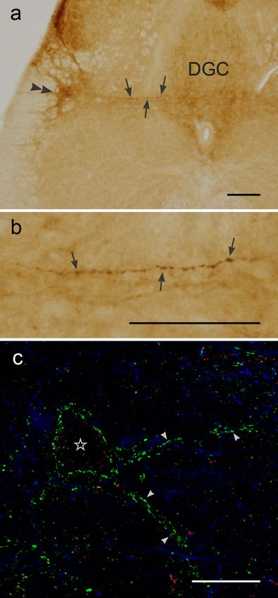
Page 19/20Distribution of mGluR7-LI in lumbar segment L6 coronal sections. (a) Intense mGluR7-LI in the sacral
parasympathetic nucleus (SPN; double arrowhead), the dorsal gray commissure (DGC), and around the
putative dendrites extending medially from the SPN to the DGC (arrows). (b) High magnification of
putative dendrites decorated with mGluR7-LI shown in (a). (c) Triple-labeled merged images of mGluR7-LI,
galanin-LI, and nNOS-LI. Putative soma (open star) and dendrites (arrowheads) are outlined with small
granules showing mGluR7-LI (green), which appears to be unrelated to galanin-LI (red) or nNOS-LI (blue).
Scale bars: 200 µm in (a) and (b); 20 µm in (c); DGC, dorsal gray commissure.
Page 20/20You can also read