Spectrum of Cervical Lesions and Cytohistological Correlation: A Study in Tertiary Care Center - IJCMR
←
→
Page content transcription
If your browser does not render page correctly, please read the page content below
ORIGINAL RESEARCH

                                                                                                                                           Section: Pathology
                                                                                                   www.ijcmr.com

Spectrum of Cervical Lesions and Cytohistological Correlation: A
Study in Tertiary Care Center
Anuradha Sharma1, Sonia Singh2

                                                                     cancer, suggesting that sexually transmitted infections like
  ABSTRACT                                                           HIV, Chlamydia, gonorrhea, and syphilis are of etiological
  Introduction: Cancer of the cervix is a global health              importance in the disease. Potentially oncogenic venereal
  problem and clinical cytopathology brings about detection          infections include Herpes simplex type 2, cytomegalovirus,
  and diagnosis of disease at stages earlier than possible before    human papilloma virus, HPV 16 and 18 account for more
  which can be further confirmed by histopathology technique.        than 70% of all cervical cancers.3
  The objective of the present study was to evaluate accuracy of     As the cervix is relatively easily accessible organ, the
  cervical cytology in the diagnosis of cervical lesions including
                                                                     logistics for screening cervical cancer are also simple.
  both neoplastic and non neoplastic.
                                                                     Screening programmes have reduced the incidence and
  Material and Methods: The study comprised of 100 patients
  with abnormal Pap smear cytology. A detailed clinical history,
                                                                     mortality from cancer cervix in many developed countries. It
  general physical and systemic examination was conducted            is well accepted that Pap smear has been the most effective
  and cervical biopsies as well as pap smears received from          cancer screening test ever introduced.4
  department of Obstetrics and Gynaecology were processed            To date the cervical cancer prevention effort worldwide
  and stained.                                                       have focused on screening sexually active women using
  Results: A total of seven cases of squamous cell carcinoma         cytological smears and treating precancerous lesion thus
  were reported majority of which were seen in the advanced          by decreasing the incidence and mortality from cervical
  age group of greater than 50. In present study 61% cases were      cancer. The diagnosis is made by screening an asymptomatic
  reported as NILM, 01% as ASCUS, 06% AGUS. LSIL was                 population. Tests in use are cervical cytology, and
  diagnosed on cytology in 20% patients, whereas 04% patients        histopathological examination of the biopsy materials added
  had HSIL and 07% patients revealed SCC, and remaining
                                                                     by various techniques such as cervicography and assessment
  01 patient (01%) with adenocarcinoma were diagnosed on
                                                                     of HPV DNA typing.5
  cytological evaluation. The histopathological findings in
  100 cases confirmed 72% cases of chronic cervicitis, 15% as        MATERIAL AND METHODS
  CIN-1, 03% of CIN-2, 01% CIN-3, 01% adenocarcinoma and
                                                                     This was a prospective study done over a period of one
  08%were diagnosed as invasive squamous cell carcinoma.
  Cytohistological correlation of 100 cases revealed an overall
                                                                     year. The present study was conducted in the Department of
  sensitivity of 95.60% and a specificity of 77.78%.                 Pathology consisting of 100 cases with cervical lesions. Pap
  Conclusion: Pap smear test was found to be equally sensitive       smears and biopsies were collected from all the patients for
  to histopathological examination for the early detection of        the study and correlation was done. A detailed clinical history,
  different cervical lesions.                                        general physical and systemic examination was conducted
                                                                     and cervical biopsies as well as pap smears received from
  Keywords: Cancer Cervix,           Pap   Smear,    Histological    department of Obstetrics and Gynaecology were processed
  Correlation, Early Diagnosis                                       and stained. The results were recorded and compared.
                                                                     The patients presenting with discharge per vaginum, bleed
                                                                     per vaginum and hypertrophied uterine cervix were included
INTRODUCTION                                                         in the study. The cytological data obtained was compared with
Disease and health are reflected accurately in both tissue and       the histopathological diagnosis. Statistical data pertaining to
cellular patterns which can be studied under histopathology          the sensitivity, specificity and positive predictive evaluation
and cytopathology respectively. Clinical cytopathology               of Pap smear and biopsies in diagnosing cervical lesions was
brings about detection and diagnosis of disease at stages
earlier than possible before.1                                       1
                                                                      Tutor, Department of Pathology, 2Assistant Professor, Department
Cancer cervix is a global health problem. It is the second
                                                                     of Pathology, Dr Radhakrishnan Government Medical College,
most common cancer among women in the world and ranks
                                                                     Hamirpur, Himachal Pradesh, India
as the first most frequent cancer among women in India and
in the other developing countries. The worldwide incidence           Corresponding author: Dr. Sonia Singh, H. No 561, Sector 8,
of cervical cancer is approximately 510,000 new cases                Faidabad, Haryana, India
annually, with approximately 288,000 deaths worldwide.2
                                                                     How to cite this article: Anuradha Sharma, Sonia Singh. Spectrum
Cervical cancer occurs early and strikes at the reproductive
                                                                     of cervical lesions and cytohistological correlation: a study in
period of a woman’s life. The incidence rises in 30-34 years         tertiary care center. International Journal of Contemporary Medical
of age and peaks at 55-65 years with median age of 38                Research 2019;6(6):F5-F10.
years. Early sexual activity, multiple sexual partners and low
socioeconomic status are major risk determinants of cervical         DOI: http://dx.doi.org/10.21276/ijcmr.2019.6.6.13

     International Journal of Contemporary Medical Research                                                                          F5
     ISSN (Online): 2393-915X; (Print): 2454-7379 | ICV: 98.46 |                           Volume 6 | Issue 6 | June 2019
Sharma, et al. Cytological Spectrum of Cervical Lesions
Section: Pathology
calculated. Relationship of age with inflammatory lesions, sil and
carcinoma
STATISTICAL ANALYSIS
Maximum numbers of cases (26) reported as NILM, were
For statistical analysis, a report of NILM, ASCUS,AGUS, in the reproductive age group of 31-40 years. LSIL was
LSIL, HSIL, Adenocarcinoma and SCC is considered seen commonly in the age group of 31 to 40 years. Out of
positive. Statistical analysis is shown in table 3. Descriptive the total patients showing HSIL (4), maximum (2) were
statistics were used for the interpretation of data. reported in the age group of more than 50 years whereas
RESULTS
The present study was based upon the cytological and
histological evaluation of cervical lesions from 100 patients.
Age distribution of the patients in the study
The age of the patients ranged from 21 to 70 years. The
maximum numbers of the patients were in third decade of
life, followed by fourth decade. The youngest patient in our
study was 25 years of age and the oldest patient was 70 years
of age. The mean age was 40.06 years.
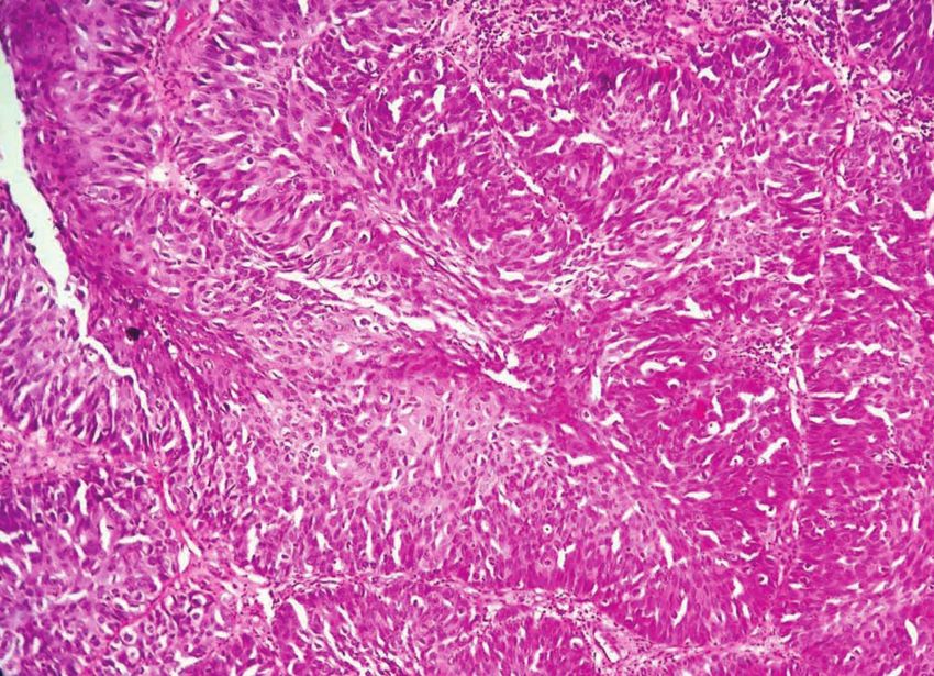
Figure-2: Basaloid variant of squamous cell carcinoma (H and E,
x 400)
Figure-1: Squamous cell carcinoma (Pap, x 400).
Cytological Diagnosis No of cases
NILM 61
ASCUS 01
AGUS 06
LSIL 20
HSIL 04
SCC 07 Figure-3: Adenocarcinoma. Smear showing cells arranged in
rosettes with, nuclear pleomorphism, hyperchromasia with coarse
Table-1: Categorisation of cytodiagnosis.
nuclear chromatin. (Pap, x400)
Cytological Diagnosis No of cases Histopathological Diagnosis
Chronic cervicitis CIN-1 CIN-2 CIN-3 Adenocar-cinoma SCC
NILM 61 61 00 00 00 00 00
ASCUS 01 01 00 00 00 00 00
AGUS 07 06 00 00 00 01 00
LSIL 20 05 15 00 00 00 00
HSIL 04 00 00 03 01 00 00
SCC 07 00 00 00 00 00 07
Total 100 73 15 03 01 01 07
Table-2: Cyto-histological correlation
Statistical Analysis Lesions (Percentage) Overall Study
NILM ASCUS AGUS LSIL HSIL ADENOCAR-CINOMA SCC
Sensitivity 83.33 00.00 00.00 93.75 66.00 100 85.71 95.60
Specificity 96.55 98.98 94.04 94.04 98.95 100 98.94 77.78
PPV 98.36 00.00 00.00 75.00 50.00 100 100 97.75
NPV 70.00 98.98 98.94 98.70 97.94 100 98.94 63.03
Diagnostic accuracy 88.00 93.00 93.94 94.00 95.00 100 99.00 94.00
Table-3: Analysis of the data
F6
International Journal of Contemporary Medical Research
Volume 6 | Issue 6 | June 2019 | ICV: 98.46 | ISSN (Online): 2393-915X; (Print): 2454-7379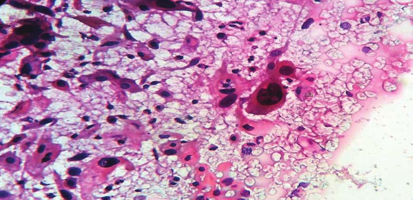
Sharma, et al.                                                                               Cytological Spectrum of Cervical Lesions

                                                                                                                                        Section: Pathology
                                                                    Degenerative changes like vacoulation of the cytoplasm,
                                                                    perinuclear halo etc. was observed. Reactive changes
                                                                    like hyperkeratosis and parakeratosis were also seen. The
                                                                    endocervical cells showed squamous metaplasia in few
                                                                    cases.
                                                                    Cases reported as ASCUS showed borderline cytological
                                                                    changes like nuclear enlargement, 2.5-3 times a normal
                                                                    intermediate cell nucleus, mild nuclear hyperchromasia,
                                                                    smooth nuclear outline and mild variation in size and shape.
                                                                    Cases reported as LSIL had nuclear size 4-6 times the size
Figure-4: Adenocarcinoma (H and E, x 100).                          of normal intermediate cell nucleus, occupying less than 1/3
                                                                    rd of the total area of the cells. Chromatin was uniformly
others were reported between 21-40 years of age. One case           distributed, but coarsely granular. Koilocytes were observed
of adenocarcinoma was reported at 40 years of age. A total          as superficial or intermediate squamous cells showing
of seven cases of squamous cell carcinoma reported on               perinuclear halo, with peripheral condensation of cytoplasm,
cytology, maximum (3) were seen in the advanced age group           wrinkled nuclear membrane and multinucleation.
of >50 years of age.                                                Smears diagnosed as HSIL showed abnormal cells of
Relationship of parity with inflammatory lesions, sil, and          variable size. In addition to superficial and intermediate cell
carcinoma                                                           types, parabasl cell were also found. Most of the cells were
The overall incidence of cervical cancer was found to               round to oval but spindle shaped elongated cells and cells
increase with increasing parity. Maximum number of cases            with bizarre shapes were also seen. Cytoplasmic staining
of HSIL, SCC and adenocarcinoma were seen in multiparous            was usually cyanophllic but some showed eosinophillia of
women, presenting mainly with post-menopausal bleeding.             the cytoplasm. The cytoplasm was sparse in some cases
Relationship of presenting symptoms with inflammatory               forming a rim around the nucleus. The nuclei were large
lesions, sil and invasive carcinoma.                                hyperchromatic with increased N: C ratio and nucleus
Discharge per vaginum (DPV) was the most common                     occupying ½-2/3 rd of the total area of the cell.
complaint of total 48% of the cases. Majority of the                Cytological smears reported as squamous cell carcinoma
cases, who presented with discharge per vaginum were                showed marked variation in size and shape with caudate
inflammatory in nature (34 cases). Patients who were                and spindle cells. The cytoplasm of some of the cells stained
diagnosed as having squamous cell carcinoma mostly                  eosinophillic. The nuclei were enlarged, irregular with
presented with post menopausal bleeding (03 cases), bleed           hyperchromatic, coarsely granular and unevenly distributed
per vaginum (02 cases) and contact bleeding(02). A single           nuclear chromatin. Most of the smears showed a dirty
case of adenocarcinoma presented with discharge per                 background due to excessive necrosis and tumor diathesis.
vaginum.                                                            Histopathological findings
Gross appearance of cervix and its relationship with                Histopathalogical evaluation of H and E stained paraffin
inflammatory lesions, sil and invasive carcinomas                   sections of cervical biopsy revealed that 72 patients had
Hypertrophied uterine cervisx (HUC) was observed in 41%             chronic cervicitis. CIN-1 was diagnosed in 15 patients
of the patients maximum of which were reported as Negative          and 04 were diagnosed to have CIN-2, CIN-3. Squamous
for intraepithelial lesions on pap smears followed by LSIL.         cell carcinoma was identified in 08 cases and 01 case was
Other per speculum findings included endocervicitis with            diagnosed as adenocarcinoma.
erosion (EE), erosion (Er), endocervicitis (E) and a visible        The histopathological findings in chronic cervicitis consisted
growth arising from cervix. Hard, nodular growth was                of basal cell hyperplasia of the ectocervical lining. The
observed in 5% of the patients, out of which three turned out       endocervix also showed basal cell hyperplasia and squamous
to be invasive squamous cell carcinoma.                             metaplasia of endocervical glands. The inflammatory
Categorization of cytodiagnosis                                     exudates consisted mainly of mononuclear cells.
In cytology, smears were examined and classified according          Cases reported as CIN-1 in cervical biopsy specimens
to The Bethesda system into NILM, ASCUS (atypical                   showed relatively regular arrangement of cells with
squamous cells of undetermined significance), LSIL (low             preserved stratification in upper two thirds of the epithelium.
grade squamous cells of undetermined significance), HSIL            The dysplastic changes consisting of aberrations in nuclear
(high grade squamous cells of undetermined significance),           morphology and disturbed arrangement and stratification
AGUS (atypical squamous cells of undetermined                       were observed mostly in lower 1/3rd of the epithelial lining.
significance), SCC (squamous cell carcinoma) and                    Cases reported as CIN-2 the section showed immature
adenocarcinoma, depicted in table-1.                                basaloid cells occupying upto two-third of the epithelial
The smears of NILM showed mainly superficial and                    thickness not extending into the upper third of the epithelium.
intermediate cells along with marked inflammatory exudates          The cases reported as CIN-3, evidence of stratification
consisting of neutrophils, lymphocytes and histiocytes.             was seen only in the upper 1/3rd of the epithelium.

      International Journal of Contemporary Medical Research                                                                     F7
      ISSN (Online): 2393-915X; (Print): 2454-7379 | ICV: 98.46 |                        Volume 6 | Issue 6 | June 2019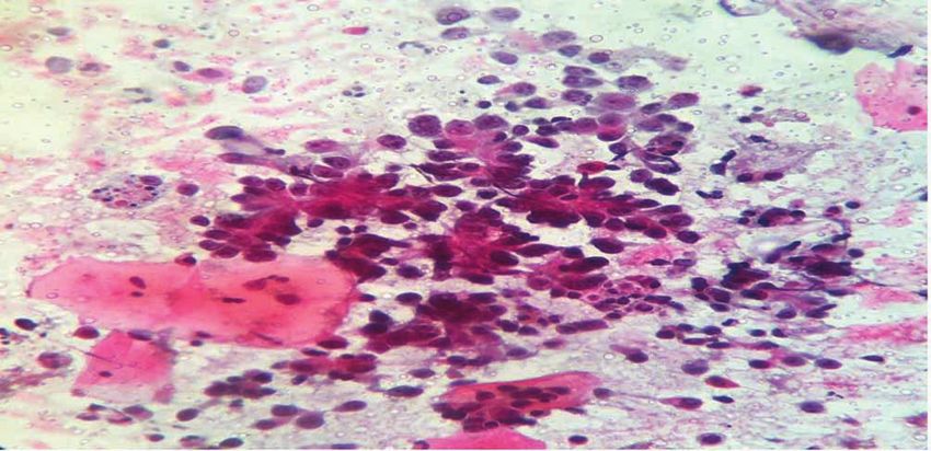
Sharma, et al.                                                                               Cytological Spectrum of Cervical Lesions
Section: Pathology

                     Nuclear abnormalities consisting of increased nuclear               well with the study of Chhabra et al6 where more than two
                     size,hyperchromatic, coarsely granular nuclear chromatin            third of the women were in the reproductive age group. It
                     was seen throughout the epithelium. Mitosis was observed            also collaborates the fact that sexually active women in the
                     in increased numbers.                                               reproductive age group have a higher chance of having an
                     Squamous cell carcinoma of the cervix showed two patterns           unhealthy cervix. In a study conducted by Mulazim Hussain
                     – Keratinizing and Non keratinizing. In the keratinizing            Bukhari et al7 peak incidence of abnormal smears were found
                     squamous cell carcinoma irregular infiltrating nests of             during age group of 30-39 years.
                     cells with cytoplasmic keratinization and epithelial pearl          Negative for intraepithelial lesion or malignancy (NILM)
                     formation was observed. Non keratinizing squamous cell              In present study, NILM accounted for maximum number
                     carcinomas did not show epithelial pearl formation. Necrosis        of 61% of cases observed in the age group of 31-40 years.
                     was a prominent feature.                                            Similarly in the study of Musmar8 inflammatory reactive
                     Cyto-histological correlation                                       smears were found to be commonest with frequency of
                     Correlation of cervical smear findings with histopathological       57.2%, whereas in the study of Pradhan B9 et al number
                     diagnosis revealed the following:                                   of inflammatory smears were 30%. In an another study
                     Of the total 61 cases diagnosed as negative for                     conducted by Mulazim H Bukhari7 the maximum number
                     intraepithelial lesion or malignancy (NILM) were confirmed          of inflammatory smears accounted for 38.3% which were
                     histopathologically in cervical biopsies as depicted in table-2.    observed in the age group of 30-39 years.
                     01 case of ASCUS was reported on pap smear turned out to            Low grade squamous intraepithelial lesion (LSIL)
                     be chronic cervicitis on histopathological examination.             It was observed that as age progresses there were sequential
                     Cytological diagnosis of LSIL was offered in 20 patients, 15        progression in the development of LSIL to HSIL and
                     out of them were confirmed to have CIN-1 on histopathology,         HSIL to squamous cell carcinoma. The result of our study
                     and rest of the 5 were found to have chronic cervicitis.            correlates well with Pradhan B et al9 who reported the mean
                     Cytological diagnosis of HSIL was offered in 04 patients.           age of LSIL as 31-40 and accounted for 31%, and Chhabra
                     Histological conformation of CIN-3 was made in 01 patient           et al6 who reported mean age of LSIL as 34.6 years. In
                     along with CIN-2 in 03 patients. 07 cases of squamous               present study the maximum number of patients presenting
                     cell carcinoma diagnosed on cytology were confirmed                 with LSIL were also in the age group of 31-40 years. The
                     histologically.                                                     present observation has been supported by fact that Cervical
                     DISCUSSION                                                          Intraepithelial Neoplasia is continuous process which begins
                                                                                         in its morphologically identifiable stage as LSIL and ends in
                     The main objective of the present study was to evaluate the
                                                                                         invasive cancer.
                     usefulness of cytology in detecting various preneoplastic
                     and neoplastic lesions of cervix, to evaluate and to interpret      High grade squamous intraepithelial lesion (HSIL)
                     the cases of epithelial lesions according to The Bethesda           HSIL is a cytological category that encompasses biologically
                     2001 classification system and correlation of cytological           different category smears, entities like moderate dysplasia
                     findings with follow-up histology sections. Carcinoma               and in situ squamous cell carcinoma, with potentially
                     uterine cervix is one of the leading causes of cancer death         different outcome. Most of the cases of squamous cell
                     among women worldwide. To detect this widely prevalent              carcinoma and high grade intraepithelial lesions in the
                     cancer at an early stage, the simplest test has been a pap          present study were reported in the age group of >50 years
                     smear. To check the sensitivity and specificity of Bethesda         and HSIL was observed in 4% of the cases. This correlates
                     system, the cytological findings have to be correlated with         well with the study of Subhalakshmi Mukopadhayay10,
                     histopathology considering it as gold standard.                     where HSIL was encountered in about 9.04% of cases in the
                     The Bethesda system for reporting cervical cytological              age group of 46-50 years.
                     diagnosis is a uniform system for reporting, and is useful          Squamous cell carcinoma
                     to provide effective communication among cytopathologist            Maximum cases of squamous cell cancer were seen in our
                     and referring physician. It also facilitates cytological and        study in patients of >50 years of age. This correlates with
                     histopathological correlation. The present study provides           other studies which are given as under in table 9. Age for
                     an analysis of patterns of Pap smear, age distribution,             detection of SCC in studies conducted by Regan et al11,
                     relationship of the lesion with different age groups, study of      Domandia et al12, Chhabra et al6 are 45-55 years, 39-48 years
                     the clinical features.                                              and 41-50 years respectively.
                     Age distribution                                                    Parity
                     In our study, 82% of the patients were in the reproductive          Another established risk factor for cervical cancer is high
                     age group. The age of the patients ranged from 21 to 70 years       parity. Chhabra et al6 observed most of the cases of squamous
                     of age with a mean of 40.06 years. The maximum patients             cell carcinoma in women who had four or more children.
                     with cervical lesions presented in the age group of 21-50           In their study, no case of squamous intraepithelial lesion or
                     years. Other studies have also shown a similar wide range of        cancer was seen in nulliparaous women. Our study was also
                     patients presenting with cervical lesions. Our study correlates     consistent with the same findings that maximum number of

                     F8
                                                                                  International Journal of Contemporary Medical Research
                                  Volume 6 | Issue 6 | June 2019 | ICV: 98.46 |                          ISSN (Online): 2393-915X; (Print): 2454-7379
Sharma, et al. Cytological Spectrum of Cervical Lesions
Section: Pathology
patients of squamous cell carcinoma were noted in women study conducted by Nawaz14, Gupta and Sondhai15 were 92%
who had three or more children in 03 patients. In a study and 74%.
conducted by Mulazim Hussain Bukhari7 premalignant In the study by Gupta and Sondhai15 100 cases were reported
and malignant lesions were most commonly observed with as CIN-2 and CIN-3 on histology and on cytology they were
multipara women (67.6%). In our study also premalignant correctly able to identify 74 HSIL cases whereas in 26 cases
and malignant lesions were seen in multipara women that a diagnosis of LSIL or below was given. These cases were
accounted a total of 66 patients. reviewed of which 16 cases were reclassified as HSIL on
Symptoms cytology while remaining 10 were the cases which showed
The most common presenting symptom in our study was persistent diagnosis of LSIL. 12/16 (75%) cases represented
discharge per vaginum in 47 cases. Most of cases who interpretative error. Sampling error was 7/10 and air drying
presented with this complaint were inflammatory in nature, 5/10 was found in under diagnosed cases.
followed by bleed per vaginum which accounted for 20 The correlation rate for CIN-2 and CIN-3 in present study is
cases. Patients who were diagnosed as having squamous 100% as compared to 92% in the study done by Nawaz et al.14
cell carcinoma mostly presented with post menopausal In study by Gupta and Sondhai15, correlation was established
bleeding. This correlated well with the study conducted in 74% of cases whereas that by Yeoh16 it was74.6%.
by Pradhan B et al9 where discharge per vaginum was the In our study for CIN-2 and CIN-3 the Sensitivity was 66%
commonest complaint in different lesions of cervix and with Specificity of 98.95%, Positive Predictive Value 50%
bleed per vaginum was found to be more specific for cervical and Negative Predictive Value of 97.94% was obtained.
malignancy. The same was observed in the study conducted Compared to the study conducted by Vaishali Jain17 the
by Mulazim Hussain Bukhari et al7 where vaginal discharge sensitivity was 42% with specificity of 36.8% and positive
and abnormal bleeding was found in 91.2%and 60.7% predictive value of 61.3% was observed.
respectively, of the neoplastic lesions. Squamous cell carcinoma (SCC)
In our study the common per speculum finding was On cytology 07 patients were diagnosed as SCC and one was
hypertrophied uterine cervix in 41% of the cases. On diagnosed as HSIL subsequently on histopathology all cases
cytology 26% of these cases were of NILM, 10% were those were diagnosed as SCC giving a correlation rate of 87.5% in
of LSIL, 02% HSIL and 3% were diagnosed as SCC. Growth our study. It is seen that correlation rate for squamous cell
was also the common presenting symptom in patients with carcinoma in our study was 87.5% with sensitivity of 85.71%,
SCC. the specificity was as high as 98.94% and with positive
predictive value of 100%. Thus a negative predictive value
Comparison of pattern of pap smear with various studies
of 98.94%, with over all diagnostic accuracy of 99.0% was
In the present study, inflammatory smears accounted for
obtained. As compared to the study conducted by Vaishali
maximum number of cases those were 61% similar to the
Jain17 the correlation rate was 83.6% with sensitivity 84%.
study of Subhalakshmi Mukhopadhyay10 where Negative
The specificity was 90.4% and positive predictive value of
for intraepithelial lesion or malignancy were found to be the
95.3% in their study. The correlation rate for squamous cell
commonest with the frequency of 64%.
carcinoma in our study is 87.5% compared to the studies
CIN-1 by Saha13, Yeoh16 and Nawaz14 in which the rate was 100%,
In our study 100 patients were exposed to biopsy where 15 60% and 97.33% respectively.
patients were diagnosed as CIN-1 on histology, on cytology
total 20 patients were reported as LSIL, remaining 05 cases CONCLUSION
which were reported as LSIL on cytology those were reported Pap smear test was found to be equally sensitive to
as chronic cervicitis on histopathology. histopathological examination for the early detection of
For CIN-1 in our study the Sensitivity of 93.75%, Specificity different cervical lesions. However, it is advised to perform
94.04%, Positive Predictive Value 75%, Negative Predictive biopsy if any abnormalities are detected in Pap smear for
Value 98.7% with a Diagnostic accuracy of 94% was correlation and confirmation. Carcinoma cervix is one
obtained. As compared to the study conducted by Saha R of the most common cancers in women and it is the most
and Thapa M13 sensitivity, specificity, positive predictive researched disease. The regular screening of population by
value, negative predictive value, diagnostic accuracy for Pap smear is a cost effective method for early detection of
CIN-1 was 60%, 93.9%, 75%, 88.6%, 86%, respectively. premalignant and malignant cervical lesions. Correlation of
The correlation rate for CIN-1 in present study is 75% as cervical cytology with cervical biopsies has been a common
compared to Saha13 study having the same correlation rate component of continuous quality improvement programme
of 75%. for accreditation purposes.
CIN-2 and CIN-3 REFERENCES
In our study of 100 patients 04 were reported as HSIL on 1. John K,Frost, MD. Gynecologic and Obstetric
smear cytology. In subsequent biopsy study these cases Cytopathology. In: E.R. Novak and J.D. Woodruff.
were reported as CIN-2 and CIN-3. So the correlation was Novak‟s Gynecological and Obstetric Pathology With
established in 100% of the cases. The correlation rates in the Clinical and Endocine Relations. Philadelphia: W.B.
International Journal of Contemporary Medical Research F9
ISSN (Online): 2393-915X; (Print): 2454-7379 | ICV: 98.46 | Volume 6 | Issue 6 | June 2019Sharma, et al. Cytological Spectrum of Cervical Lesions
Section: Pathology
Saunders; 1979:p.634.
2. K.Kaarthigeyan. Cervical cancer in India and HPV
vaccination. Indian J Med Paedr Oncol. 2012; 33:7-12.
3. Lyon J L, Gardner JW, West DW. Smoking and
carcinoma in situ of uterine cervix. Am J Public Health.
1983; 73:558-562.
4. Murphy N. Estimation of reduction in life time risk
cervical cancer through one life time screening.
Neoplasm 1993; 40:2558.
5. Mettin C, Dodd GD. The American Cancer Society
guidelines for the cancer related check-up. An update
Int'l J Cancer. 1991; 41:279-82.
6. Chhabra Y, Behera BG, Khalkho J et al.
Cytomorphological study of PAP smears for
precancerous and cancerous lesions. Journal of
Cytology. 2003; 20: 64-67.
7. Mulazim Hussain Bukhari, Kanwal Saba, Samina
Qamar, Muhammad Muddasar Majeed, Shahida Niazi,
et al. Clinicopathological importance of Papanicolaou
smears for the diagnosis of premalignant and malignant
lesions of the cervix. J Cytol. 2012; 29: 20–25.
8. Musmar SG. Patterns and factors affecting pap smear
test in Nablus, A retrospective study. Middle East
Journal of Family Medicine. 2004; 4:4.
9. Pradhan B, Pradhan SB, Mital VP. Correlation of
PAP smear findings with clinical findings and cervical
biopsy. Kathmandu University Medical Journal. 2007;
5:461-467.
10. Subhalakshmi Mukopadhayay. Evaluation of the
category high-grade squamous intraepithelial lesion in
The Bethesda System for reporting cervical cytology.
Journal of cytology. 2013; 30:33-35.
11. Regan JW, Seidemann IL and Patten SF. Developmental
stages of in situ carcinoma of uterine cervix. An
analytical study of cells. Acta Cytol.1953; 6:538-546.
12. Domandia M H. Role of vaginal cytology for early
detection of cancer. The Congress Proceedings of 6th
Asian Congress Of Obstet And Gynaec. 1974; 95.
13. Saha R, Thapa M. Correlation of cervical cytology with
cervical histology, Kathmandu univ Med J (KUMJ).
2005; 3: 222-224.
14. Nawaz FH, Aziz AB, Perwez S, Rizvi JH. Prevalence
of abnormal papanicolaou smears and cytohistological
correlation. Asia–Pacific Journal of clinical oncology.
2005; 1: 128- 132.
15. Gupta Sanjay, Sodhani P. Why is high grade squamous
intraepithelial neoplasia under-diagnosed on cytology
in a quarter of cases? Analysis of smear characteristics
in discrepant cases. Indian Journal of Cancer. 2004; 41:
104-108.
16. Yeoh GPS and Chan KW. The accuracy of Papnicolaou
smears predictions. Cytohistological Correlation of 283
cases. HKMJ. 1997; 3: 373-376.
17. Vaishali Jain and AS Vyas. Cervical Neoplasia-Cyto-
Histological Correlation (Bethesda System) A Study of
276 Cases. J Cytol Histol. 2010; 1:106.
Source of Support: Nil; Conflict of Interest: None
Submitted: 09-04-2019; Accepted: 17-05-2019; Published: 16-06-2019
F10
International Journal of Contemporary Medical Research
Volume 6 | Issue 6 | June 2019 | ICV: 98.46 | ISSN (Online): 2393-915X; (Print): 2454-7379You can also read