Sonic Hedgehog Is a Member of the Hh/DD-Peptidase Family That Spans the Eukaryotic and Bacterial Domains of Life - MDPI
←
→
Page content transcription
If your browser does not render page correctly, please read the page content below
Journal of
           Developmental
           Biology

Review
Sonic Hedgehog Is a Member of the
Hh/DD-Peptidase Family That Spans the
Eukaryotic and Bacterial Domains of Life
Henk Roelink        ID

 Department of Molecular and Cell Biology, University of California, 16 Barker Hall, 3204,
 Berkeley, CA 94720, USA; Roelink@berkeley.edu; Tel.: +510-642-5126

 Received: 11 May 2018; Accepted: 7 June 2018; Published: 8 June 2018

 Abstract: Sonic Hedgehog (Shh) coordinates Zn2+ in a manner that resembles that of peptidases.
 The ability of Shh to undergo autoproteolytic processing is impaired in mutants that affect the Zn2+
 coordination, while mutating residues essential for catalytic activity results in more stable forms of
 Shh. The residues involved in Zn2+ coordination in Shh are found to be mutated in some individuals
 with the congenital birth defect holoprosencephaly, demonstrating their importance in development.
 Highly conserved Shh domains are found in parts of some bacterial proteins that are members of
 the larger family of DD-peptidases, supporting the notion that Shh acts as a peptidase. Whereas this
 Hh/DD-peptidase motif is present in Hedgehog (Hh) proteins of nearly all animals, it is not present
 in Drosophila Hh, indicating that Hh signaling in fruit flies is derived, and perhaps not a good model
 for vertebrate Shh signaling. A sequence analysis of Hh proteins and their possible evolutionary
 precursors suggests that the evolution of modern Hh might have involved horizontal transfer of a
 bacterial gene coding of a Hh/DD-peptidase into a Cnidarian ancestor, recombining to give rise to
 modern Hh.

 Keywords: Sonic Hedgehog; bacterial Hedgehog; Drosophila Hedgehog; Zn2+ peptidase; DD-peptidase;
 Hedgehog evolution

1. The Sonic Hedgehog Pro-Protein Gives Rise to the Mature Ligand after an Autoproteolytic
Cleavage Event
     The Hedgehog (Hh) gene was first identified in the now famous developmental
Drosophila melanogaster screen performed by Christiane Nüsslein-Volhard and Eric Wieshaus
in the late 1970s. The screen used a technique known as “saturation mutagenesis” to isolate the
genes involved in the formation of the Drosophila body plan [1]. Hh mutant larvae have a solid
lawn of denticles on the cuticle rather than stripes of denticles on the anterior half of each segment,
hence the name “hedgehog”. Like other segment polarity genes found in this screen, Hh genes are also
widely conserved among animals, and mammals have three Hh paralogs (Sonic, Indian, and Desert
Hedgehog) that, like in Drosophila, play central roles in development [2].
     Sonic Hedgehog (Shh) coordinates a Zn2+ metal ion with H141, D148, and H183
(mouse numbering, Figure 1) residues that are typical for Zn2+ peptidases [3], such as the
bacterial peptidase, thermolysin [4]. Like many peptidases, the Shh undergoes an intramolecular
auto-processing reaction resulting in cleavage between G198 and C199. As a consequence of this
cleavage event, the N-terminal product of this cleavage (ShhN), is modified with cholesterol [5,6].
Subsequently Shh is modified by N-terminal acylation [7–9], rendering ShhNchol obligatory membrane
bound. Secretion of this form of Shh requires Disp1and Scube2 [10] and ADAM-type metalloproteases,
yielding a form that is stripped of its lipid modifications and active in signaling [11,12] (Figure 1).

J. Dev. Biol. 2018, 6, 12; doi:10.3390/jdb6020012                                    www.mdpi.com/journal/jdb
J. Dev. Biol. 2018, 6, 12                                                                                                 2 of 11

Whereas      ShhN harbors the Zn2+ peptidase motif, the carboxyterminal domain has similarities
   J. Dev. Biol. 2018, 6, x FOR PEER REVIEW                                                                         2 of 11
                                                                                                                            to
self-splicing bacterial inteins [13]. These inteins typically cleave before a cysteine residue through
thebacterial
      resolution       of [13].
                 inteins    a thio-ester   intermediate
                                 These inteins             [6]. The
                                                typically cleave  beforeG198/C199      site ofthrough
                                                                           a cysteine residue    cleavage theisresolution
                                                                                                                 consistent
with
   of athis    idea. intermediate
         thio‐ester      Single amino[6].  acid
                                              Thechanges
                                                  G198/C199in C-terminal
                                                              site of cleavagedomains    can prevent
                                                                                  is consistent          auto-processing,
                                                                                                 with this  idea. Single
   amino acid
resulting     in thechanges    in C‐terminal
                        perdurance     of thedomains    can prevent
                                                Shh pro-protein        auto‐processing,
                                                                   [14].                   resultingof
                                                                           Similarly, mutations      in the
                                                                                                        the perdurance
                                                                                                             residues that
   of   the   Shh             2+
                      pro‐protein    [14].  Similarly,  mutations    of  the   residues
directly mediate Zn coordination prevent the autoproteolytic processing of the Shh [15]   that directly    mediate   Znand
                                                                                                                         2+

   coordination
thermolysin           prevent the[16].
                   pro-proteins     autoproteolytic
                                         There thusprocessing
                                                      appear to beof the Shh [15]requirements
                                                                      structural    and thermolysin   pro‐proteins
                                                                                                   in both  the ShhC  [16].
                                                                                                                         and
   There    thus    appear     to be structural   requirements   in  both   the ShhC   and   ShhN
ShhN domains for auto-processing to proceed, perhaps indicating that overlapping/complementary      domains     for auto‐
   processing to proceed,
endopeptidase          activitiesperhaps    indicating
                                   associated          thatShhN
                                                 with the   overlapping/complementary         endopeptidase
                                                                    and -C domains are involved                 activities
                                                                                                          in autocatalytic
   associated      with    the  ShhN  and    ‐C domains   are involved    in  autocatalytic  processing.
processing. The inherent endopeptidase activity of Shh is coupled to the addition of a cholesterol         The   inherent
                                                                                                                     moiety
   endopeptidase activity of Shh is coupled to the addition of a cholesterol moiety to ShhN (ShhNChol)), 2+
to ShhN (ShhNChol) ), which affects the distribution of the ligand [17]. Interestingly, many of the Zn
   which affects the distribution of the ligand [17]. Interestingly, many of the Zn2+ peptidase catalytic
peptidase catalytic residues are not required for signaling by ShhN [18], and2+ consequently, the Zn2+
   residues are not required for signaling by ShhN [18], and consequently, the Zn coordination domain
coordination domain of Shh has been referred to as the “pseudo active” site [19,20]. Nevertheless,
   of Shh has been referred2+to as the “pseudo active” site [19,20]. Nevertheless, the importance of the
the importance         of the Zn coordination domain has become apparent as mutations of this domain
   Zn2+ coordination domain has become apparent as mutations of this domain have been associated
have been associated with congenital malformations, demonstrating their role in development.
   with congenital malformations, demonstrating their role in development.

         Figure 1. Diagram of the Sonic Hedgehog (Shh) processing/maturation steps. Shh is translated into a
      Figure 1. Diagram of the Sonic Hedgehog (Shh) processing/maturation steps. Shh is translated into
         pro‐protein, consisting of a ShhN and ShhC domain, that undergoes an autoproteolytic processing
      a pro-protein,  consisting of a ShhN and ShhC domain, that undergoes an autoproteolytic processing
         event prior to entry into the Golgi. This results in the attachment of a cholesterol moiety to the ShhN
      event prior  to entry    into the
         domain, likely shielding     theGolgi.  This resultsdomain.
                                          Zn2+ coordination    in the attachment   ofShhN
                                                                        In the Golgi, a cholesterol    moiety to the ShhN
                                                                                           chol is further lipidated by a
      domain,  likely  shielding    the  Zn 2+ coordination domain. In the Golgi, ShhN              is further lipidated
         palmitoyl chain (green bars), further forcing its membrane association. Release      chol     and shedding   areby
      a palmitoyl
         regulated processes involving Disp1, Scube2 and ADAM metalloproteases, resulting in the release ofare
                    chain   (green   bars),  further  forcing  its membrane    association.   Release    and  shedding
      regulated  processes
         Shh devoid            involving
                      of its lipid         Disp1,
                                   moieties,   withScube2
                                                    its Zn2+ and  ADAM metalloproteases,
                                                             coordination   domain exposed. resulting in the release of
                                                          2+
      Shh devoid of its lipid moieties, with its Zn coordination domain exposed.
J. Dev. Biol. 2018, 6, 12                                                                                           3 of 11

  J. Dev. Biol. 2018, 6, x FOR PEER REVIEW                                                                         3 of 11

2. Both the N- and C-Terminal Domains of Shh Are the Targets for Point Mutations Found
  2. Both the N‐ and C‐Terminal Domains of Shh Are the Targets for Point Mutations Found in
in Holoprosencephaly
  Holoprosencephaly
      SHH mutations are commonly found in holoprosencephaly, a congenital syndrome that can
       SHH mutations are commonly found in holoprosencephaly, a congenital syndrome that can be
be caused by aberrant Shh signaling [21–23] (Figure 2C). Single amino acid substitutions can be
  caused by aberrant Shh signaling [21–23] (Figure 2C). Single amino acid substitutions can be found
found in both the N- and C-domains of the Shh pro-protein but are more prevalent in ShhN
  in both the N‐ and C‐domains of the Shh pro‐protein−but               are more prevalent in ShhN (40/181 in N,
(40/181 in N, 38/266 in C, Z =−4 −3.6, p = 3.2 ×               10 4 ). Two of the Zn2+ coordination residues
  38/266 in C, Z = −3.6, p = 3.2 × 10 ). Two of the Zn2+ coordination residues (H140 and D147 (Figure 2C,
(H140 and D147 (Figure 2C, blue)), have been found to be mutated in holoprosencephalic individuals,
  blue), have been found to be mutated in holoprosencephalic individuals, indicating that they are
indicating that they are required for normal Shh function, consistent with the notion that the
  required for normal Shh function, consistent with the notion that the putative peptidase activity of
putative peptidase activity of Shh is important for signaling. The D148 equivalent is not conserved
  Shh is important for signaling. The D148 equivalent is not conserved in Drosophila Hh, indicating it is
in Drosophila Hh, indicating it is not required for binding to Ptch. Traiffort et al. showed that
  not required for binding to Ptch. Traiffort et al. showed that Shh‐H140P fails to undergo auto‐
Shh-H140P fails to undergo auto-processing, and was detected only as the Shh pro-protein [15].
  processing, and was detected only as the Shh pro‐protein [15]. This indicates that the perdurance of
This indicates that the perdurance of the Shh pro-protein might contribute to holoprosencephaly.
  the Shh pro‐protein might contribute           to holoprosencephaly. It further shows that the correct Zn2+
                                            2+ coordination is necessary for processing the Shh pro-protein
Itcoordination
   further shows     that  the correct   Zn
                  is necessary for processing the Shh pro‐protein into ShhNChol. The face of ShhN opposite
into  ShhN
  to the Zn2+Chol . The facedomain
              coordination       of ShhN    opposite tobythe
                                         is dominated            Zn2+α‐helix
                                                             a large    coordination    domain
                                                                               (Figure 2A).  This is dominated
                                                                                                  helix            byina
                                                                                                         is enriched
large  α-helix  (Figure     2A).  This helix   is  enriched   in point   mutations    found
  point mutations found in holoprosencephalic individuals (Figure 2C, dark green). Two tested in holoprosencephalic
individuals
  mutations, (Figure
                SHH‐W117G2C, darkandgreen).   Two were
                                         W117R,      testedunable
                                                            mutations,    SHH-W117G
                                                                      to undergo           and W117R,[15],
                                                                                     auto‐processing      were further
                                                                                                                 unable
toemphasizing
    undergo auto-processing        [15], further    emphasizing    the  structural  requirements
                   the structural requirements of the N‐domain in auto‐processing. Similarly, severalof the  N-domain
inmutations
   auto-processing.      Similarly,domain
              in the C‐terminal     several prevent
                                              mutations    in the C-terminal
                                                        processing              domain prevent
                                                                     [14,15], emphasizing          processing
                                                                                             the central         [14,15],
                                                                                                          role that this
emphasizing
  domain playsthe     central rolethe
                   in processing     that
                                       Shhthis   domain plays
                                             pro‐protein.   The Shhin mutations
                                                                       processingfound
                                                                                    the Shh    pro-protein. The that
                                                                                          in holoprosencephaly      Shh
mutations    found    in holoprosencephaly        that thus  likely affect  Shh   function  indicate  critical
  thus likely affect Shh function indicate critical roles for both the N‐terminal and C‐terminal domains       roles for
both  the N-terminal      and C-terminal    domains     in auto-processing,    leaving  the precise
  in auto‐processing, leaving the precise mechanisms and events by which the Shh pro‐protein matures mechanisms     and
events  by which the Shh pro-protein matures unresolved.
  unresolved.

        Figure 2. Salient features of the Shh protein. (A) Crystal structure of ShhN. The Zn2+ (steel) and Ca2+
      Figure 2. Salient features of the Shh protein. (A) Crystal structure of ShhN. The Zn2+ (steel) and Ca2+ (green)
        (green) coordination domains are indicated. Residues in the large α‐helix mutated in
      coordination domains are indicated. Residues in the large α-helix mutated in holoprosencephaly are labeled
        holoprosencephaly are labeled in green. (B) Legend. (C) Lineup of mouse Shh (top) and human
      in green. (B) Legend. (C) Lineup of mouse Shh (top) and human (bottom) Shh. Point mutations resulting
        (bottom) Shh. Point mutations resulting in single amino acid changes in Shh found in
      in single amino acid changes in Shh found in holoprosencephalic individuals are shown above         the lineup.
        holoprosencephalic individuals are shown above the lineup. Residues mediating Ca2+ and Zn2+
      Residues mediating Ca2+ and Zn2+ coordination are labeled in blue and green respectively. Green dots
        coordination are labeled in blue and green respectively. Green dots indicate conserved residues
      indicate conserved residues between Rhizobium Hh and mouse/human Shh.
        between Rhizobium Hh and mouse/human Shh.
J. Dev. Biol. 2018, 6, 12                                                                              4 of 11

3. Shh Has All the Hallmarks of a DD-Peptidase
       The notion that Shhs are pseudo-proteases is primarily based on studies claiming that the Zn2+
coordination site is required to maintain the correct and stable Shh structure and for Shh to bind to
Ptch1 [24], but it does not mediate protease activity [18]. This rejection of the Shh protease function was
based one experiment with a negative outcome, which used simple biochemical assays using artificial
peptide substrates, common peptidase inhibitors, and E. coli expressed non-lipidated ShhN mutants
that were not derived from the Shh pro-protein. Under such experimental conditions, even testing
established proteases would possibly fail to reveal their proteolytic activities. A possibly more useful
approach would be to more precisely determine the effects of Robotnikinin, a small molecule that
binds to the Shh Zn2+ coordinating domain [25] at high nanomolar concentrations [26], in cells that
lack Ptch function.
       In Zn2+ peptidases, the E177 equivalent abstracts a proton from the catalytic water at the
Zn coordination domain, which is followed by a nucleophilic attack of the OH− on the peptide
    2+

backbone. Shh-E177A is, therefore, predicted to be impaired for the intrinsic Zn2+ peptidase activity.
Analysis of this mutant has revealed two interesting properties. First, Shh-E177A is unable to mediate
signaling from the notochord to the overlying neural tube (in trans), but is more capable than Shh of
inducing the Hh response when expressed in the developing neural tube (likely in cis) [27]. Second,
purified ShhN-E177A is more stable in solution than ShhN, indicating a cannibalistic peptidase activity
that is intrinsic to ShhN. This activity is inhibited by the binding of one or two Ca2+ ions [4] to ShhN
(Figure 2A), using a binding motif that is conserved among Hh proteins and present in thermolysin.
It appears that interfering with the putative Zn2+ peptidase activity either via the Zn2+ coordination
domain or E177 has negative consequences for Shh signaling during development, indicating a role
for this peptidase activity-associated residue for normal Shh function.
       The conservation of the Zn2+ coordinating, Ca2+ coordinating and other catalytic residues
throughout evolution supports the possibility that Hhs are not pseudo-proteases but, the properly
matured form with the endogenous substrate, may indeed act as peptidases. Furthermore, a structural
analysis of interactions between Shh and its Hh interacting protein (Hhip) showed that they
resemble molecular interactions between matrix metalloproteases (MMPs) and the tissue inhibitor of
metalloproteinase (TIMP). In both cases, a loop present in Hhip and TIMP interacts closely with the
ShhN-coordinated Zn2+ , thus blocking catalytic activity. This striking similarity between unrelated
protease/inhibitor pairs further supports the notion that ShhN is a Zn2+ peptidase; not only is an
active site present, but interactions of this site with Hhip and possibly, Ptch1 also resemble established
metalloprotease/inhibitor interactions [19].
       As is typical for a protease active site, the Zn2+ in ShhN sits at the bottom of a cleft and is exposed
to solvent and not hidden inside of the molecule. This configuration is conserved in the mature form
of the well-characterized protease, thermolysin [3]. ShhN homologs in bacteria are characterized by a
conserved Zn2+ coordination motif which defines a family of prokaryotic proteins that are characterized
by the DD-peptidase fold [28]. The signature peptidase fold is a central, five-stranded, antiparallel
β-sheet, separating the Zn2+ coordination domain from several α-helices, as found in ShhN (Figure 2A).
Members of this family include murein endopeptidase (penicillin resistant enzymes) and peptidase
M15 (bacterial D-alanyl-D-alanine carboxypeptidases (DD-peptidase), the target for penicillin) [28].
Both these peptidases play critical roles in bacterial cell wall modification. Shh and peptidase M15 share
the two histidine residues and an aspartic acid residue that mediate Zn2+ coordination (H141 D148,
H183 for Shh). The overall Zn2+ coordination motif is also found in lysostaphin, another peptidase
that cleaves peptide bonds in bacterial peptidoglycan, and is referred to as the “LAS” (lysostaphin,
D-alanyl-D-alanine carboxypeptidase, Shh) arrangement [29]. DD-peptidases characteristically contain
an H-X(6)-D and an H-X-H motif that coordinate Zn2+ into an accessible catalytic cleft. Based on all of
these criteria, Shh is a member of this class of peptidases [28], further supporting the notion that Shh can
function as a protease, perhaps even targeting a glycoprotein. Characterized bacterial DD-peptidases
use a catalytic mechanism in which the acyl-linked peptide-enzyme intermediate is resolved by a
J. Dev. Biol. 2018, 6, x FOR PEER REVIEW 5 of 11
J. Dev. Biol. 2018, 6, 12 5 of 11
peptidoglycan chain, thus cross‐linking the peptidoglycan chains in the bacterial cell wall, and it
remains to be
nucleophilic determined
attack if Shh has
of the adjacent a similar activity.
peptidoglycan chain, thus cross-linking the peptidoglycan chains in
the bacterial cell wall, and it remains to be determined if Shh has a similar activity.
4. Several Bacterial Species Have Highly Conserved HhN Domains
4. Several Bacterial Species Have Highly Conserved HhN Domains
Remarkably, several species of bacteria carry highly conserved Hedgehog proteins with
Remarkably,
unknown function several species
(Figures of bacteria
3 and carry highly conserved
4 and Supplementary file). An Hedgehog proteins145
approximately with unknown
amino acid
function (Figures 3 and 4 and Supplementary file). An approximately
domain comprising the bulk of ShhN (Figure 3, green box) and containing the typical DD‐peptidase 145 amino acid domain
comprising
motif has over the50%
bulkidentity
of ShhN to(Figure 3, greenproteins
some bacterial box) and containing
(Figure the this
4B), and typical
classDD-peptidase motif
of protein domains
has
willover 50% identity
be referred to some All
to as bacHhs. bacterial proteins
the residues (Figure in
involved 4B),
Znand this
2+ and Caclass of protein domains
2+ coordination will be
are identical, as
referred 2+ 2+
is E177, to as bacHhs.
a residue All thefor
required residues involved in Zn
non‐autonomous and Ca coordination
Shh signaling. In bacteria, are the identical, as is E177,
Hh/DD‐peptidases
adomain
residueisrequired for non-autonomous
the C‐terminal part of larger Shh signaling.
proteins. In bacteria,
Homologs of thethe Hh/DD-peptidases
N‐terminal domains ofdomain is the
the bacterial
C-terminal part of larger proteins. Homologs of the N-terminal domains
Hh proteins can be found to be associated with different subtypes of Zn peptidases throughout of the bacterial
2+ Hh proteins can
be found(Figure
to be associated withbacterial
differentspecies,
subtypes 2+
of Zn some peptidases throughout bacteria (Figure 4A).
bacteria 4A). In a few including Rhizobium and Bradyrhizobium species,
In
thea few bacterialpart
N‐terminal species, including
preceding the some Rhizobium anddomain
Hh/DD‐peptidase Bradyrhizobium species,
is predicted tothe N-terminal
contain three
part preceding the
transmembrane Hh/DD-peptidase
regions, placing the domain
Hh domain is predicted
in the to contain three
periplasmic transmembrane
space (Figure 4A).regions,
In this
placing the Hh
configuration, thedomain in the periplasmic
third transmembrane domain space (Figure bacHh
of Rhizobium 4A). Inlines
thisupconfiguration,
perfectly with the signalthird
transmembrane
sequence of Shh. domain
No clearof Rhizobium
homologsbacHh of thelines
Hh up perfectlyPatched
receptors, with theand signal sequence ofare
Smoothened, Shh.present
No clearin
homologs
any of theseof bacteria,
the Hh receptors, Patched and
or their Rhizobium Smoothened,
Legume are present
hosts, further in anythe
supporting of idea
thesethat
bacteria,
bacHhs or serve
their
Rhizobium Legume
as peptidases, rather hosts,
thanfurther supporting
as ligands. the ideaofthat
The similarity ShhN bacHhs
to its serve as peptidases,
bacterial counterparts rather
is as than
high as
as
ligands. The similarity of ShhN to its bacterial counterparts is as high as to
to other distant metazoan Hhs, and higher than its similarity to the Cnidarian hedgling N‐domain and other distant metazoan Hhs,
and higher HhN
Drosophila than its similarity
(Figure 4B). to the Cnidarian
Given the high hedgling
degree ofN-domain
similarityand Drosophila
between Shh HhN
and its(Figure 4B).
bacterial
Given the highit degree
counterparts, is likelyofthatsimilarity
they sharebetween Shhfunction.
a specific and its bacterial counterparts,
The relatively it is likely
small number that they
of sequenced
share a specific
bacteria with a function. The relatively
highly conserved HhNsmalldomainnumber
(I foundof sequenced bacteria
about a dozen) dowith
not ashare
highly conserved
any obvious
HhN domain (Iorfound
characteristics aboutniches,
ecological a dozen)anddo thenot sharefunction
precise any obvious of thischaracteristics
bacHh peptidase or ecological niches,
activity remains
and the precise function of this bacHh peptidase activity remains unknown.
unknown.
Figure 3. HhN domains are present in bacteria. Lineup of several hypothetical proteins in bacteria,
Figure 3. HhN domains are present in bacteria. Lineup of several hypothetical proteins in bacteria,
Hedglingand
Hedgling andmetazoan
metazoanHhs.Hhs.The
Theconserved
conserveddomain
domain(HhN,
(HhN, green
green box,
box, about
about 175175 residues)
residues) is flanked
is flanked by
by other
other sequences
sequences in the
in the various
various species.
species. In bacteria
In bacteria (yellow
(yellow box)box) the HhN
the HhN domain
domain is theisC-terminal
the C‐terminalend
end
of theofhypothetical
the hypothetical proteins.
proteins. The HhN
The HhN domain
domain is theisN-terminal
the N‐terminal part
part of of Hedgling
Hedgling and(red
and Hhs Hhsbox).
(red
box). The blue and red lines indicate medium and high levels of conservation. Accession
The blue and red lines indicate medium and high levels of conservation. Accession numbers of the numbers of
the aligned proteins are in the supplementary
aligned proteins are in the Supplementary file. file.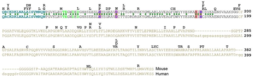
J. Dev. Biol. 2018, 6, 12                                                                                                    6 of 11
 J. Dev. Biol. 2018, 6, x FOR PEER REVIEW                                                                                   6 of 11

      Figure 4.
     Figure   4. The  Hh/DD‐peptidase
                    The Hh/DD-peptidase      family. (A) Diagram
                                                 family.            of the structure
                                                            (A) Diagram                 of the DD‐peptidases
                                                                              of the structure                  in bacteria
                                                                                                   of the DD-peptidases
      andbacteria
     in    animals.and
                     In bacteria,
                          animals. the peptidases
                                         In bacteria,are the
                                                         the C‐terminal
                                                              peptidasesdomains
                                                                             are theofC-terminal
                                                                                          larger proteins. In all cases,
                                                                                                       domains            the
                                                                                                                   of larger
      peptidase    domain   is predicted     to be located   in the  periplasmic     space.  In
     proteins. In all cases, the peptidase domain is predicted to be located in the periplasmic Rhizobium   Hh,   the   third
      transmembrane
     space.             domain
               In Rhizobium     Hh,(red)
                                      theisthird
                                            at thetransmembrane
                                                   same position asdomain
                                                                       the signal   sequence
                                                                                 (red)         in metazoan
                                                                                         is at the           Hh (red).
                                                                                                    same position      as (B)
                                                                                                                           the
      Lineup    of  the  Hh/DD‐peptidase         domains    of   bacteria   (green    background),
     signal sequence in metazoan Hh (red). (B) Lineup of the Hh/DD-peptidase domains of bacteria       Hedgling     (salmon
      background)
     (green           and HhsHedgling
              background),      (purple background).       Mutationsand
                                            (salmon background)         in the
                                                                             HhsZn(purple
                                                                                   2+ coordinating/DD‐peptidase motif
                                                                                             background). Mutations in
      defining
     the  Zn2+ residues    in Hedgling and Drosophila
                 coordinating/DD-peptidase                    Hh are indicated
                                                     motif defining   residues in  in Hedgling
                                                                                       bold. The and
                                                                                                  blue Drosophila
                                                                                                        columns indicate
                                                                                                                      Hh are
      residues  involved  in  Zn 2+ coordination and define the DD‐peptidase motif;       2+  the green
     indicated in bold. The blue columns indicate residues involved in Zn coordination and define the    columns    indicate
      residues involved
     DD-peptidase          in the
                       motif;  Ca2+green
                                     binding.   Varroaindicate
                                           columns     is typical for all non‐Drosophilid
                                                                residues   involved in Ca2+   insects. Aligned
                                                                                                binding.  Varroasequences
                                                                                                                   is typical
      are in the supplementary      file.
     for all non-Drosophilid insects. Aligned sequences are in the Supplementary file.
J. Dev. Biol. 2018, 6, 12                                                                           7 of 11

5. What Are the Possible Substrates for the Shh Peptidase Activity?
     The general properties of bacterial DD-peptidases and lysostaphins as modifiers of the bacterial
cell wall are likely shared with the bacHhs. The peptidoglycans that are a major component of
the bacterial periplasmic space have some similarities to the proteoglycans that are common in the
insect and vertebrate extracellular matrix. Both bacterial cell wall peptidoglycans and animal matrix
proteoglycans are large molecules in which polypeptides are covalently attached to chains of glycans.
In particular, the matrix heparin sulfate proteoglycans (HSPGs) bind Shh and can both negatively
and positively affect the Shh response [30–32]. Furthermore, mutations in ext genes that code for
glycosyltransferases that catalyze glycosaminoglycan in addition to the core protein disrupt Hh
signaling in vertebrates [33] and insects [34]. It is thus possible that functional conservation between
bacHhs and Shh is reflected in the ability of Shh to cleave or modify proteoglycans, thus affecting the
Shh response or distribution, independent of binding to the canonical receptors.
     Although any Shh antagonist could be a possible target for Shh peptidase activity, both Hhip and
Ptch1 are unlikely substrates, as they have the characteristics of metalloprotease inhibitors [19].

6. Drosophila Hh Is Not a Member of the Hh/DD-Peptidase Family
      Drosophila Hh (dmHh) has been the guide molecule for all Hh signaling since the discovery
that it is necessary for embryogenesis [1]. However, dmHh is unusual in that is does not have the
core sequences H-X(6)-D, H-X-H that define the Hh/DD-peptidase motif, but instead, dmHh has
H-X(6)-T, H-X-Y. Furthermore, the catalytically important E177 (mouse numbering) is mutated in
dmHh into a valine residue. The absence of the Hh/DD-peptidase motif is unique to all sequenced
Drosophilids; searching with the Drosophila sequence that encompasses the DD-peptidase motif
(ESLHYEGRAVTIATSDRDQSKYGMLARLAVEAGFDWVSYVSRRHIYCSVKS) places Drosophilids as
an outgroup to all other arthropods and deuterostomes. Drosophilids are the outlier because Hh proteins
in all other protostomes and deuterostomes retain the conserved Hh/DD-peptidase Zn2+ coordination
motif. Searching with a hypothetical dmHh peptide that conforms to the Hh/DD-peptidase motif
and E177 does no longer uniquely group with the Drosophilids, and has similar homology to several
vertebrate and insect Hhs. Given the high degree of HhN conservation between bacteria and eukaryotes
(Figure 3), the most plausible explanation for this is that Drosophilids have lost some of the activities
associated with the ancestral Zn2+ coordination domain. The loss of Zn2+ coordinating residues in
conjunction with the E177 homolog is no surprise in light of the likely loss of peptidase activity in
Drosophila Hh. The observation that vertebrate Shh is active in Drosophila embryos [35], but Drosophila
Hh is not active in vertebrates, further supports the notion that some aspects of Hh signaling present in
most animals are lost in fruit flies, but that both proteins can bind to Ptch. These observations question
to what extent the lessons learned in Drosophila embryos regarding Hh processing and signaling
can be directly applied to vertebrates, or even other arthropods, as all these animals maintain an
intact Hh/DD-peptidase domain. The study of Hh signaling in another insect, such a Tribolium [36],
might help to resolve how Drosophila Hh signaling is impacted by the loss of the DD-peptidase motif
in the ligand.

7. The Presence of bacHhs Suggests an Alternative Hypothesis Regarding the Evolution of
Modern Hh
     Hh genes are present in Cnidarians (corals and jellyfish), but not in sponges and protozoa [37].
However, another class of proteins that contain an N-terminal HhN-like domain is the Hedgling
proteins that can be found in Cnidarians, sponges and Choanoflagellates [38], which are protozoans.
In these Hedgling proteins, the HhN domain is followed by a large C-terminal domain that is related
to cell adhesion molecules. A plausible evolutionary path is that the HhN domain of Hedgling was
recombined to a Hog domain that is present in most genomes (Figure 5 green arrows). However,
the HhN domain of Hedgling does not contain a complete Hh/DD-peptidase motif, whereas, it is
likely that its putative ancestral form in the last universal common ancestor (LUCA) had an intact
J. Dev. Biol. 2018, 6, 12                                                                                      8 of 11

J. Dev. Biol. 2018, 6, x FOR PEER REVIEW                                                                       8 of 11
Hh/DD-peptidase domain. Although this evolutionary path from a LUCA DD-peptidase via Hedgling
to  Hh could
 could         have conceivably
        have conceivably           occurred
                            occurred  withoutwithout    horizontal
                                               horizontal           gene (Figure
                                                            gene transfer transfer5,(Figure 5, greenitarrows),
                                                                                     green arrows),     would
ithave
   would   have several
       required   required   several recombination
                          recombination    events. Theevents.    The
                                                        first event   first have
                                                                    would   evententailed
                                                                                   would the
                                                                                           have   entailed the
                                                                                               recombination
recombination     of theHh
 of the bacterial‐like   bacterial-like
                            domain from Hha domain    from atoDD-peptidase
                                             DD‐peptidase                     to form
                                                                form the Hedgling      the Hedgling
                                                                                     protein, and the protein,
                                                                                                       second
and   the second   event  would   have   involved   recombination    of the  HhN   domain
 event would have involved recombination of the HhN domain of Hedgling into modern Hh.      of Hedgling    into
modern
 FollowingHh.this
              Following   thisevolutionary
                   potential   potential evolutionary   path, the Hh/DD-peptidase
                                             path, the Hh/DD‐peptidase                motifhave
                                                                             motif would    would   have
                                                                                                 been     been
                                                                                                        lost in
lost in Hedgling   and  re-established
 Hedgling and re‐established in Hh.     in Hh.

       Figure 5. Models for the evolution of modern Hh proteins. The presence of both Hhs and Hedglings
      Figure 5. Models for the evolution of modern Hh proteins. The presence of both Hhs and Hedglings in
       in Cnidarians support the model that the HhN of Hh domain arose via a recombination of the
      Cnidarians support the model that the HhN of Hh domain arose via a recombination of the Hedgling
       Hedgling N‐domain. As Hedgling is present in at least some protozoa, it is plausible that it was
      N-domain. As Hedgling is present in at least some protozoa, it is plausible that it was derived from
       derived from a DD‐peptidase in the last universal common ancestor (LUCA). Alternatively, a Hh/DD‐
      a DD-peptidase in the last universal common ancestor (LUCA). Alternatively, a Hh/DD-peptidase
       peptidase could have entered a Cnidarian ancestor from a bacterium via horizontal gene transfer,
      could have entered 2+a Cnidarian ancestor from a bacterium via horizontal gene transfer, never losing
       never losing the Zn coordination in the process. Modern Hh was present in the Urbilaterian (the last
      the Zn2+ coordination in the process. Modern Hh was present in the Urbilaterian (the last common
       common ancestor of protostomes and deuterostomes) and was retained in most of its offspring. The
      ancestor of protostomes and deuterostomes) and was retained in most of its offspring. The loss of the
       loss of the Hh/DD‐peptidase motif in Drosophilids is derived, as is the loss of all Hh in some bilaterian
      Hh/DD-peptidase motif in Drosophilids is derived, as is the loss of all Hh in some bilaterian protostomes
       protostomes (such as roundworms).
      (such as roundworms).

      The presence of highly conserved HhN protein domains in bacteria suggests an alternate
      The presence
 hypothesis,        of horizontal
              in which  highly conserved    HhN protein
                                   bacHh//DD‐peptidase       domains
                                                          gene  transferinfrom
                                                                            bacteria suggeststoan
                                                                                a bacterium         alternate
                                                                                                 a Cnidarian
hypothesis,  in which  horizontal  bacHh//DD-peptidase    gene  transfer  from  a bacterium   to
 ancestor was followed by recombination of the HhN domain with a Cnidarian Hog domain, resulting a Cnidarian
ancestor  was followed
 in a modern            by recombination
               Hh gene (Figure             of theThis
                                5, red arrows).   HhN  possible    with a Cnidarian
                                                         domainevolutionary     path Hog domain,
                                                                                      would         resulting
                                                                                             have required
in a modern    Hh gene (Figure 5, red arrows).   This  possible  evolutionary   path  would  have
 only one recombination event and would not have involved the subsequent loss and re‐establishment  required
only  one
 of the   recombination event
        Hh/DD‐peptidase        andas
                          domain,   would   not have
                                      is indicated forinvolved   the subsequent
                                                        the evolutionary          loss
                                                                           path via    and re-establishment
                                                                                     Hedgling.  As the more
 parsimonious explanation it is perhaps a more plausible path for the evolution of modern Hh. The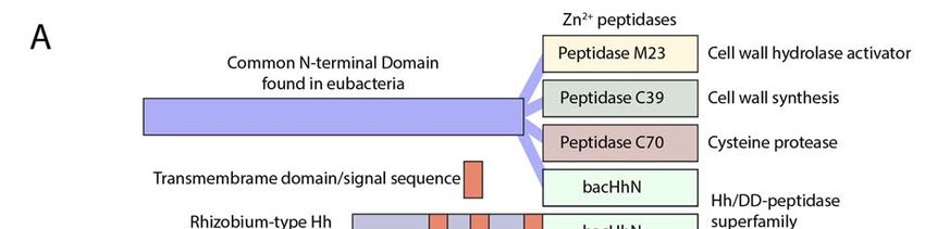
J. Dev. Biol. 2018, 6, 12                                                                                           9 of 11

of the Hh/DD-peptidase domain, as is indicated for the evolutionary path via Hedgling. As the
more parsimonious explanation it is perhaps a more plausible path for the evolution of modern Hh.
The higher conservation between ShhN and bacterial Hh/DD-peptidases than between ShhN and the
N-domain of Hedgling lends further support to the horizontal gene transfer model.

8. Conclusions
     Although the Zn2+ coordination domain of Shh is often referred to as its “pseudo catalytic”
domain [19,39,40], the remarkable similarity of most Hhs to bacterial Hh/DD-peptidases further
supports the notion that Shh functions as a peptidase during development. Some mutations found in
holoprosencephaly patients break the Hh/DD-peptidase motif, and negatively affect Hh signaling,
possibly by preventing autoproteolytic cleavage, indicating that the intrinsic Zn2+ peptidase activity of
Shh is critical to its function. The lack of the Hh/DD-peptidase domain in Drosophila Hh demonstrates
that it is evolutionary derived, and perhaps not the best model for Hh signaling in animals with
the ancestral Hh/DD-peptidase motif, like humans and mice. Finally, the presence of HhN protein
domains in some bacteria supports an alternate pathway for the evolution of Hh, via horizontal gene
transfer from bacteria into an Urbilaterian ancestor, resulting in the modern Hh which has retained
ancestral peptidase activity.

Supplementary Materials: The following are available online at http://www.mdpi.com/2221-3759/6/2/12/s1.
Funding: This research was funded by NIGMS grant number R01GM117090.
Conflicts of Interest: The author declares no conflict of interest.

References
1.    Nüsslein-Volhard, C.; Wieshaus, E. Mutations affecting segment number and polarity in Drosophila. Nature
      1980, 287, 795–801. [CrossRef] [PubMed]
2.    Echelard, Y.; Epstein, D.J.; St-Jacques, B.; Shen, L.; Mohler, J.; McMahon, J.A.; McMahon, A.P. Sonic hedgehog,
      a member of a family of putative signaling molecules, is implicated in the regulation of CNS polarity. Cell
      1993, 75, 1417–1430. [CrossRef]
3.    Hall, T.M.; Porter, J.A.; Beachy, P.A.; Leahy, D.J. A potential catalytic site revealed by the 1.7-A crystal
      structure of the amino-terminal signalling domain of Sonic hedgehog. Nature 1995, 378, 212–216. [CrossRef]
      [PubMed]
4.    Rebollido-Rios, R.; Bandari, S.; Wilms, C.; Jakuschev, S.; Vortkamp, A.; Grobe, K.; Hoffmann, D. Signaling domain
      of Sonic Hedgehog as cannibalistic calcium-regulated zinc-peptidase. PLoS Comput. Biol. 2014, 10, e1003707.
      [CrossRef] [PubMed]
5.    Porter, J.A.; Ekker, S.C.; Park, W.J.; von Kessler, D.P.; Young, K.E.; Chen, C.H.; Ma, Y.; Woods, A.S.; Cotter, R.J.;
      Koonin, E.V.; et al. Hedgehog patterning activity: Role of a lipophilic modification mediated by the
      carboxy-terminal autoprocessing domain. Cell 1996, 86, 21–34. [CrossRef]
6.    Porter, J.A.; Young, K.E.; Beachy, P.A. Cholesterol modification of hedgehog signaling proteins in animal
      development. Science 1996, 274, 255–259. [CrossRef] [PubMed]
7.    Taylor, F.R.; Wen, D.; Garber, E.A.; Carmillo, A.N.; Baker, D.P.; Arduini, R.M.; Williams, K.P.; Weinreb, P.H.;
      Rayhorn, P.; Hronowski, X.; et al. Enhanced potency of human Sonic hedgehog by hydrophobic modification.
      Biochemistry 2001, 40, 4359–4371. [CrossRef] [PubMed]
8.    Pepinsky, R.B.; Zeng, C.; Wen, D.; Rayhorn, P.; Baker, D.P.; Williams, K.P.; Bixler, S.A.; Ambrose, C.M.;
      Garber, E.A.; Miatkowski, K.; et al. Identification of a palmitic acid-modified form of human Sonic hedgehog.
      J. Biol. Chem. 1998, 273, 14037–14045. [CrossRef] [PubMed]
9.    Chamoun, Z.; Mann, R.K.; Nellen, D.; von Kessler, D.P.; Bellotto, M.; Beachy, P.A.; Basler, K. Skinny hedgehog,
      an acyltransferase required for palmitoylation and activity of the hedgehog signal. Science 2001, 293, 2080–2084.
      [CrossRef] [PubMed]
10.   Tukachinsky, H.; Kuzmickas, R.P.; Jao, C.Y.; Liu, J.; Salic, A. Dispatched and scube mediate the efficient
      secretion of the cholesterol-modified hedgehog ligand. Cell Rep. 2012, 2, 308–320. [CrossRef] [PubMed]
J. Dev. Biol. 2018, 6, 12                                                                                                     10 of 11

11.   Roelink, H.; Porter, J.A.; Chiang, C.; Tanabe, Y.; Chang, D.T.; Beachy, P.A.; Jessell, T.M. Floor plate and motor
      neuron induction by different concentrations of the amino-terminal cleavage product of sonic hedgehog
      autoproteolysis. Cell 1995, 81, 445–455. [CrossRef]
12.   Ohlig, S.; Farshi, P.; Pickhinke, U.; van den Boom, J.; Höing, S.; Jakuschev, S.; Hoffmann, D.; Dreier, R.;
      Schöler, H.R.; Dierker, T.; et al. Sonic hedgehog shedding results in functional activation of the solubilized
      protein. Dev. Cell 2011, 20, 764–774. [CrossRef] [PubMed]
13.   Hall, T.M.; Porter, J.A.; Young, K.E.; Koonin, E.V.; Beachy, P.A.; Leahy, D.J. Crystal structure of a Hedgehog
      autoprocessing domain: Homology between Hedgehog and self-splicing proteins. Cell 1997, 91, 85–97.
      [CrossRef]
14.   Tokhunts, R.; Singh, S.; Chu, T.; D’Angelo, G.; Baubet, V.; Goetz, J.A.; Huang, Z.; Yuan, Z.; Ascano, M.;
      Zavros, Y.; et al. The full-length unprocessed hedgehog protein is an active signaling molecule. J. Biol. Chem.
      2010, 285, 2562–2568. [CrossRef] [PubMed]
15.   Traiffort, E.; Dubourg, C.; Faure, H.; Rognan, D.; Odent, S.; Durou, M.-R.; David, V.; Ruat, M. Functional
      characterization of sonic hedgehog mutations associated with holoprosencephaly. J. Biol. Chem. 2004, 279, 42889–42897.
      [CrossRef] [PubMed]
16.   Inouye, K.; Kusano, M.; Hashida, Y.; Minoda, M.; Yasukawa, K. Engineering, expression, purification, and
      production of recombinant thermolysin. Biotechnol. Annu. Rev. 2007, 13, 43–64. [PubMed]
17.   Zeng, X.; Goetz, J.A.; Suber, L.M.; Scott, W.J.J.; Schreiner, C.M.; Robbins, D.J. A freely diffusible form of Sonic
      hedgehog mediates long-range signalling. Nature 2001, 411, 716–720. [CrossRef] [PubMed]
18.   Fuse, N.; Maiti, T.; Wang, B.; Porter, J.A.; Hall, T.M.; Leahy, D.J.; Beachy, P.A. Sonic hedgehog protein signals not
      as a hydrolytic enzyme but as an apparent ligand for patched. Proc. Natl. Acad. Sci. USA 1999, 96, 10992–10999.
      [CrossRef] [PubMed]
19.   Bosanac, I.; Maun, H.R.; Scales, S.J.; Wen, X.; Lingel, A.; Bazan, J.F.; de Sauvage, F.J.; Hymowitz, S.G.;
      Lazarus, R.A. The structure of SHH in complex with HHIP reveals a recognition role for the Shh pseudo
      active site in signaling. Nat. Struct. Mol. Biol. 2009, 16, 691–697. [CrossRef] [PubMed]
20.   Maun, H.R.; Wen, X.; Lingel, A.; de Sauvage, F.J.; Lazarus, R.A.; Scales, S.J.; Hymowitz, S.G. Hedgehog
      pathway antagonist 5E1 binds hedgehog at the pseudo-active site. J. Biol. Chem. 2010, 285, 26570–26580.
      [CrossRef] [PubMed]
21.   Odent, S.; Atti Bitach, T.; Blayau, M.; Mathieu, M.; Aug, J.; Delezo de, A.L.; Gall, J.Y.; Le Marec, B.; Munnich, A.;
      David, V.; et al. Expression of the Sonic hedgehog (SHH) gene during early human development and phenotypic
      expression of new mutations causing holoprosencephaly. Hum. Mol. Genet. 1999, 8, 1683–1689. [CrossRef] [PubMed]
22.   Hehr, U.; Pineda-Alvarez, D.E.; Uyanik, G.; Hu, P.; Zhou, N.; Hehr, A.; Schell-Apacik, C.; Altus, C.; Daumer-Haas, C.;
      Meiner, A.; et al. Heterozygous mutations in SIX3 and SHH are associated with schizencephaly and further expand
      the clinical spectrum of holoprosencephaly. Hum. Genet. 2010, 127, 555–561. [CrossRef] [PubMed]
23.   Roessler, E.; El-Jaick, K.B.; Dubourg, C.; Vélez, J.I.; Solomon, B.D.; Pineda-Alvarez, D.E.; Lacbawan, F.; Zhou, N.;
      Ouspenskaia, M.; Paulussen, A.; et al. The mutational spectrum of holoprosencephaly-associated changes
      within the SHH gene in humans predicts loss-of-function through either key structural alterations of the ligand
      or its altered synthesis. Hum. Mutat. 2009, 30, E921–E935. [CrossRef] [PubMed]
24.   Day, E.S.; Wen, D.; Garber, E.A.; Hong, J.; Avedissian, L.S.; Rayhorn, P.; Shen, W.; Zeng, C.; Bailey, V.R.; Reilly, J.O.; et al.
      Zinc-dependent structural stability of human Sonic hedgehog. Biochemistry 1999, 38, 14868–14880. [CrossRef]
      [PubMed]
25.   Hitzenberger, M.; Schuster, D.; Hofer, T.S. The Binding Mode of the Sonic Hedgehog Inhibitor Robotnikinin,
      a Combined Docking and QM/MM MD Study. Front. Chem. 2017, 5, 76. [CrossRef] [PubMed]
26.   Stanton, B.Z.; Peng, L.F.; Maloof, N.; Nakai, K.; Wang, X.; Duffner, J.L.; Taveras, K.M.; Hyman, J.M.; Lee, S.W.;
      Koehler, A.N.; et al. A small molecule that binds Hedgehog and blocks its signaling in human cells. Nat. Chem. Biol.
      2009, 5, 154–156. [CrossRef] [PubMed]
27.   Himmelstein, D.S.; Cajigas, I.; Bi, C.; Clark, B.S.; Van Der Voort, G.; Kohtz, J.D. SHH E176/E177-Zn(2+)
      conformation is required for signaling at endogenous sites. Dev. Biol. 2017, 424, 221–235. [CrossRef] [PubMed]
28.   Van Heijenoort, J. Peptidoglycan hydrolases of Escherichia coli. Microbiol. Mol. Biol. Rev. 2011, 75, 636–663.
      [CrossRef] [PubMed]
29.   Bochtler, M.; Odintsov, S.G.; Marcyjaniak, M.; Sabala, I. Similar active sites in lysostaphins and D-Ala-D-Ala
      metallopeptidases. Protein Sci. 2004, 13, 854–861. [CrossRef] [PubMed]
J. Dev. Biol. 2018, 6, 12 11 of 11
30. Carrasco, H.; Olivares, G.H.; Faunes, F.; Oliva, C.; Larraín, J. Heparan sulfate proteoglycans exert positive
and negative effects in Shh activity. J. Cell. Biochem. 2005, 96, 831–838. [CrossRef] [PubMed]
31. Capurro, M.I.; Xu, P.; Shi, W.; Li, F.; Jia, A.; Filmus, J. Glypican-3 inhibits Hedgehog signaling during
development by competing with patched for Hedgehog binding. Dev. Cell 2008, 14, 700–711. [CrossRef]
[PubMed]
32. Witt, R.M.; Hecht, M.-L.; Pazyra-Murphy, M.F.; Cohen, S.M.; Noti, C.; van Kuppevelt, T.H.; Fuller, M.;
Chan, J.A.; Hopwood, J.J.; Seeberger, P.H.; et al. Heparan sulfate proteoglycans containing a glypican 5 core
and 2-O-sulfo-iduronic acid function as Sonic Hedgehog co-receptors to promote proliferation. J. Biol. Chem.
2013, 288, 26275–26288. [CrossRef] [PubMed]
33. Siekmann, A.F.; Brand, M. Distinct tissue-specificity of three zebrafish ext1 genes encoding proteoglycan
modifying enzymes and their relationship to somitic Sonic hedgehog signaling. Dev. Dyn. 2005, 232, 498–505.
[CrossRef] [PubMed]
34. Bellaiche, Y.; The, I.; Perrimon, N. Tout-velu is a Drosophila homologue of the putative tumour suppressor
EXT-1 and is needed for Hh diffusion. Nature 1998, 394, 85–88. [PubMed]
35. Krauss, S.; Concordet, J.P.; Ingham, P.W. A functionally conserved homolog of the Drosophila segment
polarity gene hh is expressed in tissues with polarizing activity in zebrafish embryos. Cell 1993, 75, 1431–1444.
[CrossRef]
36. Villarreal, C.M.; Darakananda, K.; Wang, V.R.; Jayaprakash, P.M.; Suzuki, Y. Hedgehog signaling regulates
imaginal cell differentiation in a basally branching holometabolous insect. Dev. Biol. 2015, 404, 125–135.
[CrossRef] [PubMed]
37. Adamska, M.; Matus, D.Q.; Adamski, M.; Green, K.; Rokhsar, D.S.; Martindale, M.Q.; Degnan, B.M. The
evolutionary origin of hedgehog proteins. Curr. Biol. 2007, 17, R836–R837. [CrossRef] [PubMed]
38. Fairclough, S.R.; Chen, Z.; Kramer, E.; Zeng, Q.; Young, S.; Robertson, H.M.; Begovic, E.; Richter, D.J.;
Russ, C.; Westbrook, M.J.; et al. Premetazoan genome evolution and the regulation of cell differentiation in
the choanoflagellate Salpingoeca rosetta. Genome Biol. 2013, 14, R15. [CrossRef] [PubMed]
39. Pettigrew, C.A.; Asp, E.; Emerson, C.P. A new role for Hedgehogs in juxtacrine signaling. Mech. Dev. 2014,
131, 137–149. [CrossRef] [PubMed]
40. Ochi, H.; Pearson, B.J.; Chuang, P.-T.; Hammerschmidt, M.; Westerfield, M. Hhip regulates zebrafish muscle
development by both sequestering Hedgehog and modulating localization of Smoothened. Dev. Biol. 2006,
297, 127–140. [CrossRef] [PubMed]
© 2018 by the author. Licensee MDPI, Basel, Switzerland. This article is an open access
article distributed under the terms and conditions of the Creative Commons Attribution
(CC BY) license (http://creativecommons.org/licenses/by/4.0/).You can also read