Solving the enigma of posterolateral tibial plateau fractures, the clue protocol
←
→
Page content transcription
If your browser does not render page correctly, please read the page content below

Acta Orthop. Belg., 2021, 87, 125-136                                                   ORIGINAL STUDY

  Solving the enigma of posterolateral tibial plateau fractures, the clue protocol

                 Radwan G. Metwaly, Zeiad M. Zakaria, Mohamed A. Elgebeily, Hany El Zahlawy

                  From the Faculty of Medicine, Orthopedics Department, Ain Shams University, Cairo, Egypt

The study aim is to evaluate functional and radio-                                 INTRODUCTION
logical outcomes following a suggested protocol based
on the four-column classification for management of                 Through the last decade, tibial plateau fractures
posterolateral column tibial plateau fractures.                  have shown dramatic evolution in their diagnostic
A prospective cohort study was performed in level                prerequisites together with new surgical approaches,
I academic center on 42 patients with mean age
                                                                 reduction techniques and fixation implants being
of 36 years (22-59). Eleven patients had isolated
posterolateral column fractures whereas 31 patients
                                                                 implemented (1,2). This could be attributed to the
had associated columns fractures. According to the               three-column theory based on three-column CT
suggested protocol, all cases of isolated posterolateral         classification presented by Luo et al (3).
column fracture started treatment via arthroscopic                  Increased attention towards the coronal plane
evaluation of soft tissue injuries (menisci and liga-            fractures as predictors of reduction loss increased the
ments), arthroscopically assisted reduction and inter-           use of the posterior, posteromedial and posterolateral
nal fixation by rafting screws followed by ORIF if               approaches (2-6). With further understanding of the
plating was needed. If associated with other columns             column principle, better functional outcome and
fractures, columns were fixed sequentially in an                 overall results have been recorded (2).
anti-clockwise direction starting from anteromedial                 Posteromedial coronal fractures have previously
column.                                                          been studied well in the literature (either isolated
Average follow up was 26 months. Mean time to union
                                                                 or in bicondylar plateau fractures) with substantial
was 16.3 (12-22) weeks. No radiological evidence of loss
of coronal or sagittal alignment was detected at final
                                                                 agreement on the necessity of using posteromedial
follow up. Five patients had an average depression of            approach with anti-shear buttress plating of these
5 millimeters that did not need further intervention             fragments to avoid postoperative collapse (4,5,7).
at this short-term follow up. Mean KOOS was 81 (72-
88). The average knee range of motion was (0° - 127°).
One patient had temporary common peroneal nerve                  n Radwan G. Metwaly, AP
injury, one patient had deep infection and two had               n Zeiad M. Zakaria, PhD
superficial wound infection.                                     n Mohamed A. Elgebeily, PhD
implementing the suggested protocol gives good to                n Hany El Zahlawy, PhD
excellent radiological and functional results as regard            Faculty of Medicine, Orthopedics Department, Ain Shams
posterolateral tibial plateau fracture. A larger study             University, Cairo, Egypt.
group with longer follow up is needed.                           Correspondence : Hany El Zahlawy, Address : 18 Rabaa
                                                                   Housing Project, El Nasr city, Cairo, Egypt. Phone :
Keywords : Tibial plateau fractures ; postero-lateral              +201002508641, Fax : +20224509378.
column ; protocol.                                                 Email : hzahlawy@hotmail.com
                                                                   ©
                                                                     2021, Acta Orthopædica Belgica.

No benefits or funds were received in support of this study.
None of the authors have a conflict of interest.                             Acta Orthopædica Belgica, Vol. 87 - 1 - 2021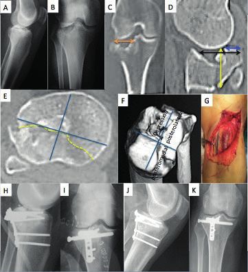
126                             r.g. metwaly, z.m. zakaria, m.a. elgebeily, h. el zahlawy

   On the other hand, the enigma of posterolateral
tibial plateau fractures as regards their best
fixation technique is still unsolved. Different new
classifications have been introduced and many
modifications of the posterolateral approach were
described (8-11). Unfortunately, none of these
previously described gates are completely innocent.
Innovating more biological and minimally invasive
techniques minimizes the complications associated
with such extensive approaches. However, this
should not be on the expense of biomechanical
stability or increased risk of neurovascular com-
promise (4,12,13).
   Arthroscopically assisted reduction and internal
fixation (ARIF) of tibial plateau fractures have                        Fig 1. — Diagram of patient selection in methodology.
shown good to excellent results even with long
term follow ups (14). It allows direct visualization of
the articular fracture surface with its more precise
                                                                                PATIENTS AND METHODS
reduction in comparison to image intensifier alone,
minimal soft tissue disruption and most importantly                     Between June 2015 and December 2018, a
it can address the associated meniscal and liga-                     prospective cohort clinical study was conducted
mentous injuries especially the lateral meniscus                     in a level I academic center on patients with tibial
tears, with their repair providing better functional                 plateau fractures. Informed consent was obtained
outcomes (15,16).                                                    from all individual participants included in the
   This study suggests a protocol based on precise                   study. All procedures performed in this study were
diagnosis for managing posterolateral plateau                        in accordance with the ethical standards of the
fractures (either isolated or associated with other                  institutional and national research committee. Poly-
columns injuries). Choosing the proper approach,                     traumatized patients, those with associated ipsila-
reduction technique and fixation implant facilitates                 teral limb fractures, open fractures, pathological
the postoperative rehabilitation, gives better func-                 fractures including stress insufficiency fractures and
tional outcome and prevents the progression of                       skeletally immature patients were excluded from
osteoarthritis.                                                      the study.

  Table 1. — Number of patients in the study regarding mechanism of the trauma, different types and whether isolated or combined
                                                   with other column(s) fracture
                                    Isolated posterolateral column             Combined posterolateral + other column(s)
                                                (n=11)                                          (n=31)
Mechanism of trauma                  Low energy       High energy             Low energy                       High energy
                                          8                 3                      8                               23
                                               χ2=2.273                                       χ2=7.258
                                               p=0.132                                       p=0.007**
Definitive surgery mean (days)            2                 5                      7                               10
3D CT classification                Split      Split      Depressed + Anterolateral + Posteromedial + Anteromedial Combination
                                             depressed
                                      2          3             6          14               4                 7              6
Posterolateral type                                                       Split            Split depressed           Depressed
                                                                            7                     11                     13
**p
solving the enigma of posterolateral tibial plateau fractures, the clue protocol                                     127

   From 122 tibial plateau fractures being treated,                      of soft tissue condition with a mean gap of 8 days
83 were included in this study. 3D-CT scan was                           between external and internal fixation. Four-column
done for the patients to analyze the fracture pattern.                   classification was used to plan the definitive surgical
Based on the four-column classification proposed                         approach, reduction technique and implants needed
by Chang SM et al (17), 42 patients (34.4%) had                          for fracture fixation.
posterolateral column involvement ; 11 with isolated                        The basic principles for fixation of intra-arti-
column fracture while 31 had their posterolateral                        cular fractures were applied in all cases to allow
column fracture associated with other columns                            early range of motion. Anatomical reduction and
fractures (figure 1).                                                    rigid stability of articular surface was followed by
   Mechanism of injury was recorded as either high-                      alignment reduction and relative stability of meta-
energy trauma (road traffic injury, fall from height                     physeal fracture with or without bone grafting. The
more than three meters and heavy blunt object                            suggested protocol as shown in (figure 2) was used
trauma) or low-energy trauma (fall from height less                      to plan the needed approaches and fixation implants.
than three meters, sports injury or slip on ground).                     In details, the protocol we propose is as follows :
A staged procedure as opposed to a single staged                            1. Posterolateral approach with posterior buttress
surgery was performed for the high-energy injuries                       plating with or without bone graft is done in :
to optimize the soft tissue condition for better                            ● Isolated posterolateral plateau fracture, split or
wound healing in 26 patients (table 1).                                        split depressed type with the height and width
   All patients were classified on initial X-rays                              of the cortical fragment more than 15mm (in
according to Schatzker. If a staged procedure was                              order to have a surface area which accepts at
proposed, 3D-CT scan was postponed till after                                  least two screws in the horizontal limb of the
initial reduction and traction with external fixation                          plate).
for better orientation of fracture fragments. The                           ● Isolated posterolateral plateau fracture, split
definitive fixation was postponed till improvement                             or split depressed type with the height and

Fig 2. — The “clue” protocol suggested to solve the enigma of posterolateral tibial plateau fractures (a) the height is the vertical
distance in the sagittal plane from the plateau surface till the distal extension of the cortical fragment (b) the width is the horizontal
distance in the coronal plane (both criteria of height and width are needed for decision making).
MIPPO = minimally invasive percutaneous plate osteo-synthesis. BG = bone grafting. PL = posterolateral. ARIF = arthroscopically
assisted reduction and internal fixation.

                                                                                      Acta Orthopædica Belgica, Vol. 87 - 1 - 2021
128 r.g. metwaly, z.m. zakaria, m.a. elgebeily, h. el zahlawy
width of the cortical fragment less than 15mm
but the extent of articular fragment of the split
segment more than 15 mm.
2. Per cutaneous rafting screws is done in :
● Isolated posterolateral plateau fracture, split
type with the height and width of the cortical
fragment less than 15mm and the extent of
articular fragment of the split segment also
less than 15 mm.
● Isolated posterolateral plateau fracture, split-
depression type with the height and width
of the cortical fragment less than 15mm and
the extent of articular fragment of the split
segment also less than 15 mm provided there
is no comminution (less than 3 fragments) and Fig 3. — The sequence of column fixation in associated
columns fracture of tibial plateau starting from the anteromedial
the extent of the depressed area in the plateau
fragment.
is less than its one third.
● Isolated posterolateral plateau fracture,
depression type and the depression is less than 7. Combined anterolateral and posterolateral
5mm. columns fractures : Extended anterolateral approach
● Isolated posterolateral plateau fracture, is used and rim plating for posterolateral fragment
depression type and the depression is more fixation (considered as if isolated depression or
than 5mm but extent of depression is less than split depression type with extent in the plateau more
one third of the plateau or not comminuted than one third)
(less than three fragments). When dealing with posterolateral column injury,
3. Extended anterolateral approach with posterior sequence of fracture reduction and fixation was
buttress plating with or without bone graft is done dictated by the diagnosis, whether it was isolated or
in the following conditions : combined injury. When combined with anteromedial
● Isolated posterolateral plateau fracture, column injury, anteromedial approach was utilized
split depressed or depression types and the according to the protocol. The whole procedure was
depression is more than 5mm but extent of performed in the supine position unless additional
depression is more than one third of the plateau posterior or posterolateral approaches were needed.
or comminuted (more than three fragments) Reduction of the anteromedial column was ini-
● Associated posterolateral with anterolateral tially done to obtain a stable leveled plateau (the
fractures cornerstone of reduction) followed by posterolateral
4. Extended anterolateral approach and postero- column fixation in the supine position when using
medial approach are done in multiple combinations rafting screws only. In cases needing buttress
of fracture patterns. plating via the additional approaches, the patients
5. Posterior approach is done in posterolateral were repositioned prone. In cases with combined
associated with posteromedial fractures or alterna- anterolateral column affection, all the procedure was
tively, we can use separate incisions : posteromedial completed again in the supine position, however the
and posterolateral approaches. reduction was performed from posterior to anterior
6. If anteromedial fracture is present in asso- using the extended anterolateral approach. With
ciation with posterolateral fracture then they are posteromedial column affection the procedure
managed as isolated posterolateral fracture and could be done either in supine position only using
medial minimally invasive percutaneous plate posteromedial approach or was completed in prone
osteosynthesis (MIPPO) position when posterolateral plating was needed
Acta Orthopædica Belgica, Vol. 87 - 1 - 2021solving the enigma of posterolateral tibial plateau fractures, the clue protocol 129
Fig 4. — Arthroscopic view of torn posterior horn of lateral Fig 5. — The railway creation with bone impactor for elevation
meniscus (PHLM) associated with posterolateral plateau of depressed plateau fracture.
fracture.
after arthroscopic articular depression reduction and widen the lateral joint space, an anterior cruciate
preliminary raft screws fixation (figure 3). ligament (ACL) guide system was used to insert
After anesthesia, patients were placed in a two guide wires from the anteromedial proximal
supine position on a radiolucent orthopedic table. tibia 10 cm from joint line towards the edge of the
With formal sterilization and draping of the depressed plateau to create a railway for the bone
affected limb, the calf was exposed to monitor graft impactor which was inserted after creating
edema and increased compartment pressure during a bone window in the anteromedial cortex (figure
arthroscopy. The contralateral iliac bone graft site 5). In cases with associated anteromedial column
was prepared to be harvested in cases where large fractures, the already present split in the medial
bone defects were found. Tourniquet was inflated cortex was used as a window for passage of the
150 mmHg above systolic pressure with antibiotic impactor. After adequate elevation of the depression
administration prior to its inflation. via the impactor was confirmed arthroscopically,
Knee arthroscopy was initially performed for a guide wire for rafting screw was inserted from
assessment of fracture lines, locations of articular lateral to medial to prevent collapse. In cases with
surface depression and associated meniscal/liga- comminuted depressed surface (plateau islands)
mentous injuries. It was performed in all isolated this step was repeated. The number and size of the
posterolateral fractures however it wasn’t the rafting screws were decided according to the surface
rule in all cases of the combined fractures. Pump area and degree of comminution. Rafting screws
pressure was controlled not to be over 40mmHg fixation was followed by reassessment of the joint
and an outflow superolateral portal was used. The space without varus support to confirm complete
joint cavity was washed removing blood clots and elevation of the depressed fragment(s) (figure 6).
partial synovectomy was done to clearly visualize Lateral meniscus tears were sutured with all-
the amount of depression and identify any meniscal inside meniscus implant Cinch® (Arthrex, Naples,
tears (figure 4). FL). Bony avulsions of ACL ligament from tibia
After orienting with the depressed area in the were transfixed into bone via no.5 Fiberwire®
posterolateral column, with the knee flexed not (Arthrex, Naples, FL) over bony tunnels in the
more than 90° and supported in varus position to anteromedial cortex. Medial meniscus tears were
Acta Orthopædica Belgica, Vol. 87 - 1 - 2021130 r.g. metwaly, z.m. zakaria, m.a. elgebeily, h. el zahlawy Fig 6. — Isolated posterolateral tibial plateau fracture (A, B) : preoperative X-rays with double line sign (C, D, E) : CT scan of split-depressed posterolateral fracture with fracture segment
solving the enigma of posterolateral tibial plateau fractures, the clue protocol                             131

second day with quadriceps muscle strengthening,                       The overall split posterolateral tibial plateau
anti-inflammatory anti-edema measures, non-                         fracture subtype was nine patients of which 3
weight bearing mobilization with crutches till six                  patients needed buttress plating while the rest could
weeks after which gradual assisted weight bearing                   be fixed with rafting screws only (these six fractures
progressed till full fracture union, knee range of                  were fulfilling the proposed criteria for screw only
motion limited to 30° in first three weeks, increased               fixation in split fractures). No bone grafting was
gradually to 90° at six weeks, after which the range                done for such a fracture whether when isolated or in
of motion was progressed to obtain full range within                association with other columns fracture.
the next six weeks.                                                    For the depressed subtype, 19 patients were
   Patients were evaluated regarding radiological                   included. Plating was done for 6 patients from
union time, range of motion, KOOS score and                         extended anterolateral approach while 13 patients
presence of any complications.                                      were fixed with screws. Bone grafting was done in
                                                                    15 patients. The 4 patients without grafting had a
                         RESULTS                                    pre-reduction depression of less than 5mm so screws
                                                                    were enough to fill the defect. This was statistically
   From the forty-two patients included in this                     significant as regard the need of bone grafting in
study, thirty-three were males and only nine                        depressed fractures to add stability for fixation with
patients were females. The mean age of the patients                 screws only specifically with ASIF techniques to fill
was thirty-six ranging from (22-59). As regard the                  the reduction pathway.
mechanism of injury, twenty-six fractures were                         Fourteen patients had a split-depressed subtype
due to high-energy mechanisms while sixteen were                    with statistically significant occurrence in association
due to low energy mechanisms with a statistically                   with other columns fracture. 10 patients were fixed
significant difference between the isolated and                     with plate while 4 had been fixed with screws only.
associated posterolateral plateau fractures with a                  Bone grafting was done in three patients. On the
higher incidence of associated plateau injuries in                  contrary of depressed only fractures, bone grafting
high energy trauma.                                                 in split-depressed fractures was not necessary and

                                Table 2. — Different fixation methods and the need for bone graft

                   Split subtype                        Split depressed                          Depressed
                          9                                    14                                   19
          Raft screws          Plating          Raft screws           Plating          Raft screws         Plating
               6                  3                  4                  10                 13                 6
                     X2=1.000                              X2=2.571                              X2=2.579
                     p=0.317                                p=0.109                               p=0.108
          Bone graft           No graft         Bone graft           No graft          Bone graft          No graft
                -                 9                  3                  11                 15                 4
                   ……………..                                 X2=4.571                              X2=6.368
                                                           p=0.033*                               p=0.012*
      *p
132 r.g. metwaly, z.m. zakaria, m.a. elgebeily, h. el zahlawy
Table 4. — KOOS score and average time of union whether fracture is isolated or combined
Parameters Fracture type
Isolated Combined
p value Significance
n=11 n=31
(Mean±SD) (Mean±SD)
KOOS 82.00±3.61 80.96±3.45 0.404 NS
Time of union 16.64±2.20 16.16±2.58 0.590 NS
NS=non-significant.
Table 5. — Patients’ data with loss of reduction after fixation
Fracture type Isolated/combined Gender Age Mechanism of Method of Application KOOS Time to
injury fixation of bone graft union
1 Split depressed Combined Male 45 Slippage Rafting _______ 83 17
screws
2 Depressed Combined Male 25 Road traffic Buttress Applied 82 18
plate
3 Depressed Combined Female 31 Hit by heavy Buttress Applied 80 14
object plate
4 Split depressed Combined Male 25 Road traffic Buttress ________ 74 15
plate
5 Split depressed Combined Male 29 Road traffic Buttress ________ 72 18
plate
this was also statistically significant but for the No compartment syndrome was recorded in any
favor of no bone grafting. (Table 2) of the cases. Single case of temporary common
All the patients had been followed for a mini- peroneal nerve palsy occurred where a posterolateral
mum of 24 months (average of 26 months). 16 approach was used that was improved after 4 months
patients had lateral meniscus tears that were with complete regain of function. 5 patients had loss
sutured with all inside technique, 3 with avulsed of reduction in the form of depression (2-5 mm) that
ACL that were repaired with transosseous sutures did not need further intervention in this short term
over bony tunnels and 3 had medial meniscus tears follow up, no special complaint as regard the pain
with only 1 case that needed repair while the other nor the function were recorded (table 5). There was
2 cases had small radial tears that were excised. no evidence of loss of the coronal or the sagittal
Fracture union was evidenced at a mean of 16.3 alignment at last follow up. One patient had deep
(12-22) weeks with no additional bone grafting. infection (in which posteromedial and extended
The mean KOOS score was 81 (72-88) at the last anterolateral approaches were done) where serial
follow up. The average knee range of motion was debridement was done with retained implants and
(0°-127°) with an average loss of 8° degrees of negative pressure wound therapy was used after
flexion in comparison to contralateral knee. There eradication of infection. Two patients had superficial
was no statistical significant difference as regard infection treated with extended antibiotics with no
the different three types of posterolateral column need for surgery. The patients returned to work in an
fractures in terms of functional and radiological average of 4 months postoperatively. Participation in
results whether occurring alone or in combination recreational sports activities started after 8 months.
with other columns fracture (Tables 3 & 4).
Comparing isolated and associated posterolateral DISCUSSION
column fractures, there was no statistical significant
difference as regard the average time of union and Many classification systems had been described
function (Tables 3 & 4). for tibial plateau fractures but none is ideal as regard
Acta Orthopædica Belgica, Vol. 87 - 1 - 2021solving the enigma of posterolateral tibial plateau fractures, the clue protocol 133
the inter-observer reliability and the prediction of different PLF modalities that has the least harmful
postoperative outcome. The inter-observer reliability risk (8,11,12,29-33).
increases with CT scan based classifications (17- The extended anterolateral approach described
21). An update of the three-column theory into four by Cho JW et al. (34) allows good visualization
columns had been suggested dividing the posterior of the PLF in the supine position. Combined
column into posteromedial and posterolateral anterolateral column fracture can be managed
columns (22-24). from the same approach. Other columns can be
In the past few years, different parameters also managed without changing patient position.
for mapping of plateau fractures and descriptive This approach carries the disadvantage of uneasy
criteria for the morphological characteristics of placement of posterior buttress plate that gives
the posterolateral fragment fractures (PLF) were better biomechanical stability specifically in split
illustrated including its height, angle and surface and split-depressed subtypes.
area (24-25). To our knowledge none linked these Chen HW et al. (35) proposed a treatment
different morphological characteristics to a clinical guide for posterior tibial plateau fractures based on
guide to choose the best surgical approach and different five types including both posteromedial
fixation implant. and posterolateral fragments. This classification was
PLF gained attention because of difficult ap- depending on retrospective analysis of 39 cases that
proaches and reduction techniques. Although many all had been approached from posterior, in contrast
surgical approaches are available for posterolateral to this study, they ignored the presence of associated
fragment, none is ideal in providing maximum other columns fractures and did not link the fixation
visualization and easy reduction and application implant with the fracture pattern.
of fixation implant that provides best mechanical Cho JW et al. (34) proposed a treatment algorithm
stability (26-28). for posterolateral fractures either as isolated or
In our series the incidence of PLF (whether iso- associated with other columns that was very
lated or combined with other columns) was 34.4%. beneficial. They were dependent on two approaches ;
Which is comparable to other published studies in extended anterolateral and the posterolateral ap-
literature (15%-44.2%). This incidence increased in proaches ; they dealt with the posterolateral fracture
comparison to that previously thought because of as if it is all the same and did not explain the
the use of CT scan (21,24,25) . different morphological patterns as depressed, split,
Surgical approaches for PLF can be divided into or split depressed and their influence on the choice
posterior, posteromedial and posterolateral with or of the approach and the fixation implant. They
without fibular head osteotomy. These approaches preferred the use of the newly introduced rim plates.
allow direct reduction of the fracture fragment In the current study the morphological pattern of
but with high risk of injury to vital neurovascular the fracture was identified and the management was
structures including the popliteal neurovascular applied according to the algorithm in (figure 2).
structures, common peroneal nerve, inferior lateral Meniscal and ligamentous injuries mainly
geniculate artery and anterior tibial vessels that lateral meniscal tears and ACL disruption affect
cross the interosseous ligament from posterior to postoperative functional outcome and need to be
anterior about five centimeters from the joint line addressed to accelerate rehabilitation programs.
limiting the distal extension of these approaches. ARIF showed good to excellent long-term out-
Associated anterior columns fractures cannot comes with more precise diagnoses, and provide
be accessed from these approaches and usually safe management when combined with the bio-
requires changing of patient position from prone to mechanically suitable implant (15,16,36). This goes
supine which prolong the operative time. Different hand in hand in the current study as arthroscopic
modifications of the posterolateral approaches were examination was done to all the patients and ideal
described. Unfortunately, there is no specific criteria soft tissue management was done to improve the
upon which surgeon can choose the approach in long term outcome for the patients.
Acta Orthopædica Belgica, Vol. 87 - 1 - 2021134 r.g. metwaly, z.m. zakaria, m.a. elgebeily, h. el zahlawy
From the previously mentioned problems the soft tissue injuries. Larger fragments will need to
need for a treatment algorithm for management of be buttressed with a plate that can be approached
tibial plateau fractures urge especially with PLF through a posterolateral approach after performing
either isolated or combined. The authors developed arthroscopic assessment and meniscal and liga-
this treatment protocol trying to guide surgeons mentous injuries management with exchange of
when faced with tibial plateau fractures with PLF patient position from supine to prone that facilitate
either isolated or in association with other column reduction of the fracture. Comminuted fractures
injuries. “Do no further harm” was the message and those with a depression > 1/3 tibial plateau
from this algorithm in order to choose the least can be buttressed from lateral or posterior with the
traumatic approach that provide the appropriate use of rim plates through an extended anterolateral
visualization for reduction and allow for fixation approach with or without posteroanterior rafting
that provide stability till fracture union. screws.
The morphological characteristics of the postero- For depressed fractures, the comminution and
lateral fragment were based on the work of Sohn the extent into the plateau are the main parameters
HS et al. (26). In cases of split and split-depressed determining the fixation implant. And they can be
fracture subtypes, the choice of fragment size dealt with as in split-depressed fractures.
(extent of articular surface in the split fragment) In cases of associated columns fractures, we
either more than or less than 1.5 cm was based on started our reduction from anteromedial column
morphological criteria found in literature describing that is the cornerstone upon which building up
the average surface area of PLF of about 14±6% of our plateau can be achieved in a manner of anti-
the plateau. Together with putting in consideration clockwise rotation. Posteromedial and posterolateral
that fragments height (cranio-caudal dimension) columns, posterior approach was used. Anterolateral
less than 1.5cm means that it is within the flare of the and posterolateral columns, extended anterolateral
posterolateral cortex, so plating of these fragments approach was used. Anteromedial and posterolateral
might not be necessary and applying a buttress plate columns, medial MIPPO (minimally invasive
will require a skewed contouring of the plate which percutaneous plate osteosynthesis) together with
is difficult and might annoy the patient flexion range treating the posterolateral as if isolated. And if
and rub on the soft tissue. more than two columns affected. Posteromedial
Again for the width (medio-lateral dimension) approach together with extended anterolateral
means that it represents a small surface area for the approaches were the gates to deal with the whole
transverse limb in the T-plate, otherwise using a one circumference of the plateau. This goes with many
third tubular or straight plate will lead to fixation of studies published in the literature supporting such a
the fragment with only one screw, so again plating protocol (1,6,7,9,12,27,34,35).
might not be necessary (25). That was comparable to In a series of seven cases done by Cho JW et
our detailed algorithm in the current study. al. (34) the union of fracture was achieved in all
For multifragmentary comminuted articular sur- the cases at a mean of 12 weeks after surgery (8-
face we aimed at the best rafting technique through 18 weeks) with no loss of reduction in their follow
plating that could maintain stability of the fragments up and the patients reported functional outcomes
till union together with or without bone grafting (37). using the Lysholm score (average, 88.7 ; range, 72-
Therefore, in cases of isolated PLF, small sized 95) with no evidence of complications related to
split fragments with height, width and extent less their approach. That was comparable to our cohort
than 1.5 cm or those split depressed with less except in the presence of complications as 1 of our
than three fragments not extending towards the patients developed temporary common peroneal
anterolateral column (solving the enigma of posterolateral tibial plateau fractures, the clue protocol 135
debridement was done with retained implants and 2. Wang Y, Luo CF, Zhu Y, et al. Updated Three-Column
negative pressure wound therapy was used after Concept in surgical treatment for tibial plateau fractures –
A prospective cohort study of 287 patients. Injury. 2016 ;
eradication of infection. 2 patients had superficial
47(7) : 1488-1496.
infection treated with extended antibiotics with 3. Luo CF, Sun H, Zhang B, et al. Three-column fixation for
no need for surgery. This increased incidence of complex tibial plateau fractures. J. Orthop Trauma. 2010 ;
complications was attributed to the relatively higher 24 : 683-92
number of patients in our series (42) in comparison 4. Bhattacharyya T, McCarty LP 3rd, Harris MB, et al.
to the 7 cases in the other study. The posterior shearing tibial plateau fracture : treatment
and results via a posterior approach. J Orthop. Trauma.
In a prospective cohort study of 287 patients
2005 ; 19 : 305-310.
done by Wang Y et al. (2), the mean time to union 5. Kim CW, Lee CR, An KC, et al. Predictors of reduction
was 13.5 weeks (range : 10-28), average functional loss in tibial plateau fracture surgery : Focusing on posterior
knee society score was 93.0 (range : 80-95). The coronal fractures. Injury. 2016 ; 47 : 1483-1487.
average range of motion of the fractured knees 6. Kokkalis ZT, Iliopoulos ID, Pantazis C. What’s new in
was 1.5-121.5°. Twelve patients suffered from the management of complex tibial plateau fractures? Injury.
superficial skin infection, one limited skin necrosis 2016 ; 47 : 1162-1169.
7. Weaver MJ, Harris MB, Strom AC, et al. Fracture pattern
and two developed wound dehiscence that all healed and fixation type related to loss of reduction in bicondylar
without operative management. These results tibial plateau fractures. Injury. 2012 ; 43(6) : 864-9.
were comparable to our series. 2 patients of their 8. Chen HW, Liu GD, Ou S, et al. Open reduction and
study had intra operative vascular injury repaired internal fixation of posterolateral tibial plateau fractures
primarily and 13 cases (4.5%) had temporarily through fibula osteotomy-free posterolateral approach. J.
common peroneal nerve injury compared to our Orthop. Trauma. 2014, 28(9) : 513-7.
9. Musahl V, Tarkin I, Kobbe P, et al. New trends and
series (2.4%) and none of our patients developed
techniques in open reduction and internal fixation of
vascular injury. fractures of the tibial plateau. J. Bone Joint Surg. Br. 2009 ;
The main limitation of this study is the small 91 : 426-433.
number of patients especially in the isolated 10. Stannard JP, Brown SL, Robinson JT, et al. Recon-
postero-lateral fragment group (11 patients) and struction of the postero-lateral corner of the knee.
the short term follow up which could not detect the Arthroscopy. 2005 ; 21 : 1051-1059.
11. Tao J, Hang DH, Wang QG, et al. The posterolateral
development of posttraumatic arthritis taking in
shearing tibial plateau fracture : treatment and results via
consideration that 5 patients had loss of reduction a modified posterolateral approach. Knee. 2008 ; 15 : 473-
accuracy in the form of depression of equal or less 479.
than 5mm. They had no special complaint as regard 12. Carlson DA. Posterior bicondylar tibial plateau fractures.
pain or function. Actually this might not progress J. Orthop. Trauma. 2005 ; 19 : 73-78.
into arthritis especially after repair of lateral 13. Frosch KH, Balcarek P, Walde T, et al. A new postero-
lateral approach without fibula osteotomy for the treatment
meniscal tear that protects the articular surface.
of tibial plateau fractures. J. Orthop. Trauma. 2010 ; 24(8) :
The suggested protocol may provide a clue to 515-20.
solve the enigma of planning a posterolateral tibial 14. Yi-Sheng Chan, Chih-Hao, Yang-Pin Lo, et al. Arthro-
plateau fracture whether isolated or associated scopy-Assisted Surgery for Tibial Plateau Fractures : 2- to
with other columns fractures as regard the proper 10-Year Follow-up Results. Arthroscopy. 2008 ; 24(7) :
approaches and fixation implants depending on 760-768.
proper diagnosis based on the CT four column 15. Elabjer E, Benčić I, Ćuti T, et al. Tibial plateau fracture
management : arthroscopically-assisted versus ORIF
classification without adding further harm to the procedure - clinical and radiological comparison. Injury.
patient. 2017 ; 48 Suppl 5 : S61-S64.
16. Hartigan DE, McCarthy MA, Krych AJ, et al. Arthro-
REFERENCES scopic-assisted reduction and percutaneous fixation of tibial
plateau fractures. Arthrosc. Tech. 2015 ; 2 ; 4(1) : e51-5.
1. Prat-Fabregat S, Camacho-Carrasco P. Treatment 17. Chang SM, Hu SJ, Du SC, et al. Four-quadrant/column
strategy for tibial plateau fractures : an update. EFORT classification of tibial plateau fractures. Int. Orthop. 2018 ;
Open. Rev. 2017 ; 1 : 225-232. 42(3) : 725-727.
Acta Orthopædica Belgica, Vol. 87 - 1 - 2021136 r.g. metwaly, z.m. zakaria, m.a. elgebeily, h. el zahlawy
18. Maripuri SN, Rao P, Manoj-Thomas A, et al. The 28. Musahl V, Tarkin I, Kobbe P, et al. New trends and
classification systems for tibial plateau fractures : how techniques in open reduction and internal fixation of
reliable are they? Injury. 2008 ; 39(10) : 1216-21. fractures of the tibial plateau. J. Bone Joint Surg. Br. 2009 ;
19. Millar SC, Arnold JB, Thewlis D, et al. A systematic 91 : 426-433.
literature review of tibial plateau fractures : What classi- 29. Stannard JP, Brown SL, Robinson JT, et al.
fications are used and how reliable and useful are they? Reconstruction of the postero-lateral corner of the knee.
Injury. 2018 ; 49(3) : 473-490. Arthroscopy. 2005 ; 21 : 1051-1059.
20. Taşkesen A, Demirkale İ, Okkaoğlu MC, et al. Intra- 30. Yu GR, Xia J, Zhou JQ, et al. Low-energy fracture of
observer and interobserver reliability assessment of tibial posterolateral tibial plateau : treatment by a posterolateral
plateau fracture classification systems. Eklem Hastalik prone approach. J. Trauma Acute Care Surg. 2012 ; 72 :
Cerrahisi. 2017 ; 28(3) : 177-81. 1416-1423.
21. Hu YL, Ye FG, Ji AY, et al. Three-dimensional 31. Sun H, Luo CF, Yang G, et al. Anatomical evaluation
computed tomography imaging increases the reliability of of the modified posterolateral approach for posterolateral
classification systems for tibial plateau fractures. Injury. tibial plateau fracture. Eur. J. Orthop. Surg. Traumatol.
2009 ; 40(12) : 1282-5. 2013 ; 23 : 809-818.
22. Krause M, Frosch KH. Response to the letter-to-the-editor 32. Huang YG, Chang SM. The posterolateral approach for
by Dhillon et al. “Simple four column classification can plating tibial plateau fractures : problems in secondary
dictate treatment for intra articular tibial plateau fractures hardware removal. Arch. Orthop. Trauma Surg. 2012 ;
much better than ten segment classification”, Injury. 2017 ; 132 : 733-734.
48(10) : 2369-2370. 33. Chang SM, Zheng HP, Li HF, et al. Treatment of isolated
23. Martínez-Rondanelli A, Escobar-González SS, Henao- posterior coronal fracture of the lateral tibial plateau
Alzate A, et al. Reliability of a four-column classification through posterolateral approach for direct exposure and
for tibial plateau fractures. Int. Orthop. 2017 ; 41(9) : 1881- buttress plate fixation. Arch. Orthop. Trauma Surg. 2009 ;
1886. 129 : 955-962.
24. Krause M, Menzdorf L, Preiss A, et al. Are there four 34. Cho JW, Samal P, Jeon YS, et al. Rim Plating of
tibial plateau columns? Yes there are, as illustrated by a Posterolateral Fracture Fragments (PLFs) Through a
postero-lateral apple-bite fracture. Response to a letter-to- Modified Anterolateral Approach in Tibial Plateau
the-editor. Int. Orthop. 2018 ; 42(2) : 443-446. Fractures. J. Orthop. Trauma. 2016 ; 30 : e362-e368.
25. Xiang G, Zhi-Jun P, Qiang Z, et al. Morphological 35. Chen HW, Chen CQ, Yi XH. Posterior tibial plateau
characteristics of posterolateral articular fragments in tibial fracture : a new treatment-oriented classification and sur-
plateau fractures. Orthopedics. 2013 ; 1 ; 36(10) : e1256- gical management. Int. J. Clin. Exp. Med. 2015 ; 8(1) : 472-
61. 9.
26. Sohn HS, Yoon YC, Cho JW, et al. Incidence and fracture 36. Warner SJ, Garner MR, Schottel PC, et al. The Effect
morphology of posterolateral fragments in lateral and of Soft Tissue Injuries on Clinical Outcomes After Tibial
bicondylar tibial plateau fractures. J. Orthop. Trauma. Plateau Fracture Fixation. J. Orthop. Trauma. 2018 ; 32(3) :
2015 ; 29(2) : 91-7. 141-147.
27. Zhang W, Luo CF, Putnis S, et al. Biomechanical analysis 37. Sun H, Zhu Y, He QF, et al. Reinforcement strategy
of four different fixations for the posterolateral shearing for lateral rafting plate fixation in posterolateral column
tibial plateau fracture. Knee. 2012 ; 19(2) : 94-8. fractures of the tibial plateau : The magic screw technique.
Injury. 2017 ; 48(12) : 2814-2826.
Acta Orthopædica Belgica, Vol. 87 - 1 - 2021You can also read