Snake venom gene expression is coordinated by novel regulatory architecture and the integration of multiple co-opted vertebrate pathways
←
→
Page content transcription
If your browser does not render page correctly, please read the page content below
Downloaded from genome.cshlp.org on July 2, 2022 - Published by Cold Spring Harbor Laboratory Press

Research

Snake venom gene expression is coordinated by novel
regulatory architecture and the integration of multiple
co-opted vertebrate pathways
Blair W. Perry,1,2 Siddharth S. Gopalan,1 Giulia I.M. Pasquesi,3 Drew R. Schield,4
Aundrea K. Westfall,1 Cara F. Smith,5 Ivan Koludarov,6 Paul T. Chippindale,1
Mark W. Pellegrino,1 Edward B. Chuong,3 Stephen P. Mackessy,5 and Todd A. Castoe1
1
 Department of Biology, University of Texas at Arlington, Arlington, Texas 76019, USA; 2School of Biological Sciences, Washington
State University, Pullman, Washington 99164, USA; 3Department of Molecular, Cellular, and Developmental Biology, University of
Colorado, Boulder, Colorado 80309, USA; 4Department of Ecology and Evolutionary Biology, University of Colorado, Boulder,
Colorado 80309, USA; 5School of Biological Sciences, University of Northern Colorado, Greeley, Colorado 80639, USA; 6Animal
Venomics Group, Justus Liebig University, Giessen, 35390, Germany

        Understanding how regulatory mechanisms evolve is critical for understanding the processes that give rise to novel pheno-
        types. Snake venom systems represent a valuable and tractable model for testing hypotheses related to the evolution of nov-
        el regulatory networks, yet the regulatory mechanisms underlying venom production remain poorly understood. Here, we
        use functional genomics approaches to investigate venom regulatory architecture in the prairie rattlesnake and identify
        cis-regulatory sequences (enhancers and promoters), trans-regulatory transcription factors, and integrated signaling cascades
        involved in the regulation of snake venom genes. We find evidence that two conserved vertebrate pathways, the extracellular
        signal-regulated kinase and unfolded protein response pathways, were co-opted to regulate snake venom. In one large ven-
        om gene family (snake venom serine proteases), this co-option was likely facilitated by the activity of transposable elements.
        Patterns of snake venom gene enhancer conservation, in some cases spanning 50 million yr of lineage divergence, highlight
        early origins and subsequent lineage-specific adaptations that have accompanied the evolution of venom regulatory archi-
        tecture. We also identify features of chromatin structure involved in venom regulation, including topologically associated
        domains and CTCF loops that underscore the potential importance of novel chromatin structure to coevolve when dupli-
        cated genes evolve new regulatory control. Our findings provide a model for understanding how novel regulatory systems
        may evolve through a combination of genomic processes, including tandem duplication of genes and regulatory sequences,
        cis-regulatory sequence seeding by transposable elements, and diverse transcriptional regulatory proteins controlled by a
        co-opted regulatory cascade.
        [Supplemental material is available for this article.]

The evolution of novel traits necessitates the evolution of novel                        Snake venom systems provide an ideal system to understand
gene regulatory architecture, which may involve the evolution                       how novel regulatory systems evolve and function owing to the
of novel regulatory features, co-option or “rewiring” of existing                   tractable size of gene families that comprise venom, as well as
networks and trans-activating regulatory molecules, or a combina-                   the direct relationships between venom gene (VG) expression,
tion of both (Babu et al. 2004; Teichmann and Babu 2004;                            phenotype, and fitness (Casewell et al. 2012; Rokyta et al. 2015;
Wagner and Lynch 2010). Understanding the processes by which                        Holding et al. 2016; Zancolli and Casewell 2020). At the core of
novel regulatory systems evolve to drive novel functions can pro-                   these venom systems is a highly specialized secretory organ: the
vide valuable insight into the fundamental question of how novel                    snake venom gland (Kochva et al. 1980; Mackessy 1991;
regulatory architectures arise, and provide new perspectives on                     Mackessy and Baxter 2006). Within the venom gland, multiple
the processes and constraints that govern adaptation. However,                      VG families contribute proteins to venom (Mackessy 2021).
understanding the features that coordinate gene regulation is par-                  Some VG families are arranged in tandemly arrayed gene clusters,
ticularly challenging in eukaryotes because it involves the simul-                  and most VGs are thought to have evolved through tandem dupli-
taneous action of cis- and trans-regulatory factors, chromatin                      cation of genes with other physiological functions, followed by
state, and three-dimensional interactions of chromatin, including                   neo- or subfunctionalization to venom-specific roles and expres-
the precise coordination of enhancers and promoters (Cremer and                     sion in the venom gland (Fry et al. 2008; Casewell et al. 2013;
Cremer 2001; Ragoczy et al. 2003; Misteli 2007; Delaneau et al.                     Vonk et al. 2013; Hargreaves et al. 2014b). Despite an extensive
2019).                                                                              body of literature on snake venom systems, the mechanistic under-
                                                                                    pinning of the regulation of snake VGs and the evolutionary

Corresponding author: todd.castoe@uta.edu
Article published online before print. Article, supplemental material, and publi-   © 2022 Perry et al. This article, published in Genome Research, is available under
cation date are at https://www.genome.org/cgi/doi/10.1101/gr.276251.121.            a Creative Commons License (Attribution-NonCommercial 4.0 International), as
Freely available online through the Genome Research Open Access option.             described at http://creativecommons.org/licenses/by-nc/4.0/.

32:1–16 Published by Cold Spring Harbor Laboratory Press; ISSN 1088-9051/22; www.genome.org                                         Genome Research                1
                                                                                                                                       www.genome.org
Downloaded from genome.cshlp.org on July 2, 2022 - Published by Cold Spring Harbor Laboratory Press

Perry et al.

origins of their regulatory architecture remain poorly understood.              main unknown. Additionally, the roles of chromatin structure
To date, a small number of studies have identified several transcrip-           and enhancer–promoter architecture in regulating snake venom
tion factors (TFs) and signaling pathways that may play a regulato-             systems, as well as the evolutionary origins of these features,
ry role in at least some species and some VG families, although no              have remained largely unexplored.
studies have identified the scope of the regulatory system that                      Here, we leverage inferences from diverse functional ge-
broadly regulates snake venom expression, or the evolutionary or-               nomics data to investigate the architecture and evolution of rattle-
igins of this regulatory system (Yamanouye et al. 2000; Kerchove                snake venom regulatory systems. Using integrated analyses of
et al. 2004, 2008; Hargreaves et al. 2014b; Junqueira-de-Azevedo                RNA sequencing (RNA-seq), assay for transposase-accessible
et al. 2015; Zancolli and Casewell 2020; Margres et al. 2021).                  chromatin using sequencing (ATAC-seq), chromatin immunopre-
These studies identified a set of TFs, including AP-1, NF-κB, and               cipitation sequencing (ChIP-seq) for insulators (CTCF), open
FOX-, NF1-, and GRHL-family members, for which identifiable                     promoters (H3K4me3), open enhancers (H3K27ac), and Hi-C
binding sites occur in the promoters of various VGs (Luna et al.                chromatin contact data, we investigate novel regulatory regions
2009; Nakamura et al. 2014; Schield et al. 2019; Margres et al.                 underlying VG expression and use these inferences to address
2021). Other studies have implicated the high-level regulatory in-              broad questions about the mechanistic architecture of snake ven-
volvement of alpha- and beta-adrenergic receptors and signaling                 om regulatory systems, as well as the evolutionary origins of this
through the ERK/MAPK pathway in initiating the venom produc-                    architecture.
tion cascade (Yamanouye et al. 2000; Kerchove et al. 2008); how-
ever, subsequent studies have performed little to investigate
putative venom TFs in light of higher-level regulatory networks.                Results
Recently, a study characterized regulatory pathways activated dur-
ing venom production in the prairie rattlesnake and found evi-                  VGs are highly expressed in the snake venom gland
dence for high activation of stress response pathways, including                The venom proteome of the prairie rattlesnake is dominated by
the unfolded protein response (UPR) (Perry et al. 2020). It remains             snake venom serine proteases (SVSPs), snake venom metallopro-
an open question, however, whether stress response mechanisms                   teinases (SVMPs), phospholipase A2 (PLA2), and peptide myotox-
may also play some role in in the regulation of VGs. Although                   ins (Fig. 1A), which derive from tandemly arrayed multicopy
these prior studies provide suggestions regarding regulatory mech-              gene families (Fig. 1B; Saviola et al. 2015; Schield et al. 2019).
anisms that may be involved in venom systems, the precise details               The myotoxin cluster is not assembled in the prairie rattlesnake ge-
of venom regulation, its constitutive components, and the degree                nome and was excluded from this study. A smaller fraction of the
to which distinct VGs are regulated by common mechanisms re-                    prairie rattlesnake venom proteome is composed of additional

     A                                  B

     C

Figure 1. Venom gene (VG) expression and structure in the prairie rattlesnake. (A) Proportions of venom protein components in prairie rattlesnake venom
(adapted from Schield et al. 2019). (B) Structure of tandemly duplicated VG families, with genes colored by relative gene expression at 1DPE. Relative ex-
pression is scaled separately per family to accentuate expression variation within families. (C) VGs with significant up-regulation in the 1DPE venom gland
(DESeq2 log2foldChange > 0 and IHW-corrected P-value < 0.05) compared with expression in all nonvenom tissues (stomach, pancreas, and liver).

2    Genome Research
         www.genome.org
Downloaded from genome.cshlp.org on July 2, 2022 - Published by Cold Spring Harbor Laboratory Press

                                                                                             Evolution of venom gene regulatory networks

proteins and peptides encoded by genes in small tandem arrays                   identified known TFs with elevated expression in the venom gland
(i.e., two genes) (Fig. 1B, “CRISPs”) or as individual genes                    compared with nonvenom tissues, resulting in 111 candidate TFs
(Saviola et al. 2015). For analyses, we grouped VGs by SVMP,                    (IHW-corrected P-value < 0.05 and Log2FoldChange > 1).
SVSP, and PLA2 families and a category of “other” VGs that com-                       Separately, we identified super-enhancer (SE) regions through-
prise the minor fraction of the venom proteome (Fig. 1C).                       out the genome using H3K27ac ChIP-seq data (see Methods). SEs
Within the major VG families, most paralogs are highly expressed                are long regions of elevated H3K27ac histone marker signal (consis-
in the venom gland compared with nonvenom gland tissues (Fig.                   tent with enhancer-associated open chromatin) that represent
1C). Other paralogs do not show venom gland–specific expression                 “hotspots” of regulatory activity and tend to occur near genes cen-
(i.e., SVMP 11) and likely do not contribute to secreted venom. Of              tral to tissue-specific functions, including TFs (Hnisz et al. 2013;
the “other” VGs, nine were significantly up-regulated in the ven-               Whyte et al. 2013). SE-associated genes (genes located within or ad-
om gland compared with nonvenom tissues (Fig. 1C, “other”);                     jacent to an SE; see Methods) in the venom gland are enriched for
“other” VGs not significantly up-regulated in the venom gland                   genes coding for TFs and venom components (Supplemental Fig.
are excluded from subsequent analyses.                                          S2). Of the 81 total SE-associated TFs identified, 36 are also up-reg-
                                                                                ulated in the venom gland relative to nonvenom tissues
                                                                                (Supplemental Fig. S1). Our combined set of up-regulated and/or
Candidate TFs associated with venom regulation                                  SE-associated TFs is composed primarily of protein-binding TFs
Because many TF binding-site (TFBS) motifs are short sequences,                 (Supplemental Table S2), of which 72 have known TFBS motifs in
naive searches of known TFBSs across large stretches of genomic se-             the nonredundant JASPAR database (Sandelin et al. 2004).
quences are likely to identify many putative TFBS motif sequences                     A subset of our candidate TFs has been implicated in venom
that are not functionally relevant or bound by the associated TF.               regulation in previous studies and/or is involved in broad mecha-
To focus on TFs and TFBSs with evidence of a functional role in                 nisms previously suggested to be involved in venom regulation
the venom gland and potentially in VG regulation, we identified                 (Fig. 2A). Several SE-associated candidate TFs are involved in adren-
a set of candidate TFs using two independent approaches (Fig.                   ergic receptor binding activity, and additional candidate TFs (e.g.,
2A; Supplemental Figs. S1, S2; Supplemental Table S2). First, we                FOS and JUN) are components of the AP-1 TF complex (Fig. 2A;

     A                                                  B

Figure 2. Candidate transcription factors (TFs) underlying VG regulation. (A) Candidate TFs involved in venom regulation, with the first three columns
representing three approaches for TF identification, and the last three columns indicating TF membership in functional categories. (B) Interactions between
candidate TFs and ERK (homolog of human MAPK1) based on STRINGdb. TFs with direct interactions with ERK are shown in dark blue, and TFs that interact
with these are shown in light blue. Node sizes are scaled by the number of interactions with other TFs in the network. TFs also involved in the unfolded
protein response (UPR) are highlighted in red.

                                                                                                                            Genome Research              3
                                                                                                                               www.genome.org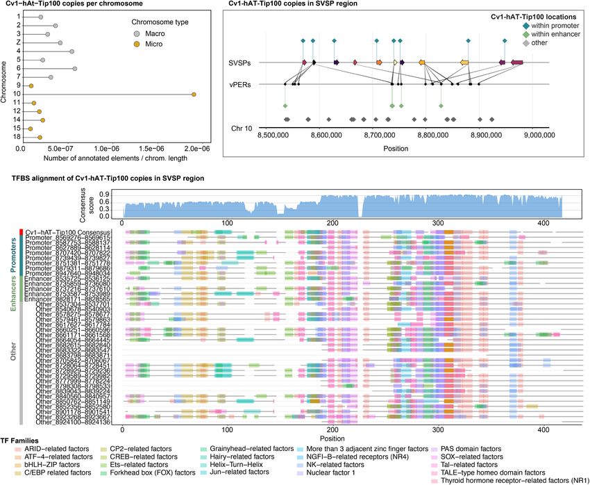
Downloaded from genome.cshlp.org on July 2, 2022 - Published by Cold Spring Harbor Laboratory Press

Perry et al.

Luna et al. 2009). An additional SE-associated candidate TF (EHF)              Supplemental Fig. S3). Genome-wide, ∼41% of promoters are
was previously suggested to play a role in regulating venom PLA2               inferred to be open in the postextraction venom gland, and genes
genes in Protobothrops (Nakamura et al. 2014). Other candidate                 with open promoters are more highly expressed (Supplemental
TFs have been identified as potential regulators of VGs in separate            Fig. S3A). This result is also observed for VGs when analyzed to-
genomic studies, including FOX-family TFs, GRHL1, NFIA and                     gether (Supplemental Fig. S3B). Within specific VG families,
NFIB, XBP1 (Fig. 2A; Margres et al. 2021; Schield et al. 2019).                SVMPs with open promoters are more highly expressed than those
EHF, NFIA, GRHL1, FOS, and JUN are both up-regulated and SE-as-                without (Mann–Whitney U test, P < 0.05) (Supplemental Fig. S3C),
sociated in the prairie rattlesnake venom gland, providing addi-               and all PLA2 gene promoters overlap with an H3K4me3 peak (Sup-
tional evidence for a role in venom regulation.                                plemental Fig. S3E). SVSPs and the combined group of “other” VGs
      Our results also highlight evidence for the co-option of two             with open promoters do not show significantly higher expression
conserved vertebrate pathways—ERK and UPR signaling—in the                     than those without open promoters (Supplemental Fig. S3D,F).
regulation of venom. Direct interactions with ERK were identified
for 19 candidate TFs (Fig. 2A,B) based on the STRINGdb database
                                                                               VG promoters are bound by TFs linked to ERK and UPR
(Szklarczyk et al. 2019), and 31 additional TFs had known interac-
                                                                               signaling pathways
tions with these 19 TFs (i.e., second-degree interactions with ERK;
Fig. 2B). This is validated by KEGG pathway analysis of the full can-          We identified TFBSs for candidate TFs with enrichment in VG pro-
didate TF set indicating overrepresentation of TFs involved in the             moters compared with the promoters of nonvenom genes. A total
ERK/MAPK signaling pathway (enrichment ratio = 2.4937, FDR =                   of 11, nine, and nine candidate TFBSs are significantly enriched in
0.044238). Additionally, 10 candidate TFs are members of the                   SVMP, SVSP, and PLA2 promoters, respectively, compared with
UPR pathway, including ATF6, BHLHA15, and CREB3L2, which                       nonvenom gene promoters (Supplemental Fig. S4; Supplemental
are both up-regulated and SE-associated in the venom gland (Fig.               Table S3). Several TFs are enriched in two of the three families
2A). As illustrated by the interaction network based on the                    (JUN, CREB3L2, ELK4, TFAP4, ARNT, FIGLA); none are enriched
STRINGdb, many of these UPR-associated TFs also interact with                  in promoters of all three (Supplemental Fig. S4). Using ATAC-seq
ERK and ERK-associated TFs (Fig. 2B).                                          footprinting scores calculated for each ATAC-seq sample with
                                                                               TOBIAS (Bentsen et al. 2020), we assessed whether specific candi-
                                                                               date TFBSs in VG promoters showed evidence of being bound by a
Promoter chromatin state and venom expression                                  protein (i.e., TF) in the postextraction venom gland. A TFBS was
We used H3K4me3 ChIP-seq data to investigate the relationships                 considered “bound” if the average footprint score across the
between gene expression and open promoters at 1DPE (Fig. 3;                    TFBS exceeded the bound threshold determined by TOBIAS

    A                                                B                                             C

Figure 3. Functional genomic landscape of major VG clusters in the venom gland. (A–C ) VG clusters with gene expression indicated by color (brighter
colors are more highly expressed) and inferred interactions with putative venom enhancer regions (vPERs). For the H3K27ac ChIP-seq, H3K4me3 ChIP-seq,
and ATAC-seq layers, normalized read density is shown, and peaks are highlighted by black bars at the top of each plot. Super-enhancers (SEs) are denoted
by yellow bars if present. Replicated ATAC-seq data are shown at bottom.

4       Genome Research
         www.genome.org
Downloaded from genome.cshlp.org on July 2, 2022 - Published by Cold Spring Harbor Laboratory Press

                                                                                             Evolution of venom gene regulatory networks

BINDetect (P-value < 0.001) in at least two of the three ATAC-seq                     As may be expected given the tandemly duplicated nature of
replicates. In PLA2 promoters, TBX3 is inferred to be bound in pro-             these VG arrays, promoter regions within each of the three VG
moters of the two highly expressed paralogs, PLA2A1 and PLA2B1                  families show a high degree of similarity (Supplemental Fig. S5),
(Fig. 4), but not in the lowly expressed PLA2C1 promoter. In                    and similarity is generally most evident between promoters of
SVMPs, only the promoter of the most highly expressed paralog                   the most highly expressed genes of each family. Inferred TFBS po-
(SVMP 6) contains TFBSs with consistent evidence of being bound                 sitions in promoter alignments are largely consistent among gene
(JUN and FOXL1; Fig. 4). The promoters of the two most highly ex-               family members, suggesting that TFBSs are conserved to a degree
pressed SVSPs, SVSP 5 and SVSP 7, both contain bound TFBSs for                  among paralogs within VG clusters (Supplemental Fig. S5).
ATF4 and JUN, with SVSP 5 also containing bound TFBSs for                             Only three of the “other” VGs have an ATAC-seq peak within
ARNT, CREB3L2, IRX2, and SOX10 (Fig. 4).                                        their inferred promoter region (CTL, VEGF1, and VEGF2)
      Multiple TFs putatively binding to VG promoters are involved              (Supplemental Fig. S6). Because these genes do not inhabit multi-
in, or are regulated downstream from, the ERK and UPR signaling                 gene arrays, candidate TFBSs were identified simply by scanning
pathways. These include TBX3, which is bound only in the two                    for potential binding motifs rather than by tests of enrichment.
most highly expressed PLA2s, as well as CREB3L2, CREB3L1,                       All three of these “other” VGs are found to have putatively bound
JUN, and ATF4, which show evidence of bound TFBSs in one or                     TFBSs for NFATC1, NFIX, MEIS1, and ELF5, with additional TFBSs
more of the three families (Fig. 4). These findings implicate central           inferred to be bound in one or two of these three promoters
roles of ERK and UPR signaling in the regulation of venom.                      (Supplemental Fig. S6).
                                                                                                                Analysis of potential novel TFBS
                                                                                                          motifs in regions of VG promoters with
                                                                                                          elevated ATAC-seq footprint scores iden-
                                                                                                          tified one motif (Supplemental Table S4).
                                                                                                          This motif is only present in promoters of
                                                                                                          the SVMP genes and shows similarity to
                                                                                                          the TFBSs of JUN, which is enriched in
                                                                                                          SVMPs and inferred to be bound in the
                                                                                                          SVMP 6 promoter (Supplemental Table
                                                                                                          S4; Fig. 4).

                                                                                                            Candidate TFBSs in promoters of
                                                                                                            nonvenom paralogs
                                                                                                            We scanned promoter sequences for all
                                                                                                            annotated nonvenom paralogs for the
                                                                                                            presence of TFBSs implicated in their cor-
                                                                                                            responding VG family (Supplemental
                                                                                                            Fig. S7; Supplemental Methods). Across
                                                                                                            the three families, two TFBSs are en-
                                                                                                            riched in the promoters of both the
                                                                                                            venom and corresponding nonvenom
                                                                                                            paralogs (ATF4 and ZBTB26 in SVSP and
                                                                                                            paralogs) (Supplemental Fig. S7B). A larg-
                                                                                                            er number of TFBSs implicated in VG
                                                                                                            regulation are present in one or more
                                                                                                            nonvenom paralogs of a given VG family
                                                                                                            despite not being enriched, and each
                                                                                                            group of nonvenom paralogs are en-
                                                                                                            riched for multiple TFBSs not implicated
                                                                                                            in regulation of related VGs (Supplemen-
                                                                                                            tal Fig. S7A).

                                                                                                            Roles of enhancers in regulating venom
                                                                                                            expression
                                                                                                            We used the activity-by-contact (ABC)
                                                                                                            model (Fulco et al. 2019) to identify puta-
                                                                                                            tive enhancer regions (PERs) and target
                                                                                                            genes using contact information (Hi-C),
                                                                                                            chromatin accessibility (ATAC-seq), and
Figure 4. TF binding sites (TFBSs) in VG promoter regions. TFBSs with the evidence for being bound in       histone modifications (H3K27ac ChIP-
promoter regions of the three major VG families. Diamonds indicate TFBSs inferred to be bound in at least   seq). We identified 1329 PERs across the
two out of three ATAC-seq replicates based on ATAC-seq footprinting analysis. Size of the diamond cor-
                                                                                                            genome associated with 837 genes in
responds to the number of bound TFBSs inferred in a given promoter (larger = more bound sites). Log-
scaled gene expression is shown above, and membership to relevant functional categories and TF families     the venom gland (Supplemental Table
is shown on the right. Genes for which no bound TFBSs were identified in the promoter are not shown.        S5), with an average of 1.87 inferred

                                                                                                                          Genome Research            5
                                                                                                                             www.genome.org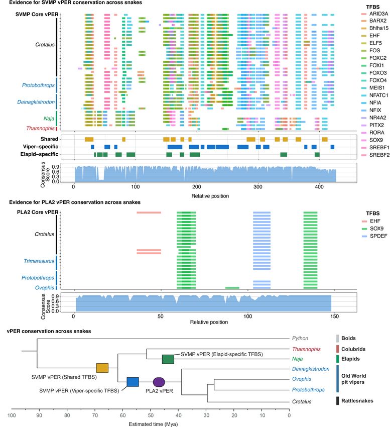
Downloaded from genome.cshlp.org on July 2, 2022 - Published by Cold Spring Harbor Laboratory Press

Perry et al.

PERs per gene. PERs are most commonly located in intergenic re-          vPERs aligns reasonably well (Supplemental Fig. S17B). We explore
gions (∼70% of all PERs), and the median distance between PERs           SVSPs further below. Alignments of SVMP, SVSP, and PLA2 vPERs
and associated genes was 9331 bp. VGs are targeted by a total of         were manually trimmed to the most consistently homologous re-
51 PERs (venom PERs [vPERs]). vPERs tend to be located relatively        gions; a consensus sequence for these was generated to represent
close to target genes with a median distance of 5614 bp. The four        a representative “core” enhancer sequence for each VG family
most highly expressed paralogs in the SVMP cluster are each              (Supplemental Fig. S17).
associated with an individual vPER located ∼4 kb upstream of                  De novo motif analysis identified one unannotated motif
the TSS (Fig. 3A). vPERs in the SVSP cluster are more variable in        that is enriched in putatively bound regions of vPERs compared
terms of the number of vPERs per gene and their location relative        with regions in nonvenom gene PERs (Supplemental Table S4), al-
to their target gene (Fig. 3B). All but one PLA2 vPERs are inferred to   though this motif is not present in all vPERs. This motif shows se-
regulate multiple genes, with two vPERs inferred to regulate both        quence similarity to ETS-related factor TFBS motifs (Supplemental
highly expressed PLA2 genes (PLA2A1 and PLA2B1) (Fig. 3C).               Table S4); several candidate TFs within this TF family are enriched
Thirteen vPERs are associated with seven “other” VGs (Supple-            and putatively bound in all three major venom families (Fig. 5A).
mental Figs. S8–S14).

Patterns of enhancer TFBSs and links to ERK and UPR signaling            VG enhancer sequences are conserved across species
A greater number of TFs were found to be enriched for TFBSs              Consistent with evidence that vPERs are relevant regulatory se-
in vPERs compared to promoters, with 22, 29, and four TFs en-            quences, we found high-similarity hits to prairie rattlesnake core
riched in SVMP, SVSP, and PLA2 vPERs, respectively (Fig. 5A;             vPER sequences in other venomous snake species, suggesting
Supplemental Table S2). Two TFBSs are found to be enriched in            that vPER sequences for SVMP and PLA2 are conserved among dis-
vPERs of all three families (SOX9 and RORA), and 11 enriched             tantly related lineages (Fig. 6A,B; Supplemental Figs. S18, S19;
TFBSs are found in both SVMP and SVSP vPERs (Fig. 5A). Based             Supplemental Table S6). Many of these orthologous sequences
on ATAC-seq footprinting scores, putatively bound TFBSs are iden-        show substantial sequence similarity within pit vipers, including
tified in five SVMP vPERs, including vPERs associated with three of      conservation of predicted TFBSs among species spanning at least
the four most highly expressed SVMP paralogs (Fig. 5A,B). Several        ∼40 million yr (MY) of divergence (Fig. 6; Kumar et al. 2017). We
of these TFs, including ELF5, ARID3A, NFIX, and MEIS1, are also          also identified similar sequences to the SVMP core vPER in a cobra
inferred to be bound in more than half of the identified SVSP            (Naja naja) and a rear-fanged colubrid (Thamnophis sirtalis), sug-
vPERs (Fig. 5A). SOX9 is present and bound in all three PLA2             gesting a degree of conservation of this enhancer sequence across
vPERs, whereas RORA and SPDEF are each bound in two vPERs                ∼70 MY of divergence (Fig. 6A,C). Many TFBS positions in BLAST
(Fig. 5A). A TFBS for EHF, which was previously implicated in            hits to the SVMP vPER were present in the majority of both viperid
PLA2 regulation (Nakamura et al. 2014), is putatively bound in a         and elapid sequences (“shared” in Fig. 6A; Supplemental Fig. S19).
single PLA2 vPER inferred to target both of the highly expressed         Other TFBSs are specific to either viperid or elapid sequences, sug-
PLA2 genes (PLA2A1 and PLA2B1) (Fig. 5A). Eight vPERs targeting          gesting independent gain (or differential retention) of TFBSs in
five of the “other” VGs are found to have candidate TFBSs with ev-       these distinct lineages (“viper-specific” and “elapid-specific” in
idence of being bound (Supplemental Fig. S15). TBX3 is inferred to       Fig. 6A; Supplemental Fig. S19). These include several TFs with di-
be bound to all eight of these vPERs, and multiple additional TFs        rect interactions to ERK (EHF, FOS, FOXO3, and FOXO4) and one
associated with ERK signaling, including ELF5, EHF, JUN, ATF4,           UPR-related TF (BHLHA15; Supplemental Fig. S19). Evaluating the
and several FOX family TFs, have evidence of binding a subset of         conservation of vPERs associated with the SVSP cluster was not fea-
these vPERs. Additionally, several TFs inferred to bind to a subset      sible because of the high-copy number of this vPER sequence in
of vPERs are involved in UPR signaling (ATF4, BHLHA15, and               the rattlesnake and in other snake genomes, which we explore in
DDIT3::CEBPA) (Supplemental Fig. S15).                                   detail below.
      Multiple TFs with putatively bound TFBSs in vPERs of one or
more VG families have known associations with ERK signaling
(Fig. 5A, right). Candidate TFBSs inferred to be bound in “other”
VG vPERs and with known interactions with ERK include TBX3,              vPERs are not associated with nonvenom paralogs
which is inferred to be bound in all “other” vPERs, among others         We surveyed the prairie rattlesnake genome, and nonvenom paral-
(Supplemental Fig. S15). TFs involved in the UPR pathway are             ogs specifically, for the presence of sequences similar to vPER re-
found to primarily bind to vPERs for SVSPs (Fig. 5A), although           gions for the three major VG families (Supplemental Methods).
BHLHA15 is inferred to bind all SVMP vPERs and a vPER interact-          For PLA2s and SVMPs, BLAST only returned hits to the immediate
ing with one “other” VG vPER (CRISP 1) (Supplemental Fig. S15),          VG clusters, most of which are hits to other identified vPERs
and DDIT3 is inferred to bind to vPERs for CRISP 1 and Vespryn           (Supplemental Fig. S20A,C). In the SVMP region, one BLAST hit
(Supplemental Fig. S15).                                                 is upstream of SVMP 11 (Supplemental Fig. S20A). This hit is locat-
      Within each VG family, multiple vPERs show considerable se-        ed near the annotated nonvenom SVMP paralogs adjacent to the
quence similarity (Supplemental Figs. S16, S17). This is most evi-       SVMP cluster, although it does not occur within an ATAC-seq or
dent for SVMP vPERs, the majority of which align well and show           H3K27ac peak and therefore is unlikely to act as an enhancer in
conserved positioning of many putatively bound TFBSs                     the venom gland. For PLA2 vPERs, BLAST identified a region span-
(Supplemental Fig. S16A). Among PLA2 vPERs, two vPERs align              ning the third exon of the PLA2gIIe nonvenom paralog with high
considerably well (vPER37 and vPER38) (Supplemental Fig.                 similarity to the core PLA2 vPER sequence (Supplemental Fig.
S16B); both of these vPERs are inferred to regulate PLA2B1, and          S20C). Conversely, the SVSP core vPER BLAST search returned
vPER38 putatively regulates both PLA2A1 and PLA2B1. Less consis-         approximately 10,000 genome-wide hits (Supplemental Figs.
tent similarity is observed among SVSP vPERs, and only a subset of       S20B, S21).

6    Genome Research
       www.genome.org
Downloaded from genome.cshlp.org on July 2, 2022 - Published by Cold Spring Harbor Laboratory Press

                                                                                             Evolution of venom gene regulatory networks

           A

           B

Figure 5. TFBSs in vPERs. TFBSs with evidence for being bound in vPERs of the three major VG families. (A) Diamonds indicate TFBSs inferred to be bound
in at least two out of three ATAC-seq replicates based on ATAC-seq footprinting analysis. Size of the diamond corresponds to the number of bound TFBSs
inferred in a given promoter (larger = more bound sites). Log-scaled gene expression is shown above, and membership to relevant functional categories and
TF families is shown on the right. For vPERs that target multiple genes, the averaged gene expression of target genes is shown (pink bars). Genes for which
no bound TFBSs were identified in the promoter are not shown. (B) An example alignment of vPER regions for SVMPs. Colored bars indicate bound TFBSs,
with bar height scaled by ATAC-seq footprint score (evidence of site being bound). Bars above and below the line indicate TFBSs inferred on the forward and
reverse strand, respectively. Expression of target genes is shown on the right, and the consensus score for the alignment is shown below. SVMP vPERs with no
bound TFBSs are not shown.

SVSP regulatory sequences are associated with                                   between SVSP vPER sequences and transposable elements (TEs)
transposable elements                                                           by comparing SVSP vPER sequences to annotated TEs from the
                                                                                prairie rattlesnake (Schield et al. 2019) and other snakes
The core SVSP vPER sequence yielded numerous genome-wide                        (Pasquesi et al. 2018). Although TEs are abundant in all three major
BLAST hits against the Crotalus viridis genome, suggesting it may               VG clusters, only the SVSP region showed an elevated abundance
be related to a repetitive element. We investigated potential links             of annotated repeats compared to the chromosomal median

                                                                                                                             Genome Research              7
                                                                                                                                www.genome.org
Downloaded from genome.cshlp.org on July 2, 2022 - Published by Cold Spring Harbor Laboratory Press

Perry et al.

    A

    B

    C

Figure 6. Conservation of putative VG enhancer sequences across snakes. Alignment and conservation of TFBSs in putative VG enhancer regions (vPERs)
among species of venomous snakes for SVMPs (A) and PLA2 vPERs (B). In A, shared and lineage-specific TFBSs between viperid (Crotalus, Protobothrops, and
Deinagkistrodon) and elapid (Naja) sequences are indicated below the alignment. (C ) Tree indicating divergence of venomous snake lineages and a hypoth-
esis for the origin and conservation of SVMP and PLA2 PERs.

(Supplemental Fig. S22A). Further, multiple SVSP vPER sequences               prevalent on Chromosome 10, which houses the SVSP cluster
share high homology with the consensus sequence of an annotat-                (Fig. 7A). Cv1-hAT-Tip100 elements are abundant within the
ed DNA transposon (a DNA-hAT-Tip100; referred to hereafter as                 SVSP array, with eight of 11 SVSP promoters and five of 17 vPERs
Cv1-hAT-Tip100). This TE sequence is significantly enriched for               overlapping one or more Cv1-hAT-Tip100 elements (Fig. 7B).
overlap with SVSP promoters and vPERs compared to the genomic                 Sequence divergence estimates between genome-wide Cv1-hAT-
background (one-tailed Fisher’s exact test; P = 5.27 × 10⁻¹⁴) and is          Tip100 elements and those in the SVSP region indicate that these

8    Genome Research
        www.genome.org
Downloaded from genome.cshlp.org on July 2, 2022 - Published by Cold Spring Harbor Laboratory Press

                                                                                     Evolution of venom gene regulatory networks

    A                                                B

    C

Figure 7. DNA transposons have rewired SVMP venom cluster regulatory networks. (A) Cv1-hAT-Tip100 copies per chromosome in the prairie rattle-
snake, normalized by chromosome length. (B) SVSP gene array and vPER inferences, with Cv1-hAT-Tip100 copies shown as colored diamonds. (C)
Alignment of SVSP-local Cv1-hAT-Tip100 copies with the genome-wide consensus. TFBSs enriched in SVSP promoters or enhancers are colored based
on TF families. Faded regions represent alignment gaps.

Cv1-hAT-Tip100s were active/inserted within the last 75 MY               2019) and incorporated CTCF ChIP-seq data to investigate the role
(Supplemental Fig. S23). Multiple TFBSs predicted to be important        of CTCF binding and insulation in VG regions (Fig. 8). Only the
to SVSP regulation are present in the estimated ancestral consensus      PLA2 VG cluster falls entirely within a single TAD, with SVMP
Cv1-hAT-Tip100 sequence and are largely conserved in the SVSP            and SVSP clusters each spanning multiple TADs (Fig. 8A,B). The
region copies (Fig. 7C). There are also multiple instances in which      most highly expressed genes in the SVMP cluster (SVMP 6–10) oc-
a small number of single-base substitutions from the consensus           cupy a single TAD, which also contains nearly all H3K27ac and
have potentially led to the gain/loss of new TFBSs in SVSP Cv1-          H3K4me3 ChIP-seq peaks in this region, suggesting regulatory iso-
hAT-Tip100 elements implicated as regulatory sequences (Fig.             lation of highly expressed genes (Fig. 8B). The most highly ex-
7C). Although several repeat elements overlap to some extent             pressed SVSPs are also within a single TAD along with several
with particular vPERs or promoters in SVMP and PLA2 regions,             lowly expressed SVSPs. Two other lowly expressed SVSPs (SVSP 1
none of these repeats show consistent and broad overlap with mul-        and SVSP 2) are contained within an adjacent TAD (Fig. 8B).
tiple regulatory regions as seen in the SVSP region (Supplemental             Only the relatively small PLA2 cluster is contained within a
Fig. S22B–D).                                                            single CTCF-bound chromatin loop (Fig. 8D). Numerous chroma-
                                                                         tin loops in the SVMP and SVSP regions indicate more complex
                                                                         regulatory substructure in these clusters (Fig. 8D). Evidence for
Chromatin organization contributes to the precision of venom             bound CTCF is frequent within major VG regions despite not
regulation                                                               always occurring terminal ends of inferred chromatin loops (Fig.
We inferred topologically associated domains (TADs) and chroma-          8C,D), possibly indicating other regulatory roles of CTCF in these
tin loops using Hi-C data from 1DPE venom glands (Schield et al.         regions. Multiple “other” VGs are contained within chromatin

                                                                                                                  Genome Research           9
                                                                                                                    www.genome.org
Downloaded from genome.cshlp.org on July 2, 2022 - Published by Cold Spring Harbor Laboratory Press
Perry et al.
A
B
C
D
E
F
G
Figure 8. Chromatin structure and organization associated with VG arrays. (A) Hi-C interaction heatmap (10-kb resolution) of 2-Mb regions centered on
VG arrays. Brighter colors indicate higher contact frequency. (B) Topologically associated domains (TADs) across VG arrays. (C) CTCF ChIP-seq density, with
black bars at the top indicating ChIP-seq peaks and triangles indicating peaks centered on a verified CTCF binding site. (D) Chromatin loops inferred from
Hi-C data that span VG arrays. Red loops indicate CTCF–CTCF bound loops, defined by the presence of a CTCF ChIP-seq peak centered around a CTCF motif
within 10 kb of both chromatin loop ends. (E) Venom array genes and inferred vPER-promoter interactions. (F ) Simplified ChIP-seq and ATAC-seq data, with
points indicating the location of ChIP/ATAC-seq peaks and yellow bars indicating SEs if present. (G) Hypotheses for three-dimensional loop structures of VG
regions.
loops, many of which were inferred to be CTCF bound chromatin accessibility and genomic organization that direct the
(Supplemental Figs. S24–S32). precise regulation of snake venom production. Together, our find-
The two lowly expressed nonvenom paralogs adjacent to the ings reveal that distinct genomic processes and features, including
SVMP cluster occupy a TAD that is distinct from highly expressed tandem duplication, compact regulatory structure, and the seed-
SVMPs, and chromatin loops suggest that they are excluded from ing of nascent regulatory sequences by TEs, likely contributed to
the loop housing active SVMP genes (Fig. 8D,E). In contrast, the evolution of novel regulatory mechanisms in different VG
the nonvenom paralog in the PLA2 cluster (PLA2gIIE) resides with- families.
in the same TAD, SE, and CTCF-bound loop (Fig. 8D–F). This Our findings support a model in which different VG families
paralog also shows higher expression in the venom gland com- evolved regulatory connections to common upstream signaling
pared with nonvenom tissues, consistent with being “linked” to, networks triggered by venom depletion (ERK signaling) and fur-
or at least not entirely isolated from, venom PLA2 regulatory mech- ther enhanced by cellular stress responses (UPR signaling). This
anisms (Fig. 1B). Hypotheses are illustrated for local chromatin overarching regulatory network involves many TFs previously
structure of each venom cluster in Figure 8G based on inferences speculated to be involved in venom regulation in different gene
from TAD, chromatin loop, CTCF binding, and gene expression families and species, and links preliminary findings from indepen-
data sets. dent studies with newly identified mechanisms to explain the
global regulation of a venom system (Fig. 9A). Additionally,
many TFs identified (e.g., AP-1 components, CREB3, and FOX fam-
Discussion ily TFs) are “pioneer” TFs that regulate local chromatin accessibil-
Key discoveries in this study frame a model of the regulatory archi- ity and recruit histone modifying proteins, and may therefore
tecture underlying snake venom systems and provide an example directly regulate chromatin structure and accessibility required
of how multiple genomic mechanisms may together establish a for the expression of VGs (Biddie et al. 2011; Zaret and Carroll
novel regulatory system for a polygenic trait. Our findings provide 2011; Fane et al. 2017; Jacobs et al. 2018; Khan and Margulies
evidence for evolutionary co-option of the ERK and UPR pathways, 2019).
acting through a suite of TFs that bind promoters and newly dis- We primarily focused on larger VG families because within-
covered enhancer sequences, in the regulation of venom expres- family comparisons enabled more powerful inferences of the im-
sion (Fig. 9A,B). We also provide new evidence for patterns of portance of signaling pathways and TFBSs regulating venom.
10 Genome Research
www.genome.orgDownloaded from genome.cshlp.org on July 2, 2022 - Published by Cold Spring Harbor Laboratory Press
Evolution of venom gene regulatory networks
A
B
Figure 9. Model of venom regulatory network. (A) Hypothesis regulatory network that controls venom regulation through high-level regulatory action of
ERK signaling. Red arrows indicate enhancer–promoter interactions. (B) Illustration of hypothesized positive-feedback loop in which venom production
activates the UPR, which in turn increases venom production through up-regulation and binding of UPR TFs that target VGs (red arrows).
However, we find that many “other” VGs that do not occur in large The evolution of novel enhancers that recruit existing regula-
multigene arrays also show evidence for regulation by ERK- and tory networks has been shown as a mechanism that can enable the
UPR-related TFs (Supplemental Fig. S15), suggesting that more evolution of new phenotypes (Rebeiz and Tsiantis 2017;
than 10 gene families have been independently integrated into Thompson et al. 2018). Here, we provide the first identification
this common regulatory network (e.g., Fig. 9). of putative enhancer sequences involved in VG regulation that ap-
Regulation of gene expression in eukaryotes is dependent on pear to be novel and specific to venomous snakes, broadly fitting
many factors, including cis-regulatory sequences, the chromatin this model for the evolution of novelty. We also show that the reg-
state of these sequences, and the three-dimensional loop struc- ulatory sequences of VGs are relatively compact, with enhancers
tures that promote or restrict transcription (Cremer and Cremer typically occurring in close proximity to target genes (see also
2001; Cremer et al. 1993). We identified multiple TADs in VG clus- Hargreaves et al. 2014a). Tandem duplication of VGs appears to
ters, indicating a degree of isolation between and interaction with- have often included the duplication of these nearby novel enhanc-
in subsets of genes in the two larger families (SVMP and SVSP), er sequences (this is particularly apparent in the SVMP cluster) (Fig.
whereas the entire PLA2 cluster occupies the center of a large 3), resulting in the propagation of duplicated genes that are “pre-
TAD and may be isolated from adjacent genes by a CTCF loop. wired” with these enhancers. Accumulating evidence for high
No inferred CTCF-insulated loops spanned the entire SVMP and structural diversity in venom regions between populations and
SVSP clusters, but we found evidence for chromatin loops and species suggests that ectopic recombination and gene conversion
bound CTCF sites in these regions that may play roles in directing may further reshuffle regulatory regions within VG clusters
enhancer–promoter activity. Accordingly, lowly expressed nonve- (Dowell et al. 2016; Giorgianni et al. 2020). Related to this hypoth-
nom paralogs adjacent to the SVMP cluster are physically isolated esis, we show that enhancer sequences putatively regulating PLA2s
from SVMPs via a TAD boundary and chromatin loops. are associated with, and perhaps derived from, exonic debris result-
Meanwhile, the PLA2 nonvenom paralog is contained within a ing from incomplete duplication of the nonvenom paralog
CTCF loop along with venom PLA2s and shows higher expression PLA2gIIe (Supplemental Fig. S33; see also Koludarov et al. 2020).
in the venom gland than in nonvenom tissues despite having no We also find evidence of homologous SVMP enhancer sequences
known function in secreted venom. in other elapid and viperid snakes (Fig. 6A). A subset of predicted
Genome Research 11
www.genome.orgDownloaded from genome.cshlp.org on July 2, 2022 - Published by Cold Spring Harbor Laboratory Press
Perry et al.
TFBSs is conserved between these lineages (including ERK-associ- tionary advantage to linking the activation of venom production
ated TFs EHF, FOS, and FOXO4), whereas others are lineage specific. with mechanisms to alleviate cellular and ER stress, thereby ensur-
This suggests that a portion of the SVMP enhancer evolved early in ing that venom components are produced efficiently and in the
the ancestor of elapids and viperids, then was further modified in correct conformation. Further, the involvement of TFs activated
parallel, including gains (or differential retention) of TFBSs for by the UPR in regulating VGs may suggest a degree of positive feed-
the same TFs at nonorthologous positions in both lineages back in which initial activation of venom production and resulting
(Supplemental Fig. S19). These lineage-specific differences in cellular stress activate the UPR, which in turn activates TFs that
putative SVMP enhancers are also consistent with divergent func- further contribute to the transcription of VGs; in this case, full
tionality of SVMPs in elapids and viperids (Guan et al. 2010). In up-regulation of VGs would only occur once the UPR is activated
contrast to SVMPs, viperid PLA2 enhancers are conserved in ela- and able to alleviate associated cellular stresses (Fig. 9B). Stress re-
pids, which is also consistent with the expansion of distinct PLA2 sponse pathways have also been implicated in other extreme adap-
VG lineages in elapids (PLA2 type IA) and vipers (PLA2 type IIA/ tations in snakes, including regenerative organ growth upon
IIB) (Schaloske and Dennis 2006; Zambelli et al. 2017) and suggests feeding (Andrew et al. 2017; Perry et al. 2019; Westfall et al.
independent evolution of enhancers for these two PLA2 lineages. 2022), highlighting the potential importance of stress response
In the context of eukaryotic gene regulation, prominent ex- mechanisms in the evolution of novel cellular and physiological
amples of large-scale regulatory rewiring have emphasized the traits by reducing the constraints imposed by cellular stress
roles of TEs in seeding regulatory elements, with recent studies re- (Wagner et al. 2019).
inforcing that TEs are often co-opted for the regulation of host Our findings provide new insight into the evolution and regu-
genes (Feschotte 2008; Ellison and Bachtrog 2013; Lynch et al. lation of snake venom systems. We provide the first regulatory
2015; Chuong et al. 2016, 2017). Our findings provide another model for snake venom systems and explain the mechanism
compelling example of TEs facilitating the rewiring of regulatory underlying the polygenic recruitment of multiple VG families.
networks that underlie novel traits. We show that hAT-Tip100 This work suggests that diverse genomic mechanisms led to the co-
DNA transposons contributed promoter and enhancer sequences option and integration of two conserved vertebrate regulatory path-
that appear to play integral roles in the regulation SVSPs, one of ways to coordinate the precise expression of multiple VG families.
the largest VG families in the prairie rattlesnake. In addition to The resolution of an overarching snake venom regulatory network
the roles of TEs in regulatory rewiring of SVSPs, TEs also tend to highlights the utility of venom systems as models for investigating
be abundant in SVMP and PLA2 VG regions, yet no specific TE the origins of regulatory networks in general (Casewell et al. 2012,
shows overlap with numerous regulatory sequences in these re- 2013; Post et al. 2020; Zancolli and Casewell 2020) and makes foun-
gions, which contrasts findings for SVSPs (Supplemental Fig. dational progress toward closing a long-standing gap in our under-
S22). However, TEs have likely contributed in other ways to the standing of snake venom systems and their origins.
evolution of SVMPs, PLA2s, and other venom loci by enhancing In a broader context, our findings provide perspectives on
opportunities for nonhomologous recombination and thereby how novel regulatory networks evolve and how various evolution-
promoting gene duplication (Castoe et al. 2011; Kent et al. 2017; ary genomic mechanisms impose or reduce constraints on this
Schield et al. 2021). process. The rewiring of regulatory networks through the evolu-
Beyond those derived from TEs, most regulatory elements of tion of novel enhancers and other cis-regulatory elements has
VGs are not enriched in nonvenom paralogs, although the pres- been hypothesized as an evolutionarily favorable mechanism for
ence of TFBS sequences in some nonvenom paralog promoters sug- the generation of novel regulatory networks that minimizes pleio-
gests that the “raw material” for certain functional TFBSs may have tropic impacts, in contrast to modification of trans-regulatory fac-
been present before the duplication and neofunctionalization of tors (Carroll 1995; Wray et al. 2003; Wray 2007). Our finding that
VGs. Evidence outside our work here also suggests that a subset snake venom evolution involved novel enhancer sequences with
of nonvenom paralogs may have already been partially responsive connections to existing regulatory networks (ERK and UPR) pro-
or “prewired” to ERK signaling (e.g., matrix metalloproteinases re- vides a key example reinforcing this theory. Previous work has
lated to SVMPs) (Arai et al. 2003). Other TFBSs likely evolved de also suggested that the integration of multiple gene regulatory net-
novo and were further propagated by tandem duplications retain- works can generate new regulatory architectures capable of pro-
ing compact cis-regulatory sequences. The diverse set of TFs impli- ducing integrated regulatory outputs from multiple stimuli
cated in venom regulation in this study and the overarching (Rebeiz et al. 2015). Evidence that snake venom regulation has
regulation of these and many other vertebrate TFs by ERK and co-opted ERK and UPR pathways suggests that integration of
UPR signaling suggest that there is broad sequence space across such pathways may be important in producing dynamic regulato-
which de novo mutations can produce TFBSs that are targeted by ry output that responds to both venom depletion (via ERK activa-
ERK- and/or UPR-controlled TFs. This example illustrates another tion) and cellular stress (UPR) (Fig. 9). Additionally, evidence that
potential strategy that may enable novel regulatory network evolu- snake VGs are associated with novel TADs and distinct chromatin
tion, through the co-option of existing pathways that control loops that contribute to the precision of venom regulation high-
many downstream TFs, which collectively target a broad spectrum lights chromatin conformation as a critical component of regula-
of potential TFBSs. tory complexity that may impose further constraints on the
The UPR is typically activated in response to stress on the en- evolution of novel gene regulatory networks.
doplasmic reticulum (ER) resulting from high magnitudes of pro-
tein production; the rapid replenishment of venom components
following depletion is expected to induce a high magnitude of Methods
stress on the ER, and recent studies have shown activation of the
UPR during venom production (Perry et al. 2020; Barua and Tissue sampling
Mikheyev 2021). An intriguing question regarding the involve- Venom gland (sampled 1 d following manual venom extraction),
ment of the UPR in venom regulation is whether there is an evolu- pancreas, stomach, and/or liver tissue samples were collected
12 Genome Research
www.genome.orgDownloaded from genome.cshlp.org on July 2, 2022 - Published by Cold Spring Harbor Laboratory Press
Evolution of venom gene regulatory networks
from three adult prairie rattlesnakes (C. viridis viridis) (for addition- didate TFs for subsequent analyses. GO terms and KEGG pathways
al detail, see Supplemental Table S1). All animals were housed and with overrepresentation in our candidate TF set and known inter-
sampled at the University of Northern Colorado under approved actions with ERK were assessed with WebGestalt 2019 (Liao et al.
and registered IACUC protocols. For details, see Supplemental 2019) and STRINGdb v11.0 (Szklarczyk et al. 2019), respectively.
Methods. For genes with clear homologs to human genes, human For additional details, see Supplemental Methods.
nomenclature standards were observed. For genes without human
homologs (i.e., venom genes), we have used nomenclature con- Identification of PERs and PER–gene interactions
ventions consistent with previous studies of venom genes in the
We used the ABC model v0.2 (Fulco et al. 2019) to identify PERs
Prairie Rattlesnake and related species (Schield et al. 2019; Perry
and infer PER-gene regulatory interactions in the postextraction
et al. 2020).
venom gland. vPERs were defined as PER regions inferred to inter-
act with one or more annotated VGs. To simplify downstream
RNA isolation, sequencing, and analyses analyses by determining one representative, or “core,” enhancer
Total RNA was extracted from snap-frozen tissues and sequenced sequence per VG family, we manually curated vPER alignments
on an Illumina NovaSeq using 150-bp paired-end reads. Raw within each family. For additional details, see Supplemental
RNA-seq reads were quality trimmed and mapped to the annotated Methods.
C. viridis genome (NCBI: GCA_003400415.2), and pairwise com-
parisons were conducted between venom and nonvenom tissues. TFBS prediction, enrichment analyses, and TFBS alignment
For subsequent analyses aimed at comparing the relative gene ex-
TFBS prediction and enrichment analyses were conducted using
pression between genes in the venom gland, we normalized gene
CiiDER v0.9 (Gearing et al. 2019), and TOBIAS ScoreBed was
expression counts in the three venom gland replicates to tran-
used to annotate TFBS positions with footprint scores. A given
scripts per million (TPM) and used the median TPM measure across
TFBS was considered “bound” if the footprint scores for that posi-
replicates for each gene as its expression in subsequent analyses.
tion in at least two of the three replicates exceeded the “bound”
For details, see Supplemental Methods.
threshold determined above by Tobias BINDetect. For additional
details, see Supplemental Methods.
Hi-C sequencing and analysis
Hi-C data for a C. viridis venom gland at 1 d postextraction was gen- Novel TFBS motif searches in venom regulatory sequences
erated previously (NCBI BioProject [https://www.ncbi.nlm.nih
We used de novo motif identification analyses in elevated ATAC-
.gov/bioproject/] accession number PRJNA413201) (for details,
seq footprint regions to identify novel TFBS motifs that would
see Schield et al. 2019). For details on Hi-C data processing and
not be otherwise detected by our candidate approach described
analysis, see Supplemental Methods.
above. Novel motifs were identified and annotated using MEME
v5.3.3 (Bailey and Elkan 1994) and TomTom v5.3.3 (Gupta et al.
ChIP data generation and analysis 2007) within the online MEME-ChIP tool v5.3.3 (Machanick
ChIP-seq libraries were generated for postextraction (1DPE) venom and Bailey 2011). For details, see Supplemental Methods.
gland tissue by Active Motif for bound CTCF and histone modifi-
cations H3K4me3 and H3K27ac. Basic ChIP-seq data processing Identifying potential conserved vPER sequences in other
was performed by Active Motif using their standard analysis pipe- venomous snake species
line (for details, see Supplemental Methods). SEs were determined
by merging enriched H3K27ac intervals if their inner distance was To investigate whether vPER sequences are conserved in other ven-
≤12,500 bp and by classifying merged regions with the top 5% omous snakes, we used BLASTN to search all snake nucleotide se-
strongest enrichment as SEs. quences on NCBI (via the online BLAST platform) and BLASTN
in BLAST+ v2.6.0 to search a set of existing snake genome assem-
blies (see Supplemental Methods). Resulting BLAST hits were
ATAC-seq data generation and analysis aligned, and TFBSs inferred to be bound in corresponding VG fam-
ATAC-seq libraries were prepared from snap-frozen venom gland ily enhancers were scanned using CiiDER. An approximated phy-
tissue and sequenced on an Illumina NextSeq 500 using 42-bp logeny for lineages represented in these analyses was downloaded
paired-end reads. ATAC-seq data were processed following from TimeTree (Kumar et al. 2017). For additional details, see
Harvard Informatics best practices (for additional details, see Supplemental Methods.
Supplemental Methods; https://github.com/harvardinformatics/
ATAC-seq). Raw ATAC-seq read coverage was normalized using Analyses of TEs associated with SVSP regulatory sequences
deepTools v3.1.3 (Ramírez et al. 2016) and edgeR v3.32.1
(Robinson et al. 2010), and Wiggletools 1.2.1 (github.com/ Using TE annotations from Pasquesi et al. (2018), we used Giggle
Ensembl/WiggleTools) was used to calculate the mean normaliz- v0.6.3 (Layer et al. 2018) to test whether SVSP regulatory regions
ed ATAC-seq density across samples. ATAC-seq footprinting (promoters and vPERs) were significantly enriched for overlap
analysis was conducted using TOBIAS v.0.12.4 (Bentsen et al. with any particular TE (one-tailed Fisher’s exact test; P < 0.05).
2020). For details, see Supplemental Methods. This analysis identified a DNA/hAT-Tip100 element (Cv1-hAT-
Tip100) that was enriched in the SVSP regulatory regions and gen-
erally common on Chromosome 10. A genome-wide consensus se-
Identifying candidate TFs quence for this element was generated using MAFFT (Katoh and
In brief, annotated TFs in the prairie rattlesnake genome were Standley 2013) by providing the DNA hAT-Tip100 consensus
cross-referenced with the results of differential gene expression from the repeat element library as reference. This consensus se-
analyses described above to identify TFs with evidence of up-regu- quence was manually curated and then used to calculate sequence
lation in the venom gland compared with nonvenom tissues (IHW divergence (pairwise-pi) for all Cv1-hAT-Tip100. For Cv1-hAT-
P-value < 0.05). Separately, we identified TF genes associated with Tip100 copies within the SVSP region, including those in regulato-
SEs. These two TF sets were merged to form one master set of can- ry and “other” intergenic sequences, we used CiiDER to identify
Genome Research 13
www.genome.orgYou can also read