Radiological and clinical features of screening-detected pulmonary invasive mucinous adenocarcinoma - Oxford Academic Journals
←
→
Page content transcription
If your browser does not render page correctly, please read the page content below
Interactive CardioVascular and Thoracic Surgery (2021) 1–7 ORIGINAL ARTICLE
doi:10.1093/icvts/ivab257
Cite this article as: Kim DH, Bae SY, Na KJ, Park S, Park IK, Kang CH et al. Radiological and clinical features of screening-detected pulmonary invasive mucinous ade-
nocarcinoma . Interact CardioVasc Thorac Surg 2021; doi:10.1093/icvts/ivab257.
THORACIC
Radiological and clinical features of screening-detected pulmonary
invasive mucinous adenocarcinoma
a
Dae Hyeon Kim , So Young Bae a, Kwon Joong Na a,b, Samina Parka,b, In Kyu Parka,b,*,
Downloaded from https://academic.oup.com/icvts/advance-article/doi/10.1093/icvts/ivab257/6376007 by guest on 28 December 2021
Chang Hyun Kang a,b, and Young Tae Kim a,b,c
a
Department of Thoracic and Cardiovascular Surgery, Seoul National University Hospital, Seoul, South Korea
b
Department of Thoracic and Cardiovascular Surgery, Seoul National University College of Medicine, Seoul, South Korea
c
Cancer Research Institute, Seoul National University College of Medicine, Seoul, South Korea
* Corresponding author. Department of Thoracic and Cardiovascular Surgery, Seoul National University Hospital, 101 Daehak-ro, Jongno-gu, Seoul 03080, Korea.
Tel: +82-2-20722342; fax: +82-2-7643664; e-mail: ikpark@snu.ac.kr (I.K. Park).
Received 23 March 2021; received in revised form 8 August 2021; accepted 14 August 2021
Abstract
OBJECTIVES: The current understanding of pulmonary invasive mucinous adenocarcinoma is largely based on studies of advanced stage
patients and data about early-stage invasive mucinous adenocarcinoma are sparse. We evaluated the radiological and clinical features of
screening-detected early-stage invasive mucinous adenocarcinoma (SD-IMA).
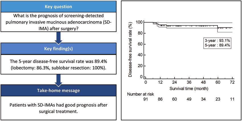
METHODS: Data from 91 patients who underwent surgical treatment for SD-IMA (2 D.H. Kim et al. / Interactive CardioVascular and Thoracic Surgery
RESULTS: Radiologically, SD-IMAs presented as a pure ground-glass nodule (6.6%), part-solid nodule (38.5%) or solid (54.9%).
Dominant locations were both lower lobes (74.7%) and peripheral area (93.4%). The sensitivity of percutaneous needle biopsy was
78.1% (25/32). Lobectomy was performed in 70 (76.9%) patients, and sublobar resection in 21 (23.1%) patients. Seventy-three (80.2%),
15 (16.5%) and 3 (3.3%) patients had pathological stage IA, IB and IIB or above, respectively. Seven patients developed recurrence, and
3 died due to disease progression. Pleural seeding developed exclusively in 2 patients who underwent needle biopsy. The 5-year
disease-free survival rate was 89.4%. The disease-free survival rates at 5 years were 86.3% in the lobectomy group and 100% in the sub-
lobar resection group.
CONCLUSIONS: SD-IMAs were mostly radiologically invasive nodules. SD-IMAs showed favourable prognosis after surgical treatment.
Keywords: Adenocarcinoma of lung • Mucinous adenocarcinoma • Early detection of cancer • Disease-free survival
Downloaded from https://academic.oup.com/icvts/advance-article/doi/10.1093/icvts/ivab257/6376007 by guest on 28 December 2021
MATERIALS AND METHODS
ABBREVIATIONS
This study was conducted in compliance with the Declaration of
C/T ratio Consolidation-to-tumour ratio Helsinki and approved by the institutional review board of Seoul
CT Computed tomography National University Hospital (Approval Number: 1909-035-1062).
DFS Disease-free survival The requirement for informed consent was waived.
IMA Invasive mucinous adenocarcinoma A total of 197 patients underwent surgery for IMA from July
IQR Interquartile range 2013 to May 2019 in our institution. We included 91 patients
MRI Magnetic resonance imaging with radiological tumoursD.H. Kim et al. / Interactive CardioVascular and Thoracic Surgery 3
nodule], consolidation-to-tumour ratio (C/T ratio), location of
tumour (peripheral/central) and distance from visceral pleura to Table 1: Patient demographics
tumour margin. The lung CT screening reporting and data system
THORACIC
category of each nodule was determined [3, 7]. The C/T ratio was Variables Total
defined as the proportion of the longest consolidation diameter (n = 91)
divided by the longest tumour diameter in the lung window
Age (years), mean ± SD 64.3 ± 10.0
setting [8]. The location of the tumour was determined by a Sex, n (%)
concentric line from the hilum and the division point was the Female 57 (62.6)
peripheral third [9]. Male 34 (37.4)
Smoking history, n (%)
Downloaded from https://academic.oup.com/icvts/advance-article/doi/10.1093/icvts/ivab257/6376007 by guest on 28 December 2021
Never smoker 67 (73.6)
Preoperative work-up and surgical management Ex-smoker 20 (22.0)
Current smoker 4 (4.4)
Pretreatment histological diagnosis was confirmed by percutane- ECOG performance status, n (%)
0 82 (90.1)
ous needle biopsy (PCNB) or intraoperative frozen section fol- 1 9 (9.9)
lowing diagnostic thoracoscopic resection. Diagnosis of primary Comorbidities, n (%) 66 (72.5)
pulmonary IMA in patients with history of previous gastrointesti- Cardiovascular disease 42 (46.2)
nal tract adenocarcinoma was made based on comprehensive Diabetes mellitus 14 (15.4)
Renal disease 1 (1.1)
pathological examination such as cytological comparison and Asthma 1 (1.1)
molecular marker studies. Disease-free interval longer than Cerebrovascular disease 3 (3.3)
5 years was also considered in differential diagnosis. Positron Previous malignancy, n (%) 24 (26.4)
emission tomography–CT (PET–CT) was performed in patients Gastrointestinal tract cancer 9 (9.9)
Breast cancer 5 (5.5)
with invasive component in the nodule or suspicious metastatic
lesions in CT scan. Brain magnetic resonance imaging (MRI) was ECOG: Eastern Cooperative Oncology Group; SD: standard deviation.
performed in patients with clinical stage IB or higher. The stan-
dard procedure for curative surgery was lobectomy; however,
sublobar resection was performed for radiologically indolent
tumours considering tumour size, C/T ratio, tumour location and Table 2: Radiological characteristics
underlying pulmonary function. The pathological parenchymal
resection margin was defined as the distance from the tumour Variables Total
edge to the nearest stapled resection margin confirmed by (n = 91)
pathologist. Location, n (%)
Right upper lobe 7 (7.7)
Right middle lobe 5 (5.5)
Statistical analysis Right lower lobe 26 (28.6)
Left upper lobe 13 (14.3)
Disease-free survival (DFS) was defined the time from date of sur- Left lower lobe 42 (46.2)
gery to the date of recurrence or death whatever comes first. Nodule type, n (%)
Pure ground-glass nodule 6 (6.6)
Patients without recurrent event or death were censored at the Part-solid nodule 35 (38.5)
date of last follow-up. The last follow-up date was 30 August Solid nodule 50 (54.9)
2020. Local recurrence was defined as recurrence in the bron- Tumour size, radiological (cm), mean ± SD 1.64 ± 0.58
chial stump or parenchymal margin. Regional recurrence was de- Solid component size (cm), mean ± SD 1.30 ± 0.70
Consolidation-to-tumour ratio,a mean ± SD 0.49 ± 0.25
fined as recurrence in the ipsilateral intrathoracic lymph node,
Distance from visceral pleural4 D.H. Kim et al. / Interactive CardioVascular and Thoracic Surgery
Downloaded from https://academic.oup.com/icvts/advance-article/doi/10.1093/icvts/ivab257/6376007 by guest on 28 December 2021
Figure 2: Heterogeneous radiological features of invasive mucinous adenocarcinomas of the lung; (A) 2.0 cm pure ground-glass nodule in the right lower lobe, (B)
1.5 cm part-solid nodule in the right middle lobe and (C) 1.5 cm sold nodule in the right lower lobe.
Radiological features
Table 3: Pathological results
The dominant sites were the lower lobes (n = 68) and almost all
nodules were located in the peripheral third of the lung (87.9%, Variables Total
(n = 91)
n = 80). The distance from the visceral pleura to tumour margin
was 1 cm or less in most tumours (93.4%, n = 85). Nineteen Tumour size, pathological (cm), mean ± SD 1.56 ± 0.84
(20.9%) patients had synchronous non-dominant nodules. pT T1mi, n (%) 1 (1.1)
The mean nodule size was 1.64 ± 0.58 cm (IQR 1.2–2.0). The T1a 22 (24.2)
T1b 41 (45.1)
type of nodules was pure ground-glass nodule in 6 (6.6%), PSN in T1c 10 (11.0)
35 (38.5%) and solid nodules in 50 (54.9%) (Fig. 2). The mean C/T T2a 16 (17.6)
ratio of PSN was 0.49 ± 0.25 (IQR 0.40–0.66). The lung CT screen- T2b 0 (0.0)
ing reporting and data system categories were 4A or 4B in most T3 1 (1.1)
pN Nx, n (%) 5 (5.5)
of cases (90.1%, n = 82). PET–CT was performed in 69 (75.8%)
N0 85 (93.4)
patients and the mean maximal standardized uptake value was N1 1 (1.1)
3.2 ± 3.0 (IQR 1.4–3.6) (Table 2). Brain MRI was performed in 42 pM M1a, n (%) 1 (1.1)
(46.2%) and no patient had brain metastasis. pStage pIA, n (%) 73 (80.2)
pIB 15 (16.5)
pIIB 2 (2.2)
Pathology and surgery pIVA 1 (1.1)
Synchronous double primary lung cancer 8 (8.8)
Visceral pleural invasion 11 (12.1)
Pretreatment histological diagnosis was attempted by PCNB in
Vascular invasion 2 (2.2)
33 patients and by intraoperative frozen section in 56 patients. Lymphatics invasion 3 (3.3)
Two patients underwent direct lobectomy due to combined
SD: standard deviation.
NMAs. In the PCNB group, a diagnosis of non-small-cell lung
cancer (NSCLC) was made in all patients (IMA—26, NMA—7). In
the frozen section group, a diagnosis of NSCLC was made in 53
Among 19 patients with non-dominant nodules, 2 had metastatic
(94.6%) patients (IMA—29, NMA or carcinoma not otherwise
IMA, 8 had synchronous NMA, 5 had inflammatory nodules and
specified—24). The frozen section results of 3 patients were false
the non-dominant nodules were not resected in 4. Adjuvant che-
negative and 1 patient underwent lobectomy afterwards. The ex-
motherapy was not performed in 3 patients, who were indicated,
tent of pulmonary resection was lobectomy in 70 patients
considering comorbidities, low chemo-sensitivity nature of IMA
(76.9%), segmentectomy in 13 (14.3%) and wedge resection in 8
and the patients’ preference.
(8.8%) patients. Systematic lymph node dissection was performed
Thyroid transcription factor-1 was positive in 50% (8/16) and
in 85 patients (93.4%). The mean number of dissected lymph
kirsten rat sarcoma viral oncogene mutations detected in 53.3%
nodes was. 8.8 ± 5.2 in N1 stations and 12.5 ± 7.3 in N2 stations.
(8/15) of IMAs. The rates of epidermal growth factor receptor
In the sublobar resection group, the mean parenchymal resection
mutation and anaplastic lymphoma kinase rearrangement were
margin distance was 2.72 ± 1.94 cm (IQR 1.25–4.35) and the
3.2% (2/62) and 4.2% (3/71), respectively.
mean margin-to-tumour ratio was 3.1 ± 2.7 (IQR 1.5–5.8). The
margin-to-tumour ratio wasD.H. Kim et al. / Interactive CardioVascular and Thoracic Surgery 5
Table 4: Data of patients with recurrence
THORACIC
PCNB Surgery Pathological stage Recurrence DFI Recurrence Survival after Current
site (months) treatment recurrence (months) status
1 - Lobectomy T2aN0M0 Brain 13.1 Radiation therapy 2.3 LD
2 + Lobectomy T2aN0M0 Contralateral lung 10.8 Operation 40 L
Radiation therapy
3 + Lobectomy T2aN0M0 Contralateral lung/node/brain 6.2 Chemotherapy 9.0 D
4 - Lobectomy T1bN0M0 Contralateral lung 59.7 Operation 15.9 L
Downloaded from https://academic.oup.com/icvts/advance-article/doi/10.1093/icvts/ivab257/6376007 by guest on 28 December 2021
5 + Lobectomy T1bN0M0 Pleura/ 9.5 Chemotherapy 5.7 D
chest wall
6 + Lobectomy T2aN1M0 Pleura 9.3 Chemotherapy 12.2 D
7 + Lobectomy T2aN0M0 Contralateral lung 17.4 No treatment 7.9 LD
D: dead; DFI: disease-free interval; L: live without disease; LD: live with disease; PCNB: percutaneous needle biopsy.
who had undergone PCNB and the disease-free intervals of these characteristics of small IMAs. Lee et al. [5] reported that IMAs
patients were 9.3 and 9.5 months, respectively. There was no present as solid nodule or PSN. No pure ground-glass nodule
pleural seeding in no-PCNB patients. Three patients experienced was noted in their study. Shimizu et al. [16] reported that 19 small
contralateral pulmonary metastasis and 1 patient experienced IMAs presented as a solid or part-solid (bubbling) nodule. No
contralateral pulmonary metastasis and nodal recurrence. There study has reported pneumonic type in small IMAs and there was
were 2 extrathoracic metastases, both of which were brain me- no pneumonic type nodule in the present study. This finding sug-
tastases (Table 4).The 2 patients with pleural seeding died 5 and gests that pneumonic type does not have a different pathogene-
12 months after recurrence, and 1 patient with brain metastasis sis but rather appears as IMA progresses. Therefore, classification
died from treatment-related adverse events. The 5-year overall of IMA into the pneumonic or solid type is not a tumour charac-
survival rate was 96.0% (Fig. 3A). teristic but rather a status of progression.
The 3- and 5-year DFS rates were 93.1% and 89.4%, respec- Radiological nodule size and C/T ratio are important selection
tively (Fig. 3B). The DFS rates at 5 years were 86.3% in the lobec- criteria for sublobar resection in small adenocarcinomas.
tomy group and 100% in the sublobar resection group, Nodules6 D.H. Kim et al. / Interactive CardioVascular and Thoracic Surgery
Downloaded from https://academic.oup.com/icvts/advance-article/doi/10.1093/icvts/ivab257/6376007 by guest on 28 December 2021
Figure 3: Kaplan–Meier curves for overall survival (A) and disease-free survival (B) (thin lines indicated the 95% confidence intervals).
recurrences in IMA are limited to the lungs, and there is no
extrapulmonary metastasis [21]. Primary failure pattern in our
study was intrapulmonary metastasis in the contralateral lung
and pleural seeding. Only 2 patients experienced extrathoracic
metastases, both in the brain.
PCNB has been reported as a risk factor for pleural seeding in
early-stage lung cancer [22–24]. Because SD-IMA is usually lo-
cated in the subpleural area and consists of mucin, which can
easily leak through the visceral pleural needle track, the risk of
pleural seeding after PCNB is much higher than in NMA. In this
study, pleural seeding exclusively developed in patients who
underwent PCNB. Therefore, the effect of PCNB on pleural recur-
rence cannot be ruled out in this study too. PCNB should be re-
served for highly selected cases in patients with subpleural
Figure 4: Kaplan–Meier curves for disease-free survival according to the extent nodule that requires surgical resection [25].
of pulmonary resection.
would not affect oncological outcomes and the current strategy
Limitations
for determining extent of resection based on the size or ratio of
the solid components can be applied in subsolid IMAs.
This study had several limitations. First, it is a retrospective study
The long-term oncological outcomes of SD-IMAs in our study
that cannot be directly applied to screening and surgical planning
also support these recommendations. In this study, most SD-
protocols. A prospective study is necessary to evaluate the rela-
IMAs were pathological stage IA (80.2%) and the 3- and 5-year
tionship between preoperative imaging results and surgical out-
DFS rates were 93.1% and 89.4%, respectively. No local recur-
comes. Second, this was a single-institution study with limited
rence occurred even among patients who underwent sublobar
number of cases. The statistical power of analyses would not be
resection. IMA is known to have a poorer prognosis than NMA
high and the results can be questionable since the number of
[2, 5, 12]. However, some studies of early-stage IMA showed no
cases and the number of events were small. To obtain a more
significant difference in DFS and overall survival between NMA
definite conclusion, a multi-centred, large population, prospec-
and NMA patients in the same stage [16, 20, 21]. In a study [5] of
tive study should be conducted.
81 solitary IMA cases in which 8% of patients underwent sublo-
bar resection, the DFS rate of patients with IMA was between
that of patients with low-grade (lepidic-predominant) NMA and CONCLUSION
that of patients with intermediate-grade (acinar/papillary-pre-
dominant) NMA, and the overall survival of patients with IMA The majority of SD-IMAs were radiologically invasive nodules in
was similar to that of patients with intermediate-grade NMA. In the periphery of the lower lobe. Patients with SD-IMAs showed
our study, the DFS of SD-IMA patients was similar to those of favourable prognosis after surgical treatment.
previous studies (79–89.2%), despite the higher proportion of
sublobar resection (23% vs 8%) [5]. Unlike NMAs, extrapulmonary
metastases are very rare in IMAs. A previous study showed that Conflict of interest: none declared.D.H. Kim et al. / Interactive CardioVascular and Thoracic Surgery 7
Author contributions [10] Moon SW, Choi SY, Moon MH. Effect of invasive mucinous adenocarci-
noma on lung cancer-specific survival after surgical resection: a
population-based study. J Thorac Dis 2018;10:3595–608.
Dae Hyeon Kim: Data curation; Formal analysis; Investigation; Resources;
[11] Motono N, Matsui T, Machida Y, Usuda K, Uramoto H. Prognostic signif-
THORACIC
Visualization; Writing—original draft; Writing—review & editing. So Young
icance of histologic subtype in pStage I lung adenocarcinoma. Med
Bae: Data curation; Methodology; Resources. Kwon Joong Na: Data curation;
Oncol 2017;34:100.
Investigation; Methodology; Resources. Samina Park: Data curation; Formal
[12] Kadota K, Nitadori J-I, Sima CS, Ujiie H, Rizk NP, Jones DR et al. Tumor
analysis; Investigation; Resources. In Kyu Park: Conceptualization; Data cura-
spread through air spaces is an important pattern of invasion and
tion; Investigation; Methodology; Project administration; Supervision;
impacts the frequency and location of recurrences after limited resection
Validation; Writing—review & editing. Chang Hyun Kang: Conceptualization;
Data curation; Investigation. Young-Tae Kim: Conceptualization; Data cura- for small stage I lung adenocarcinomas. J Thorac Oncol 2015;10:806–14.
tion; Investigation; Methodology; Resources. [13] Kim YK, Shin DH, Kim KB, Shin N, Park WY, Lee JH et al. MUC5AC and
MUC5B enhance the characterization of mucinous adenocarcinomas of
Downloaded from https://academic.oup.com/icvts/advance-article/doi/10.1093/icvts/ivab257/6376007 by guest on 28 December 2021
the lung and predict poor prognosis. Histopathology 2015;67:520–8.
[14] Park WY, Kim MH, Shin DH, Lee JH, Choi KU, Kim JY et al. Ciliated
adenocarcinomas of the lung: a tumor of non-terminal respiratory unit
Reviewer information origin. Mod Pathol 2012;25:1265–74.
[15] Nie K, Nie W, Zhang Y-X, Yu H. Comparing clinicopathological features
Interactive CardioVascular and Thoracic Surgery thanks Emmanouil Ioannis and prognosis of primary pulmonary invasive mucinous adenocarci-
Kapetanakis, Hitoshi Igai and the other, anonymous reviewer(s) for their con- noma based on computed tomography findings. Cancer Imaging 2019;
tribution to the peer review process of this article. 19:47.
[16] Shimizu K, Okita R, Saisho S, Maeda A, Nojima Y, Nakata M.
Clinicopathological and immunohistochemical features of lung invasive
REFERENCES mucinous adenocarcinoma based on computed tomography findings.
Onco Targets Ther 2017;10:153–63.
[17] Isaka T, Yokose T, Miyagi Y, Washimi K, Nishii T, Ito H et al. Detection of
[1] Dacic S. Pros: the present classification of mucinous adenocarcinomas of
tumor spread through airspaces by airway secretion cytology from
the lung. Transl Lung Cancer Res 2017;6:230–3.
resected lung cancer specimens. Pathol Int 2017;67:487–94.
[2] Cha YJ, Shim HS. Biology of invasive mucinous adenocarcinoma of the
[18] Hwang S, Han J, Choi M, Ahn M-J, Choi YS. Size of non-lepidic invasive
lung. Transl Lung Cancer Res 2017;6:508–12.
pattern predicts recurrence in pulmonary mucinous adenocarcinoma:
[3] Suzuki K, Kusumoto M, Watanabe S-I, Tsuchiya R, Asamura H.
morphologic analysis of 188 resected cases with reappraisal of invasion
Radiologic classification of small adenocarcinoma of the lung:
criteria. J Pathol Transl Med 2017;51:56–68.
radiologic-pathologic correlation and its prognostic impact. Ann Thorac
[19] Oki T, Aokage K, Nomura S, Tane K, Miyoshi T, Shiiya N et al. Optimal
Surg 2006;81:413–19.
method for measuring invasive size that predicts survival in invasive mu-
[4] Boland JM, Maleszewski JJ, Wampfler JA, Voss JS, Kipp BR, Yang P et al.
cinous adenocarcinoma of the lung. J Cancer Res Clin Oncol 2020;146:
Pulmonary invasive mucinous adenocarcinoma and mixed invasive mu-
cinous/nonmucinous adenocarcinoma—a clinicopathological and mo- 1291–8.
lecular genetic study with survival analysis. Hum Pathol 2018;71:8–19. [20] Qu Y, Zhao D, Mu J, Che N, Zhang C, Liu Z et al. Prognostic analysis of
[5] Lee HY, Cha MJ, Lee KS, Lee HY, Kwon OJ, Choi JY et al. Prognosis in primary mucin-producing adenocarcinoma of the lung: a comprehen-
resected invasive mucinous adenocarcinomas of the lung: related factors sive retrospective study. Tumour Biol 2016;37:887–96.
and comparison with resected nonmucinous adenocarcinomas. J Thorac [21] Shim HS, Zheng Z, Liebers M, Cha YJ, Ho QH, Onozato M et al. Unique
Oncol 2016;11:1064–73. genetic and survival characteristics of invasive mucinous adenocarci-
[6] Goldstraw P, Chansky K, Crowley J, Rami-Porta R, Asamura H, Eberhardt noma of the lung. J Thorac Oncol 2015;10:1156–62.
WE et al.; International Association for the Study of Lung Cancer Staging [22] Matsuguma H, Nakahara R, Kitamura T, Kondo T, Kamiyama Y, Mori K
and Prognostic Factors Committee Advisory Boards and Participating et al. Pleural recurrence after needle biopsy of the lung: an analysis in
Institutions. The IASLC lung cancer staging project: proposals for revision patients with completely resected stage I non-small cell lung cancer. J
of the TNM stage groupings in the forthcoming (eighth) edition of the Clin Oncol 2004;22:7177.
TNM classification for lung cancer. J Thorac Oncol 2016;11:39–51. [23] Kashiwabara K, Semba H, Fujii S, Tsumura S. Preoperative percutaneous
[7] McKee BJ, Regis SM, McKee AB, Flacke S, Wald C. Performance of ACR transthoracic needle biopsy increased the risk of pleural recurrence in
Lung-RADS in a clinical CT lung screening program. J Am Coll Radiol pathological stage I lung cancer patients with sub-pleural pure solid
2016;13:R25–9. nodules. Cancer Invest 2016;34:373–77.
[8] Nitadori J-I, Bograd AJ, Morales EA, Rizk NP, Dunphy MP, Sima CS et al. [24] Wang T, Luo L, Zhou Q. Risk of pleural recurrence in early stage lung
Preoperative consolidation-to-tumor ratio and SUVmax stratify the risk cancer patients after percutaneous transthoracic needle biopsy: a meta-
of recurrence in patients undergoing limited resection for lung adeno- analysis. Sci Rep 2017;7:42762.
carcinomaYou can also read