Phase-Dependent Deep Brain Stimulation: A Review - MDPI
←
→
Page content transcription
If your browser does not render page correctly, please read the page content below
brain
              sciences
Review
Phase-Dependent Deep Brain Stimulation: A Review
Lekshmy Sudha Kumari and Abbas Z. Kouzani *

                                          School of Engineering, Deakin University, Geelong, VIC 3216, Australia; lsudhakumari@deakin.edu.au
                                          * Correspondence: abbas.kouzani@deakin.edu.au

                                          Abstract: Neural oscillations are repetitive patterns of neural activity in the central nervous systems.
                                          Oscillations of the neurons in different frequency bands are evident in electroencephalograms and
                                          local field potential measurements. These oscillations are understood to be one of the key mechanisms
                                          for carrying out normal functioning of the brain. Abnormality in any of these frequency bands of
                                          oscillations can lead to impairments in different cognitive and memory functions leading to different
                                          pathological conditions of the nervous system. However, the exact role of these neural oscillations in
                                          establishing various brain functions is still under investigation. Closed loop deep brain stimulation
                                          paradigms with neural oscillations as biomarkers could be used as a mechanism to understand the
                                          function of these oscillations. For making use of the neural oscillations as biomarkers to manipulate
                                          the frequency band of the oscillation, phase of the oscillation, and stimulation signal are of impor-
                                          tance. This paper reviews recent trends in deep brain stimulation systems and their non-invasive
                                          counterparts, in the use of phase specific stimulation to manipulate individual neural oscillations. In
                                          particular, the paper reviews the methods adopted in different brain stimulation systems and devices
                                          for stimulating at a definite phase to further optimize closed loop brain stimulation strategies.

                                Keywords: brain stimulation; phase-specific brain stimulation; neural oscillations


Citation: Kumari, L.S.; Kouzani, A.Z.
Phase-Dependent Deep Brain
Stimulation: A Review. Brain Sci.         1. Introduction
2021, 11, 414. https://doi.org/
                                                Brain stimulation paradigms including electrical deep brain stimulation (DBS), opto-
10.3390/brainsci11040414
                                          genetics brain stimulation (OBS), transcranial electrical stimulation (tES), and transcranial
                                          magnetic stimulation (TMS) and repetitive transcranial magnetic stimulation (rTMS) are
Academic Editors: Allan Bieber and
                                          being increasingly employed as a therapeutic and diagnostic tools for neurological disease
Alex Green
                                          conditions such as movement disorders and psychiatric illnesses [1–5]. DBS and OBS are
Received: 5 February 2021
                                          invasive brain stimulation paradigms, where the stimulation electrode is implanted into
Accepted: 23 March 2021
                                          the brain, whereas tES and TMS are non-invasive brain stimulation (NIBS) techniques in
Published: 25 March 2021                  which the brain is stimulated without implanting any devices (e.g., electrode) inside the
                                          body [6,7]. Recently, non-invasive techniques such as rTMS are being increasingly used
Publisher’s Note: MDPI stays neutral      in the treatment of major depressive disorder (MDD) [8], and as a positive development,
with regard to jurisdictional claims in   high efficacy is being also reported in the treatment of depression, pain, and stroke [9].
published maps and institutional affil-   NIBS is also being studied as a promising treatment methodology for addictions and
iations.                                  substance-use disorders (SUDS) [10–12]. On the other hand, the invasive technique of DBS
                                          is the commonly used in the treatment of common neurological diseases like Parkinson’s
                                          disease (PD), epilepsy, tremor, etc. [13,14].
                                                Over the recent years, there have been many advancements in invasive brain stimula-
Copyright: © 2021 by the authors.
                                          tion paradigms with size-reduced, tether-less and low power devices. Most of the existing
Licensee MDPI, Basel, Switzerland.
                                          brain stimulation devices in clinical therapeutical use are ‘open-loop’, where the stimula-
This article is an open access article    tion settings are fixed and subsequent adjustments to the device settings are done manually
distributed under the terms and           during hospital visits [15]. However, this open-loop approach limits the efficacy of brain
conditions of the Creative Commons        stimulation modalities [16]. One of the main characteristics of neurological disorders is
Attribution (CC BY) license (https://     that the associated symptoms change in time and are often progressive [17]. Due to these
creativecommons.org/licenses/by/          fluctuating symptoms, often the efficacy of the system reduces with time [18]. Adding to
4.0/).

Brain Sci. 2021, 11, 414. https://doi.org/10.3390/brainsci11040414                                         https://www.mdpi.com/journal/brainsci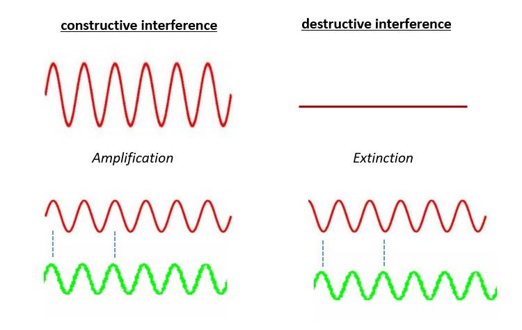
Brain Sci. 2021, 11, 414                                                                                            2 of 12

                           this, some of the other limitations of the stimulation include introducing disabling side-
                           effects, not significantly affecting some of the disease symptoms, manual programming
                           upon frequent symptom assessment, etc. [19]. All these limitations could be attributed to
                           the inherent-property of fixed stimulation settings and stimulation regardless of the state
                           of the individual in open-loop stimulation. Hence an automated approach is desirable.
                           This can be achieved in a closed-loop approach in which the stimulation parameters can
                           be adjusted in real-time depending on a feedback signal from the subject, thus making
                           it possible for changing the stimulation based on the fluctuating symptoms and further
                           paving the way for patient-tailored treatment for neurological disorders. A closed loop
                           stimulation is realized by sensing an individual’s brain signals and using it as the feedback
                           signal to the stimulation circuit. Finally, this feedback signal could help in accurately
                           adjusting the stimulation parameters for better control of disease symptom with lesser
                           side effects [20]. Promisingly, various studies on PD with closed loop DBS have presented
                           good results. For example, Arlotti et al. [21] assessed closed-loop DBS of subjects with
                           PD, reporting the stimulation as a safe and effective treatment practice for PD. In another
                           work, Swann et al., [22] demonstrated the efficacy and feasibility of closed loop DBS with
                           fully implanted devices in PD patients. Similar studies demonstrate the high efficacy of
                           closed-loop DBS [23–25]. Recently, the United States Food and Drug Administration has
                           approved Medtronic Percept, the first commercially available DBS system that can record
                           brain activity while simultaneously stimulating, to treat PD symptoms [26].
                                 For the development of closed-loop or adaptive brain stimulation strategies, the feed-
                           back signal is generated from the subject by reading different biomarkers [27] that reflect
                           the disease state of the subject. Establishing accurate biomarkers for disease states is crucial
                           for increasing the efficacy of the closed loop brain stimulation systems. Biomarkers in
                           the existing brain stimulation devices and systems are of two types: electrophysiological
                           and neurochemical. While neurochemical biomarkers indicate the state of neurotrans-
                           mitters [28], electrophysiological biomarkers (e.g., action potential (AP) as well as local
                           field potential (LFP)) provide electrical activity of the brain. The action potential or high
                           frequency neural spike is the fundamental method of communication between neurons,
                           and hence is considered an important signal for understanding the underlying neurological
                           conditions. Action potential is measured invasively using microelectrodes followed by
                           high pass filtering of the signal [29], whereas, local field potential reflects the combined
                           electrical activity of a group of adjacent neurons and is measured from the invasively
                           recorded extracellular electrophysiological activity by low pass filtering around 200 Hz [30].
                           Analyzing LFP is like interpreting rhythmic brain action from electroencephalography
                           (EEG) recordings. Moreover, as the electric field generated by the nerve cells are subject
                           to an exponential decay with distance, producing a detectable signal that needs a smaller
                           number of nerve cells to be simultaneously active in LFP than in EEG which is recorded
                           non-invasively [31].
                                 The repetitive patterns in the neural activity are observed in LFP as oscillations. These
                           neural oscillations or rhythms are produced by multiple neurons communicating with
                           each other and for allowing synchronized action during normal brain operation. Power or
                           amplitude of these neural oscillations are related to different cognitive functions [32], and
                           alterations in these oscillations is linked to the neural underpinnings of different neurologi-
                           cal diseases. In order to investigate these neural oscillations and deepen our understanding
                           of various neurological conditions, it is desirable to have an efficient closed loop brain
                           stimulation system capable of measuring and decoding the amplitude of a particular band
                           of oscillation and control the stimulation parameters based on these oscillation properties.
                           This will very well serve the therapeutical purpose of brain stimulation. Additionally, by
                           utilizing stimulation techniques like optogenetics [33], where precise control of neural cir-
                           cuits using a specific wavelength of light, closed loop brain stimulation could even be used
                           for investigating the causative mechanism of neurological disorders. For example, Pianta-
                           dosi et al. [34] employed closed loop optogenetics brain stimulation to understand the role
                           of cortico–stratial–thalamo–cortical (CSTC) neural circuits and the obsessive–compulsive
Brain Sci. 2021, 11, 414 3 of 12
disorder (OCD) in animals. This will serve both therapeutical and diagnostic purpose
of the brain stimulation. However, a major challenge in implementing such a design is
delivering the neural stimulation to alter the power or amplitude of one particular band
of oscillation in the neural signal. This is where the concept called phase-specific brain
stimulation becomes the key.
Generally, in an ON-OFF feedback control strategy, stimulation pulses are delivered
when the amplitude/power of the oscillation under consideration deviates from a certain
threshold value. Dual-threshold strategies can also be found in literature where the stim-
ulation voltage is either increased or decreased based on the upper and lower range of
oscillatory band power [24]. In either case, only the amplitude/power of the oscillation is
considered for delivering the stimulation pulses. On the contrary, in phase-specific brain
stimulation, the stimulation signal is applied considering both the threshold as well as the
instantaneous phase of the neural oscillation under observation. By taking both the ampli-
tude and phase of the LFP signals into consideration, a better method for manipulating
specific frequency bands in the neural oscillations could be achieved.
In the following sections, neural oscillations, the importance of neural oscillations
as biomarkers for neurological diseases, and significance of phase selective close-loop
brain stimulation strategies are discussed. Some of the current research work involving
the strategies being used by studies for implementing stimulation in correct phase are
explained. These works support the expectation that phase-specific closed loop brain
stimulation will demonstrate higher efficacy in modulating neural pathways.
2. Neural Oscillations as Biomarkers
Neural oscillations are the repetitive electrical activity generated spontaneously or
in response to stimuli by neurons [35]. Oscillatory activity of the neural assemblies can
be categorized as delta (0.5–3.5 Hz), theta (3.5–7 Hz), alpha (8–13 Hz), beta (18–25 Hz),
gamma (30–100 Hz), and high-frequency oscillations, HFO (100–200 Hz). There is exten-
sive evidence to suggest that these neural oscillations and the synchronization between
these neural oscillations in various cortical regions help in establishing different cognitive
phenomenon and memory functions. In addition to their role in normal brain functioning,
studies have suggested the alterations in alpha, beta, gamma, delta, and theta frequency
band activities may be associated with different neuropsychiatric disorders. These neural
oscillations and their synchrony help in establishing different cognitive phenomenon and
memory functions [36,37]. Studies have suggested the modulations in alpha, beta, gamma,
delta, and theta frequency bands in pathological brains [38–40]. Features of the neural
oscillations like location, amplitude, frequency, and phase are significant [41] and deter-
mine its effect on the neural pathway. There exist research work suggesting changes in
oscillatory dynamics in common conditions such as major depressive disorders (MDD), PD,
AD, epilepsy, Schizophrenia (SZ), and so on. Additionally, it is important to note that each
function in the brain is the result of combined actions of multiple oscillations [42], which
makes finding the apt biomarkers for these brain conditions based on changes in neural
oscillations a very complex problem to decode. In this context, a closed loop brain stimula-
tion system based on oscillation-related biomarkers provides a good opportunity to come
up with understanding of the function of the neural oscillations, and better elucidate these
cognitive disorders, their progression, and effects of medicines on the neural disorders.
Considering one of the most common neurodegenerative motor impairment diseases,
PD, the role of modulations in beta band oscillations in basal ganglia- cortical circuits has
been widely studied [43–47]. These studies suggest a direct correlation of reduction in beta
band power to bradykinesia, the motor impairment in PD. A general agreement in all these
studies is the fact that a high beta band power may contribute to motor impairment in PD,
and thus a reduction in beta band power could lead to clinical improvement in PD [15,46].
Along with beta band oscillations, there are findings that suggest a role for broad band
gamma oscillations in the range of 50 to 200 Hz in motor dysfunctions in parkinsonian
state. Studies have reported the increased activity in motor cortex resting broad bandBrain Sci. 2021, 11, 414 4 of 12
gamma [48] along with exaggerated cross-frequency coupling of broadband gamma to
the phase of the beta rhythm [49–51]. Özkurt et al. [52] reported the interaction between
high frequency oscillations bands around 250 and 350 Hz in the subthalamic nucleus (STN)
in pathophysiology of PD. A positive correlation of theta activity in STN and negative
correlation with STN beta activity with rest tremor in PD is reported in [53]. Other than the
movement disorder, there are several non-motor symptoms like cognitive impairments and
neuropsychiatric symptoms, and sleep disorders, among others associated with PD [54,55].
Attention difficulties are characteristics of cognitive impairment in PD. Bin Yoo et al. [56]
discussed enhanced activity in bilateral gamma in a bottom-up attention stream and
increased left alpha2 (10–12 Hz) connectivity in the top-down attention stream. The study
also reported a higher alpha2-gamma coupling in the right posterior parietal cortex in PD
patients than in the healthy subjects under study.
Dystonia [57,58] is yet another movement disorder caused by dysfunction of brain
regions and the communication between neurons and involves involuntary muscle contrac-
tions. Pallidal DBS is being used for the treatment of dystonia [59]. However, therapeutic
mechanism of DBS, and a proper biomarker for dystonia are still under study. Some studies,
e.g., [60–62], have suggested enhanced internal pallidum theta band activity having robust
association with symptoms in cervical dystonia (CD) [63], and in [61] theta band oscillation
is suggested as a possible biomarker for closed-loop brain stimulation in CD.
Talking about the debilitating neuropsychiatric disease, SZ, there is still more to be
understood about the origin and progression of the disease and hence a definite biomarker
for early detection and diagnosis of the disease is still under study [64,65]. Along with
other hypothesis put forward by different researchers, the role of abnormalities in neural
oscillations are also highlighted as the underlying mechanism for symptoms associated
with SZ. Synchronous gamma band activity is observed to be the mechanism for facilitation
of sensory and cognitive processes in healthy humans [66], and hence gamma oscillation
properties could function as a biomarker for the diagnosis of SZ as the defining symptoms
of SZ include cognitive and perceptual abnormalities. This is supported by the fact that
there is mounting evidence suggesting the neural oscillations in the gamma frequency
range are modified in SZ [67].
Considering the literature on the common psychiatric illness, major depressive dis-
order (MDD or clinical depression), abnormal oscillatory patterns in different frequency
bands have been widely reported. Among these, the role of alpha band oscillations in MDD
can be seen as a consistent finding [68–70]. The findings from these studies include an
elevated absolute or relative alpha power at parietal, frontal, or occipital sites. Researchers
have also reported the role of theta band rhythms in connection with MDD [71]. There is an
increasing body of evidence indicating that the lower gamma band oscillatory power can
be a prospective biomarker for MDD [72]. In one of the other major neuronal disorders, AD,
research points at a decrease in alpha, beta, and gamma oscillatory power over posterior
regions and enhancements in resting state delta and theta power [73]. Gamma oscillations
are suggested to contribute to memory encoding as well as retrieval, and hence it is not
surprising to find alterations in the gamma band oscillation in patients with AD and rodent
models of AD [74,75].
With the modulation in neural oscillation being an emerging biomarker for different
neurological diseases, a closed loop brain stimulation system with adaptive stimulation
according to the online reading of the biomarker is a very good tool to establish the
relation between these neural oscillations and different neurological conditions. This will
further enable science to decode these disease features for developing medicines and
other treatment methodologies. The concept is to read the biomarker continuously and
change the stimulation accordingly. By applying the stimulation, the aim is to target the
particular neural oscillation frequencies, and this is where phase specific stimulation comes
into picture.Brain Sci. 2021, 11, 414 5 of 13
Brain Sci. 2021, 11, 414 5 of 12
3. Closed Loop Brain Stimulation with Neural Oscillations as Biomarkers
3.On the device
Closed Loopengineering side, thewith
Brain Stimulation essential
Neuraldifference between
Oscillations a closed and an open
as Biomarkers
loop control
On system is inengineering
the device the feedbackside, mechanism. In a closed
the essential loop control
difference between system, a feed-
a closed and an
backopen
control is present, which can efficiently change the stimulation parameters
loop control system is in the feedback mechanism. In a closed loop control system, depend-
ing on the actual
a feedback and desired
control output.
is present, Using
which canextra circuitry,
efficiently the biomarkers
change the stimulation are quantita-
parameters
tively
depending on the actual and desired output. Using extra circuitry, the address
analyzed to provide precise stimulation at accurate times. This helps biomarkersone are
of the major issues associated with neural disorders, i.e., that the symptoms
quantitatively analyzed to provide precise stimulation at accurate times. This helps address do no stay
constant,
one ofandthethe condition
major issues of the patient
associated could
with varydisorders,
neural throughout i.e.,a that
period.the With the help
symptoms do no
of anstay
effective closed loop control in place, it can be ensured that the
constant, and the condition of the patient could vary throughout a period. With person is given opti-
mal the
stimulation
help of anbyeffective
continuouslyclosed monitoring
loop controltheinbiomarkers
place, it can andbemodulating
ensured that thethe
stimula-
person is
tion given
parameters accordingly. Moreover, closed loop brain stimulation,
optimal stimulation by continuously monitoring the biomarkers and modulating as mentioned be- the
fore,stimulation
helps in the diagnosis as well as study of neural disorders. This is
parameters accordingly. Moreover, closed loop brain stimulation, as mentioned important as the
scientific community
before, helps in the is diagnosis
still investigating
as well asthe cause
study of of origin
neural and progression
disorders. of multiple
This is important as the
commonly occurring
scientific community neurological diseases. Inthe
is still investigating addition
cause to of this,
originforand
untethered
progressiondevices, ex-
of multiple
tra stimulation
commonly uses up battery
occurring life unnecessarily.
neurological diseases. In addition to this, for untethered devices,
With neural oscillations as biomarkers, closed loop brain stimulation would be ben-
extra stimulation uses up battery life unnecessarily.
efitted from phase specificity.
With neural oscillations as biomarkers, closed loop brain stimulation would be bene-
fitted from phase specificity.
4. Phase Specific Stimulation
4.AsPhase Specific
pointed out inStimulation
the previous sections, manipulating the neural oscillations in LFP
could proveAs pointed out in treatment
to be a useful the previous sections,
approach formanipulating
different neural the neural oscillations
conditions. However,in LFP
could prove to be a useful treatment approach for different neural
controlling the power of an individual frequency band of oscillations in the LFP is not an conditions. However,
controlling
easily achievablethe power
task of an individual
[76]. Another important frequency
factor to band
considerof oscillations
while usinginneural
the LFP is not
oscil-
an easily
lations achievable
as biomarkers and task
high[76]. Anotherstimulation
frequency important factor to consider
is the possible sidewhile using
effects. neural
These
sideoscillations
effects are as biomarkers
mainly causedand by high frequency stimulation
the non-specificity is thefrequency
in the high possible side effects. These
stimulation.
side effects are mainly caused by the non-specificity in the high frequency
Stimulation induced side effects are due to the stimulation being not directed specifically stimulation.
Stimulation induced side effects are due to the stimulation being
to the neurological signals driving the disease symptoms [77]. Hence it is important not directed specifically
for
to the neurological signals driving the disease symptoms
the brain stimulation to be more specific to the pathological neural activity.[77]. Hence it is important for the
brain stimulation to be more specific to the pathological neural activity.
An approach to enhance the specificity of brain stimulation and modulate the oscil-
An approach
latory power in specifictobands
enhance the specificity
of neural of brain
oscillations is to stimulation
time-lock the and modulatetothe
stimulation theoscil-
latory power in specific bands of neural oscillations is to time-lock
phase of the present oscillation. Adopting the concept of constructive and destructive in- the stimulation to the
phase of the present oscillation. Adopting the concept of constructive
terference (Figure 1), stimulating at oscillatory peaks of the desired neural oscillation im- and destructive
interference (Figure 1), stimulating at oscillatory peaks of the desired neural oscillation
proves the present oscillation, while stimulating at oscillatory troughs suppresses an on-
improves the present oscillation, while stimulating at oscillatory troughs suppresses an
going oscillation [78,79]. This can be termed as phase-specific brain stimulation. As this
ongoing oscillation [78,79]. This can be termed as phase-specific brain stimulation. As
stimulation strategy is specific to a specific neural oscillation, it may cause fewer side-
this stimulation strategy is specific to a specific neural oscillation, it may cause fewer
effects as other rhythmic oscillations which are not phase-locked to the stimulation would
side-effects as other rhythmic oscillations which are not phase-locked to the stimulation
not be affected. As simple as it may sound, the challenge of realizing such a system lies in
would not be affected. As simple as it may sound, the challenge of realizing such a system
accurately predicting the oscillatory phase rapidly and delivering the apt stimulation
lies in accurately predicting the oscillatory phase rapidly and delivering the apt stimulation
pulses in real-time in a feedback loop.
pulses in real-time in a feedback loop.
Figure 1. Constructive and destructive interference—concept and illustration.
Figure 1. Constructive and destructive interference—concept and illustration.Brain Sci. 2021, 11, 414 6 of 12
Reviewing the literature on brain stimulation systems employing phase-specific stim-
ulation, we came across very few developed systems, including both invasive and non-
invasive systems. These systems are further described in the following.
Mansouri et al. [80] discussed a bench-top closed loop transcranial electromagnetic
brain stimulation platform that can interpret EEG signals in real time, predict the phase of
the underlying brain oscillations, and deliver controlled pulsed transcranial electromag-
netic stimulation output at a precise phase of the target neural oscillation. Alpha and theta
band of oscillation were the target bands in this work. Phase specificity was introduced in
the system and was implemented using an EEG system employing an Arduino develop-
ment board, and Matlab software. The authors calculated the phase delay introduced by
the signal processing to be 3.8 degrees for theta band and 57 degrees for alpha band stimu-
lation. The phase of the incoming EEG signal was determined by considering a portion
of the signal followed by the use of a 10th order elliptical filter to filter out the undesired
frequencies. After getting the frequency band of interest, the fast Fourier transform (FFT)
of the filtered signal was calculated. The FFT bin with the highest power in the desired
frequency band was considered to select the dominant frequency. Further, the timing of the
next stimulation pulse is computed from the phase and frequency values of the calculated
dominant frequency. The authors also considered the minute delays in the system or small
phase shifts that could have been introduced in the system by the filter or other signal
processing components or hardware delays. These delays in the system was accounted for
in the stimulation time by presenting an empirically calculated correction time in phase.
For example, the execution time of the Matlab code was approximately calculated to be
1 ms and was added to the pulse time to account for the delay. Stimulation is applied
using the transcranial electrical stimulator from Neuroconn. Timing of the stimulation is
communicated to the microcontroller in the Arduino board using serial USB communica-
tion. Microcontroller generated voltage waveform depending on the timing and is used to
control the stimulator which delivers a constant current output relative to the voltage.
Siegle et al. [78] used a phase-locking approach with closed loop optogenetic brain
stimulation in freely behaving mice to cause inhibition of dorsal hippocampal CA1 at
specific phases of theta band oscillations. The study reported an improved performance
when the stimulation introduced in the encoding segment was triggered by the maxima of
theta rhythm. The LFP signal was sensed using electrodes and digitally filtered between
4 and 12 Hz to record the theta band oscillations. Optogenetic stimulation was activated
once the theta power passed a threshold. Once the sensed signal was at a local minimum or
maximum, the control algorithm directed the optogenetic stimulator to deliver a 10 ms light
pulse to the invasively implanted fiber optic electrode. The study reported an average delay
between trigger and the beginning of the light pulse of 21.7 ± 7.2 ms for peak-activated
stimulation and 21.3 ± 7.4 ms for trough-activated stimulation. Accordingly, the reported
mean phase of stimulation was 96 ± 54◦ for peak-activated stimulation and −131 ± 63◦ for
trough-activated stimulation. The phase-based trough-activated stimulation demonstrated
lower precision. Another point to note is that the stimulation was 90–180 degrees away
from the target phase.
In another related work on phase-specific deep brain stimulation for movement
disorder, Cagnan et al. [81], considered the dominant phase of tremor in subjects with
essential tremor. The signals from an accelerometer were analyzed using Matlab software.
Firstly, the signals were band-pass filtered at ± 2 Hz around the tremor frequency using
4th order Butterworth filter, to get the tremor signals. Amplitude and instantaneous phase
of the tremor frequencies was calculated using Hilbert transform. Low frequency phase
locked stimulation was given at random phase with respect to the tremor phase, and the
best phase-offset was determined by trial and error. Phase-locked DBS was then introduced
during tremor episode. A symptom suppression of up to 87% was reported. Along with
increase in efficacy, by using low-frequency phase-locked stimulations, the authors were
able to power savings in the system.Brain Sci. 2021, 11, 414 7 of 12
Zarubin et al. [82] used a closed-loop transcranial alternating current stimulation
system for understanding the effects of repetitive short-time visual cortex stimulation
on the amplitude of visual alpha band oscillation when the stimulation was modified
based on the frequency and phase of those alpha band oscillations. EEG signals were
used for the experiment. For the phase prediction Hilbert transform based approach was
used. In order to reduce the complexity, quasi-stationary nature of phase dynamics for
the short time interval was assumed. For predicting the phase-dependent stimulation,
the phase information of the last 250 ms of the ongoing interval is used considering the
quasi-stationary nature. Initially, FIR filtering is employed to get the alpha band oscillations.
This was followed by Hilbert transformation to obtain instantaneous phase values. Phase
prediction was optimized by repeated search over sine waves of diverse phases. For this,
Euclidean difference (L2 norm) for vectors of immediate phase between extraction interval
and several produced sine waves was done. Further, delay due to this optimization was
calculated and compensated.
In another important work in this area, an analog feedback circuit is used to give phase-
specific stimulation. In reference [76], a closed loop transcranial electrical stimulation is
explained, where phase locked stimulation was used to enhance only the alpha oscillations
(8–15 Hz) of the subject. The closed loop circuit was realized in analog feedback circuit
instead of the usually used digital circuits. The front-end circuit, which acquires the neural
signals from the subject, consisted of a multiple feedback filter realized using the Linear
Technology LT1012 operational amplifier IC and related passive components. The front
end efficiently receives the signal and filter out the alpha band frequencies form the signal.
A pre-amplifier stage is realized using a Cereplex digitizing amplifying head stage to
give the required amplification to the filtered out alpha band signals. As the following
stage is an analog feedback circuit, the digital output from Cereplex is converted to analog
using a signal processor unit by Cerebus before feeding to the analog circuit. The analog
feedback circuit is followed by a transcranial electrical stimulator by Neuroconn GmbH.
The feedback circuit’s output controls the stimulator output current with a rate of 2 mA
per applied voltage. The calculated system delay contributed by the analog amplification
circuit and the digital components is 372◦ at 12 Hz and 360◦ at 11.6 Hz. Thus, the delay
creates a positive feedback loop for alpha band frequency amplification in the neural signal
by providing closely in-phase electrical stimulation using the Neuroconn stimulator. The
advantage of the design is that it can be easily modified for other frequency band of neural
oscillation by changing the filter design for a different passband and the total loop delay.
However, on the hardware side, for the implementation of the analog feedback circuit, the
digitized signal is converted back to analog, which requires extra circuitry.
While reviewing these systems, it is clear that these systems lack a dedicated circuit for
the phase detection and phase specific stimulation. Most of the reviewed systems work in
a bench-top setting and the software for phase detection is implemented on a separate PC.
This could be a hindrance to the miniaturization and further tether-less operation of brain
stimulation systems and there is scope of improvement which could be the development
of application specific integrated circuits specifically for the phase detection and precise
phase-specific stimulation.
5. Outlook: Closed Loop Brain Stimulation with Phase Specific Stimulation
The essential components of a closed loop brain stimulation system with phase specific
stimulation for LFP-based biomarkers are the following: a neural sensor to sense the LFP
signals from the subject, a neural stimulator for producing the stimulation patterns in the
required specifications at the right time, and a software unit to process the signals received
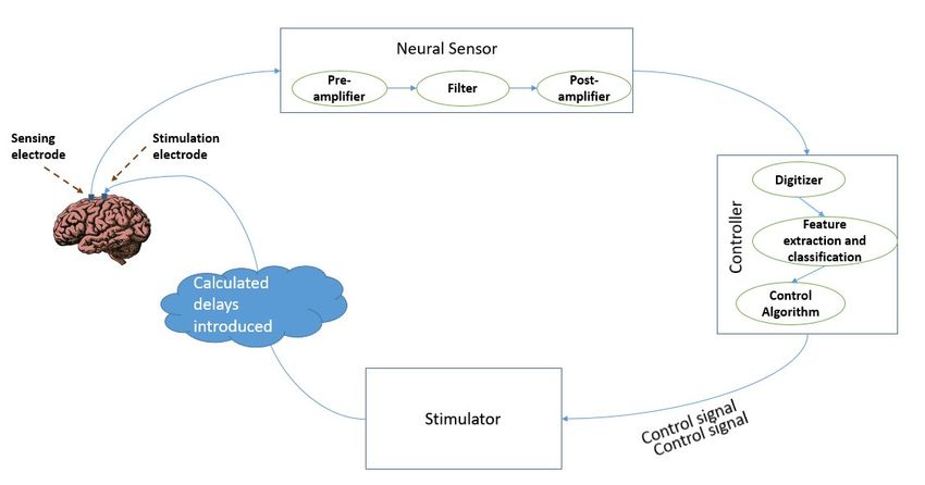
by the sensor and generate a control signal for the stimulator. A conceptual block diagram
of such a device is presented in Figure 2. The essential stages can be listed as: neural
sensing, feature extraction, classification, control, and stimulation.the LFP signals into pathological and non-pathological also have a very important role to
play in the realization of an efficient closed-loop brain stimulation system. The actual clin-
ical implementation of closed-loop DBS for rapid behavioral changes relies on accurate
and quick detection of these disease states using efficient algorithms. Algorithms ranging
Brain Sci. 2021, 11, 414 from very simple classification algorithms to sophisticated machine learning and artificial
8 of 12
intelligence-based ones can be found in literature.
Figure 2. Block diagram of phase specific deep brain stimulation.
Figure 2. Block diagram of phase specific deep brain stimulation.
Electrophysiological signals in the brain are sensed using electrodes invasively im-
While adding the attribute of phase specificity in the stimulation to the device, the
planted into the patient’s brain. A sensor circuit, comprising amplifiers and filters, condi-
controller needs to have the extra feature of calculating the phase of the ongoing signal
tions the sensed signal for further analysis. Initially, the micro amplitude range signals from
oscillations, and the delays in the network to produce the control signal for the stimulator
the electrode are amplified. This is followed by a band pass filter to filter out unwanted
to send the correct amplitude pulse at the right time and phase to the stimulation electrode
frequency signals. The cut off frequency of the band pass filter is adjusted to meet the
placed onrange
frequency the brain
of thesurface.
neural Thus, the most
oscillation significant
bands—delta requirement
(0.5–3.5 in (3.5–7
Hz), theta the realization
Hz), alphaof
such a phase-specific deep brain stimulation is the implementation
(8–13 Hz), beta (18–25 Hz), gamma (30–100 Hz), and high frequency oscillations, HFO of an algorithm for
real timeHz).
(100–200 determination of theby
This is followed phase of the uninterruptedly
a controller which extracts the recorded
featureslocal
of thefield potential
conditioned
information. A key difficulty in such a system is accurately estimating
signals after digitizing them. Features such as amplitude, phase, and power of the received the oscillatory
phase in
signals arereal time. An
extracted andefficient
based onalgorithm to control
the extracted the stimulation
features, pulse andinto
signals are classified trigger the
classes.
stimulation at the right time considering the phase of the ongoing stimulation
Following this, a control signal is generated for closing the feedback loop. A control as well as
the delays along the network needs to be realized. In the existing digital
algorithm identifies deviations, if any, of the actual stimulation output from the required signal processing
systems,
level by the time Accordingly,
of stimulation. a signal is digitized and itsadjusts
the algorithm phase isthecomputed through
stimulation a Fourier
parameters to
transform based algorithm, the target phase has long gone. A quicker
minimize the difference in actual and desired outputs with the help of the stimulation but complex method
control circuit. Thus, the algorithms and software module for real time classification of
the LFP signals into pathological and non-pathological also have a very important role
to play in the realization of an efficient closed-loop brain stimulation system. The actual
clinical implementation of closed-loop DBS for rapid behavioral changes relies on accurate
and quick detection of these disease states using efficient algorithms. Algorithms ranging
from very simple classification algorithms to sophisticated machine learning and artificial
intelligence-based ones can be found in literature.
While adding the attribute of phase specificity in the stimulation to the device, the
controller needs to have the extra feature of calculating the phase of the ongoing signal
oscillations, and the delays in the network to produce the control signal for the stimulator
to send the correct amplitude pulse at the right time and phase to the stimulation electrode
placed on the brain surface. Thus, the most significant requirement in the realization of
such a phase-specific deep brain stimulation is the implementation of an algorithm for
real time determination of the phase of the uninterruptedly recorded local field potential
information. A key difficulty in such a system is accurately estimating the oscillatory
phase in real time. An efficient algorithm to control the stimulation pulse and trigger the
stimulation at the right time considering the phase of the ongoing stimulation as well as
the delays along the network needs to be realized. In the existing digital signal processing
systems, by the time a signal is digitized and its phase is computed through a Fourier
transform based algorithm, the target phase has long gone. A quicker but complex method
involves outputting phase-locked stimulations using analog-circuitry which is still in the
early stages of development. Additionally, such an analog circuit can be implemented into
an application specific integrated circuit chip.Brain Sci. 2021, 11, 414 9 of 12
Another important factor to consider for potentially optimizing brain stimulation is
the functional connectivity of the brain. It is a well-understood fact that brain regions don’t
operate in isolation. Our brain is a network, which consists of spatially distributed, but
connected sections that work together in realizing different function [83,84]. Hence the
connectivity between the point of stimulation and other sections of the brain could greatly
affect the efficiency of DBS treatment for neurological conditions [85]. Horn et al. [86]
developed a structural and functional connectivity profile for effective STN DBS for PD
to predict the outcome efficacy of DBS. On a similar note, Fox et al. [87] predicted the
efficacy of TMS in depression by considering the functional connectivity. Hence, it can
be concluded that electrode placement is highly important in the clinical success of brain
stimulation methodologies, among others.
6. Conclusions
Phase selective stimulation is an added attribute to the deep brain stimulation paradigm
for increasing its efficacy in altering the neural oscillations in specific frequency bands.
As more and more studies are being carried out to find the link between the variations in
the energy of different frequency bands of neural oscillations and neurological disorders,
a hybrid strategy combining closed-loop DBS strategies and phase selective stimulation
could be an effective approach. Selective modulation of neural synchrony using phase-
locked stimulation offers the potential to enhance the efficacy of therapy and minimize
the side effects of the stimulation. While reviewing the literature on this approach, the
authors could not find much work done using phase selective stimulation in invasive DBS.
This paper reviewed the methodologies adopted in different brain stimulation systems and
devices for stimulating brain at a definite phase. There is also an attempt to put forward a
conceptual model incorporating the concepts of phase selective stimulation and deep brain
stimulation for standalone DBS devices.
Author Contributions: Conceptualization, L.S.K. and A.Z.K.; methodology, L.S.K. and A.Z.K.; formal
analysis, L.S.K.; investigation, L.S.K.; resources, A.Z.K.; data curation, L.S.K.; writing—original draft
preparation, L.S.K.; writing—review and editing, L.S.K. and A.Z.K.; visualization, L.S.K.; supervision,
A.Z.K.; project administration, A.Z.K.; All authors have read and agreed to the published version of
the manuscript.
Funding: This research received no external funding.
Institutional Review Board Statement: Not applicable.
Informed Consent Statement: Not applicable.
Data Availability Statement: Not applicable.
Conflicts of Interest: The authors declare no conflict of interest.
References
1. Budman, E.; Deeb, W.; Martinez-Ramirez, D.; Pilitsis, J.G.; Peng-Chen, Z.; Okun, M.S.; Ramirez-Zamora, A. Potential indications
for deep brain stimulation in neurological disorders: An evolving field. Eur. J. Neurol. 2018, 25, 434-e30. [CrossRef]
2. Lozano, A.M.; Lipsman, N.; Bergman, H.; Brown, P.; Chabardes, S.; Chang, J.W.; Matthews, K.; McIntyre, C.C.; Schlaepfer,
T.E.; Schulder, M.; et al. Deep brain stimulation: Current challenges and future directions. Nat. Rev. Neurol. 2019, 15, 148–160.
[CrossRef]
3. Cagnan, H.; Denison, T.; McIntyre, C.; Brown, P. Emerging technologies for improved deep brain stimulation. Nat. Biotechnol.
2019, 37, 1024–1033. [CrossRef] [PubMed]
4. Bestmann, S.; Walsh, V. Transcranial electrical stimulation. Curr. Biol. 2017, 27, R1258–R1262. [CrossRef] [PubMed]
5. Garcin, B.; Mesrati, F.; Hubsch, C.; Mauras, T.; Iliescu, I.; Naccache, L.; Vidailhet, M.; Roze, E.; Degos, B. Impact of transcranial
magnetic stimulation on functional movement disorders: Cortical modulation or a behavioral effect? Front. Neurol. 2017, 8, 338.
[CrossRef] [PubMed]
6. Hänselmann, S.; Schneiders, M.; Weidner, N.; Rupp, R. Transcranial magnetic stimulation for individual identification of the best
electrode position for a motor imagery-based brain-computer interface. J. Neuroeng. Rehabil. 2015, 12, 71. [CrossRef]
7. Dayan, E.; Censor, N.; Buch, E.R.; Sandrini, M.; Cohen, L.G. Noninvasive brain stimulation: From physiology to network
dynamics and back. Nat. Neurosci. 2013, 16, 838–844. [CrossRef] [PubMed]Brain Sci. 2021, 11, 414 10 of 12
8. Fitzgerald, P.B.; Hoy, K.E.; Elliot, D.; McQueen, R.N.S.; Wambeek, L.E.; Daskalakis, Z.J. Accelerated repetitive transcranial
magnetic stimulation in the treatment of depression. Neuropsychopharmacology 2018, 43, 1565–1572. [CrossRef]
9. Lefaucheur, J.-P.; Aleman, A.; Baeken, C.; Benninger, D.H.; Brunelin, J.; Di Lazzaro, V.; Filipović, S.R.; Grefkes, C.; Hasan, A.;
Hummel, F.C.; et al. Evidence-based guidelines on the therapeutic use of repetitive transcranial magnetic stimulation (rTMS): An
update (2014–2018). Clin. Neurophysiol. 2020, 131, 474–528. [CrossRef]
10. Maiti, R.; Mishra, B.R.; Hota, D. Effect of high-frequency transcranial magnetic stimulation on craving in substance use disorder:
A meta-analysis. J. Neuropsychiatry Clin. Neurosci. 2017, 29, 160–171. [CrossRef]
11. Ekhtiari, H.; Tavakoli, H.; Addolorato, G.; Baeken, C.; Bonci, A.; Campanella, S.; Castelo-Branco, L.; Challet-Bouju, G.; Clark, V.P.;
Claus, E.; et al. Transcranial electrical and magnetic stimulation (tES and TMS) for addiction medicine: A consensus paper on the
present state of the science and the road ahead. Neurosci. Biobehav. Rev. 2019, 104, 118–140. [CrossRef]
12. Diana, M.; Raij, T.; Melis, M.; Nummenmaa, A.; Leggio, L.; Bonci, A. Rehabilitating the addicted brain with transcranial magnetic
stimulation. Nat. Rev. Neurosci. 2017, 18, 685. [CrossRef]
13. Fasano, A.; Aquino, C.C.; Krauss, J.K.; Honey, C.R.; Bloem, B.R. Axial disability and deep brain stimulation in patients with
Parkinson disease. Nat. Rev. Neurol. 2015, 11, 98–110. [CrossRef]
14. Krauss, J.K.; Lipsman, N.; Aziz, T.; Boutet, A.; Brown, P.; Chang, J.W.; Davidson, B.; Grill, W.M.; Hariz, M.I.; Horn, A.; et al.
Technology of deep brain stimulation: Current status and future directions. Nat. Rev. Neurol. 2021, 17, 75–87. [CrossRef]
[PubMed]
15. Bouthour, W.; Mégevand, P.; Donoghue, J.; Lüscher, C.; Birbaumer, N.; Krack, P. Biomarkers for closed-loop deep brain stimulation
in Parkinson disease and beyond. Nat. Rev. Neurol. 2019, 15, 343–352. [CrossRef] [PubMed]
16. Hoang, K.B.; Cassar, I.R.; Grill, W.M.; Turner, D.A. Biomarkers and Stimulation Algorithms for Adaptive Brain Stimulation. Front.
Neurosci. 2017, 11, 564. [CrossRef] [PubMed]
17. Maetzler, W.; Liepelt, I.; Berg, D. Progression of Parkinson’s disease in the clinical phase: Potential markers. Lancet Neurol. 2009, 8,
1158–1171. [CrossRef]
18. Carron, R.; Chaillet, A.; Filipchuk, A.; Pasillas-Lépine, W.; Hammond, C. Closing the loop of deep brain stimulation. Front. Syst.
Neurosci. 2013, 7, 112. [CrossRef]
19. Hosain, K.; Kouzani, A.; Tye, S. Closed loop deep brain stimulation: An evolving technology. Australas. Phys. Eng. Sci. Med. 2014,
37, 619–634. [CrossRef]
20. Fleming, J.E.; Dunn, E.; Lowery, M.M. Simulation of Closed-Loop Deep Brain Stimulation Control Schemes for Suppression of
Pathological Beta Oscillations in Parkinson’s Disease. Front. Neurosci. 2020, 14, 166. [CrossRef]
21. Arlotti, M.; Marceglia, S.; Foffani, G.; Volkmann, J.; Lozano, A.M.; Moro, E.; Cogiamanian, F.; Prenassi, M.; Bocci, T.; Cortese, F.;
et al. Eight-hours adaptive deep brain stimulation in patients with Parkinson disease. Neuropsychopharmacology 2018, 90,
e971–e976. [CrossRef]
22. Swann, N.C.; De Hemptinne, C.; Thompson, M.C.; Miocinovic, S.; Miller, A.M.; Gilron, R.; Ostrem, J.L.; Chizeck, H.J.; Starr, P.A.
Adaptive deep brain stimulation for Parkinson’s disease using motor cortex sensing. J. Neural Eng. 2018, 15, 046006. [CrossRef]
[PubMed]
23. Little, S.; Beudel, M.; Zrinzo, L.; Foltynie, T.; Limousin, P.; Hariz, M.; Neal, S.; Cheeran, B.; Cagnan, H.; Gratwicke, J.; et al.
Bilateral adaptive deep brain stimulation is effective in Parkinson’s disease. J. Neurol. Neurosurg. Psychiatry 2016, 87, 717–721.
[CrossRef] [PubMed]
24. Velisar, A.; Syrkin-Nikolau, J.; Blumenfeld, Z.; Trager, M.; Afzal, M.; Prabhakar, V.; Bronte-Stewart, H. Dual threshold neural
closed loop deep brain stimulation in Parkinson disease patients. Brain Stimul. 2019, 12, 868–876. [CrossRef] [PubMed]
25. Little, S.; Tripoliti, E.; Beudel, M.; Pogosyan, A.; Cagnan, H.; Herz, D.; Bestmann, S.; Aziz, T.; Cheeran, B.; Zrinzo, L.; et al.
Adaptive deep brain stimulation for Parkinson’s disease demonstrates reduced speech side effects compared to conventional
stimulation in the acute setting. J. Neurol. Neurosurg. Psychiatry 2016, 87, 1388–1389. [CrossRef] [PubMed]
26. Medtronic, P. Green light for deep brain stimulator incorporating neurofeedback. Nat. Biotechnol. 2020, 38, 1014–1015. [CrossRef]
27. Biomarkers Definitions Working Group; Atkinson, A.J., Jr.; Colburn, W.A.; DeGruttola, V.G.; DeMets, D.L.; Downing, G.J.; Hoth,
D.F.; Oates, J.A.; Peck, C.C.; Schooley, R.T.; et al. Biomarkers and surrogate endpoints: Preferred definitions and conceptual
framework. Clin. Pharmacol. Ther. 2001, 69, 89–95.
28. Mirza, K.B.; Golden, C.T.; Nikolic, K.; Toumazou, C. Closed-Loop Implantable Therapeutic Neuromodulation Systems Based on
Neurochemical Monitoring. Front. Neurosci. 2019, 13, 808. [CrossRef]
29. Parastarfeizabadi, M.; Kouzani, A.Z. A Miniature Dual-Biomarker-Based Sensing and Conditioning Device for Closed-Loop DBS.
IEEE J. Transl. Eng. Health Med. 2019, 7, 1–8. [CrossRef]
30. David, S.V.; Malaval, N.; Shamma, S.A. Decoupling Action Potential Bias from Cortical Local Field Potentials. Comput. Intell.
Neurosci. 2010, 2010, 1–12. [CrossRef]
31. Waldert, S. Invasive vs. Non-Invasive Neuronal Signals for Brain-Machine Interfaces: Will One Prevail? Front. Neurosci. 2016,
10, 295. [CrossRef]
32. Vosskuhl, J.; Strüber, D.; Herrmann, C.S. Non-invasive brain stimulation: A paradigm shift in understanding brain oscillations.
Front. Hum. Neurosci. 2018, 12, 211. [CrossRef] [PubMed]
33. Deisseroth, K. Optogenetics. Nat. Methods 2011, 8, 26–29. [CrossRef] [PubMed]Brain Sci. 2021, 11, 414 11 of 12
34. Piantadosi, S.C.; Ahmari, S.E. Using Optogenetics to Dissect the Neural Circuits Underlying OCD and Related Disorders. Curr.
Treat. Options Psychiatry 2015, 2, 297–311. [CrossRef] [PubMed]
35. Başar, E. Brain oscillations in neuropsychiatric disease. Dialogues Clin. Neurosci. 2013, 15, 291–300. [PubMed]
36. Başar, E.; Başar-Eroglu, C.; Karakaş, S.; Schürmann, M. Gamma, alpha, delta, and theta oscillations govern cognitive processes.
Int. J. Psychophysiol. 2001, 39, 241–248. [CrossRef]
37. Düzel, E.; Penny, W.D.; Burgess, N. Brain oscillations and memory. Curr. Opin. Neurobiol. 2010, 20, 143–149. [CrossRef]
38. Singh, A. Oscillatory activity in the cortico-basal ganglia-thalamic neural circuits in Parkinson’s disease. Eur. J. Neurosci. 2018, 48,
2869–2878. [CrossRef]
39. Ahnaou, A.; Moechars, D.; Raeymaekers, L.; Biermans, R.; Manyakov, N.V.; Bottelbergs, A.; Wintmolders, C.; Van Kolen, K.; Van
De Casteele, T.; Kemp, J.A.; et al. Emergence of early alterations in network oscillations and functional connectivity in a tau
seeding mouse model of Alzheimer’s disease pathology. Sci. Rep. 2017, 7, 14189. [CrossRef]
40. Kitchigina, V.F. Alterations of Coherent Theta and Gamma Network Oscillations as an Early Biomarker of Temporal Lobe Epilepsy
and Alzheimer’s Disease. Front. Integr. Neurosci. 2018, 12, 36. [CrossRef]
41. Cole, S.R.; Voytek, B. Brain Oscillations and the Importance of Waveform Shape. Trends Cogn. Sci. 2017, 21, 137–149. [CrossRef]
42. Başar, E.; Başar-Eroğlu, C.; Güntekin, B.; Yener, G.G. Brain’s alpha, beta, gamma, delta, and theta oscillations in neuropsychiatric
diseases: Proposal for biomarker strategies. In Supplements to Clinical Neurophysiology; Elsevier: Amsterdam, The Netherlands,
2013; Volume 62, pp. 19–54.
43. Little, S.; Brown, P. The functional role of beta oscillations in Parkinson’s disease. Park. Relat. Disord. 2014, 20, S44–S48. [CrossRef]
44. Lofredi, R.; Tan, H.; Neumann, W.-J.; Yeh, C.-H.; Schneider, G.-H.; Kühn, A.A.; Brown, P. Beta bursts during continuous
movements accompany the velocity decrement in Parkinson’s disease patients. Neurobiol. Dis. 2019, 127, 462–471. [CrossRef]
45. Heinrichs-Graham, E.; Santamaria, P.M.; Gendelman, H.E.; Wilson, T.W. The cortical signature of symptom laterality in Parkin-
son’s disease. NeuroImage Clin. 2017, 14, 433–440. [CrossRef]
46. Chung, J.W.; Burciu, R.G.; Ofori, E.; Coombes, S.A.; Christou, E.A.; Okun, M.S.; Hess, C.W.; Vaillancourt, D.E. Beta-band
oscillations in the supplementary motor cortex are modulated by levodopa and associated with functional activity in the basal
ganglia. NeuroImage Clin. 2018, 19, 559–571. [CrossRef] [PubMed]
47. Woerd, E.S.T.; Oostenveld, R.; De Lange, F.P.; Praamstra, P. A shift from prospective to reactive modulation of beta-band
oscillations in Parkinson’s disease. NeuroImage 2014, 100, 507–519. [CrossRef] [PubMed]
48. Rowland, N.C.; De Hemptinne, C.; Swann, N.C.; Qasim, S.; Miocinovic, S.; Ostrem, J.L.; Knight, R.T.; Starr, P.A. Task-related
activity in sensorimotor cortex in Parkinson’s disease and essential tremor: Changes in beta and gamma bands. Front. Hum.
Neurosci. 2015, 9, 512. [CrossRef]
49. Wang, J.; Hirschmann, J.; Elben, S.; Hartmann, C.J.; Vesper, J.; Wojtecki, L.; Schnitzler, A. High-frequency oscillations in Parkinson’s
disease: Spatial distribution and clinical relevance. Mov. Disord. 2014, 29, 1265–1272. [CrossRef]
50. Swann, N.C.; De Hemptinne, C.; Miocinovic, S.; Qasim, S.; Wang, S.S.; Ziman, N.; Ostrem, J.L.; Luciano, M.S.; Galifianakis, N.B.;
Starr, P.A. Gamma Oscillations in the Hyperkinetic State Detected with Chronic Human Brain Recordings in Parkinson’s Disease.
J. Neurosci. 2016, 36, 6445–6458. [CrossRef] [PubMed]
51. De Hemptinne, C.; Swann, N.C.; Ostrem, J.L.; Ryapolova-Webb, E.S.; Luciano, M.S.; Galifianakis, N.B.; Starr, P.A. Therapeutic
deep brain stimulation reduces cortical phase-amplitude coupling in Parkinson’s disease. Nat. Neurosci. 2015, 18, 779–786.
[CrossRef]
52. Özkurt, T.E.; Butz, M.; Homburger, M.; Elben, S.; Vesper, J.; Wojtecki, L.; Schnitzler, A. High frequency oscillations in the
subthalamic nucleus: A neurophysiological marker of the motor state in Parkinson’s disease. Exp. Neurol. 2011, 229, 324–331.
[CrossRef] [PubMed]
53. Asch, N.; Herschman, Y.; Maoz, R.; Auerbach-Asch, C.R.; Valsky, D.; Abu-Snineh, M.; Arkadir, D.; Linetsky, E.; Eitan, R.; Marmor,
O.; et al. Independently together: Subthalamic theta and beta opposite roles in predicting Parkinson’s tremor. Brain Commun.
2020, 2, fcaa074. [CrossRef]
54. Chaudhuri, K.R.; Tolosa, E.; Schapira, A.H.; Poewe, W. Non-Motor Symptoms of Parkinson’s Disease; OUP: Oxford, UK, 2014.
55. Poewe, W. Non-motor symptoms in Parkinson’s disease. Eur. J. Neurol. 2008, 15, 14–20. [CrossRef] [PubMed]
56. Bin Yoo, H.; De La Concha, E.O.; De Ridder, D.; Pickut, B.A.; Vanneste, S. The Functional Alterations in Top-Down Attention
Streams of Parkinson’s disease Measured by EEG. Sci. Rep. 2018, 8, 10609. [CrossRef] [PubMed]
57. Tarsy, D.; Simon, D.K. Dystonia. N. Engl. J. Med. 2006, 355, 818–829. [CrossRef] [PubMed]
58. Albanese, A.; Di Giovanni, M.; Lalli, S. Dystonia: Diagnosis and management. Eur. J. Neurol. 2018, 26, 5–17. [CrossRef]
59. Ostrem, J.L.; Starr, P.A. Treatment of dystonia with deep brain stimulation. Neurotherapeutics 2008, 5, 320–330. [CrossRef]
[PubMed]
60. Moll, C.K.E.; Galindo-Leon, E.; Sharott, A.; Gulberti, A.; Buhmann, C.; Koeppen, J.A.; Biermann, M.; Bäumer, T.; Zittel, S.;
Westphal, M.; et al. Asymmetric pallidal neuronal activity in patients with cervical dystonia. Front. Syst. Neurosci. 2014, 8, 15.
[CrossRef] [PubMed]
61. Neumann, W.; Horn, A.; Ewert, S.; Huebl, J.; Brücke, C.; Slentz, C.; Schneider, G.; Kühn, A.A. A localized pallidal physiomarker
in cervical dystonia. Ann. Neurol. 2017, 82, 912–924. [CrossRef]
62. Neumann, W.-J.; Jha, A.; Bock, A.; Huebl, J.; Horn, A.; Schneider, G.-H.; Sander, T.H.; Litvak, V.; Kühn, A.A. Cortico-pallidal
oscillatory connectivity in patients with dystonia. Brain 2015, 138, 1894–1906. [CrossRef]Brain Sci. 2021, 11, 414 12 of 12
63. Velickovic, M.; Benabou, R.; Brin, M.F. Cervical Dystonia. Drugs 2001, 61, 1921–1943. [CrossRef]
64. Uhlhaas, P.J.; Singer, W. Oscillations and neuronal dynamics in schizophrenia: The search for basic symptoms and translational
opportunities. Biol. Psychiatry 2015, 77, 1001–1009. [CrossRef]
65. Uhlhaas, P.J.; Haenschel, C.; Nikolić, D.; Singer, W. The role of oscillations and synchrony in cortical networks and their putative
relevance for the pathophysiology of schizophrenia. Schizophr. Bull. 2008, 34, 927–943. [CrossRef] [PubMed]
66. Symond, M.B.; Harris, A.W.; Gordon, E.; Williams, L.M. “Gamma Synchrony” in First-Episode Schizophrenia: A Disorder of
Temporal Connectivity? Am. J. Psychiatry 2005, 162, 459–465. [CrossRef] [PubMed]
67. Gandal, M.J.; Edgar, J.C.; Klook, K.; Siegel, S.J. Gamma synchrony: Towards a translational biomarker for the treatment-resistant
symptoms of schizophrenia. Neuropharmacology 2012, 62, 1504–1518. [CrossRef]
68. Jaworska, N.; Blier, P.; Fusee, W.; Knott, V. Alpha power, alpha asymmetry and anterior cingulate cortex activity in depressed
males and females. J. Psychiatr. Res. 2012, 46, 1483–1491. [CrossRef] [PubMed]
69. Shim, M.; Im, C.-H.; Kim, Y.-W.; Lee, S.-H. Altered cortical functional network in major depressive disorder: A resting-state
electroencephalogram study. NeuroImage Clin. 2018, 19, 1000–1007. [CrossRef]
70. Mahato, S.; Paul, S. Electroencephalogram (EEG) Signal Analysis for Diagnosis of Major Depressive Disorder (MDD): A Review.
In Nanoelectronics, Circuits and Communication Systems; Nath, V., Mandal, J., Eds.; Lecture Notes in Electrical Engineering; Springer:
Singapore, 2019; Volume 511, pp. 323–335. [CrossRef]
71. Gheza, D.; Bakic, J.; Baeken, C.; De Raedt, R.; Pourtois, G. Abnormal approach-related motivation but spared reinforcement
learning in MDD: Evidence from fronto-midline Theta oscillations and frontal Alpha asymmetry. Cogn. Affect. Behav. Neurosci.
2019, 19, 759–777. [CrossRef]
72. Fitzgerald, P.J.; Watson, B.O. Gamma oscillations as a biomarker for major depression: An emerging topic. Transl. Psychiatry 2018,
8, 1–7. [CrossRef]
73. Jafari, Z.; Kolb, B.E.; Mohajerani, M.H. Neural oscillations and brain stimulation in Alzheimer’s disease. Prog. Neurobiol. 2020,
194, 101878. [CrossRef] [PubMed]
74. Etter, G.; van der Veldt, S.; Manseau, F.; Zarrinkoub, I.; Trillaud-Doppia, E.; Williams, S. Optogenetic gamma stimulation rescues
memory impairments in an Alzheimer’s disease mouse model. Nat. Commun. 2019, 10, 1–11. [CrossRef] [PubMed]
75. Kilias, A.; Canales, A.; Froriep, U.P.; Park, S.; Egert, U.; Anikeeva, P. Optogenetic entrainment of neural oscillations with hybrid
fiber probes. J. Neural Eng. 2018, 15, 056006. [CrossRef]
76. Widge, A.S.; Boggess, M.; Rockhill, A.P.; Mullen, A.; Sheopory, S.; Loonis, R.; Freeman, D.K.; Miller, E.K. Altering alpha-frequency
brain oscillations with rapid analog feedback-driven neurostimulation. PLoS ONE 2018, 13, e0207781. [CrossRef] [PubMed]
77. Cagnan, H.; Pedrosa, D.; Little, S.; Pogosyan, A.; Cheeran, B.; Aziz, T.; Green, A.; Fitzgerald, J.; Foltynie, T.; Limousin, P.; et al.
Stimulating at the right time: Phase-specific deep brain stimulation. Brain 2017, 140, 132–145. [CrossRef] [PubMed]
78. Siegle, J.H.; Wilson, M.A. Enhancement of encoding and retrieval functions through theta phase-specific manipulation of
hippocampus. eLife 2014, 3, e03061. [CrossRef]
79. Weerasinghe, G.; Duchet, B.; Cagnan, H.; Brown, P.; Bick, C.; Bogacz, R. Predicting the effects of deep brain stimulation using a
reduced coupled oscillator model. PLoS Comput. Biol. 2019, 15, e1006575. [CrossRef]
80. Mansouri, F.; Fettes, P.; Schulze, L.; Giacobbe, P.; Zariffa, J.; Downar, J. A Real-Time Phase-Locking System for Non-invasive Brain
Stimulation. Front. Neurosci. 2018, 12, 877. [CrossRef]
81. Cagnan, H.; Brittain, J.-S.; Little, S.; Foltynie, T.; Limousin, P.; Zrinzo, L.; Hariz, M.; Joint, C.; Fitzgerald, J.; Green, A.L.; et al.
Phase dependent modulation of tremor amplitude in essential tremor through thalamic stimulation. Brain 2013, 136, 3062–3075.
[CrossRef] [PubMed]
82. Zarubin, G.; Gundlach, C.; Nikulin, V.; Villringer, A.; Bogdan, M. Transient Amplitude Modulation of Alpha-Band Oscillations by
Short-Time Intermittent Closed-Loop tACS. Front. Hum. Neurosci. 2020, 14. [CrossRef]
83. Heuvel, M.P.V.D.; Pol, H.E.H. Exploring the brain network: A review on resting-state fMRI functional connectivity. Eur.
Neuropsychopharmacol. 2010, 20, 519–534. [CrossRef]
84. Shafi, M.M.; Westover, M.B.; Fox, M.D.; Pascual-Leone, A. Exploration and modulation of brain network interactions with
noninvasive brain stimulation in combination with neuroimaging. Eur. J. Neurosci. 2012, 35, 805–825. [CrossRef]
85. Horn, A.; Li, N.; Dembek, T.A.; Kappel, A.; Boulay, C.; Ewert, S.; Tietze, A.; Husch, A.; Perera, T.; Neumann, W.-J.; et al. Lead-DBS
v2: Towards a comprehensive pipeline for deep brain stimulation imaging. NeuroImage 2019, 184, 293–316. [CrossRef] [PubMed]
86. Horn, A.; Reich, M.; Vorwerk, J.; Li, N.; Wenzel, G.; Fang, Q.; Schmitz-Hübsch, T.; Nickl, R.; Kupsch, A.; Volkmann, J.; et al.
Connectivity predicts deep brain stimulation outcome in Parkinson disease. Ann. Neurol. 2017, 82, 67–78. [CrossRef] [PubMed]
87. Fox, M.D.; Buckner, R.L.; White, M.P.; Greicius, M.D.; Pascual-Leone, A. Efficacy of Transcranial Magnetic Stimulation Targets
for Depression Is Related to Intrinsic Functional Connectivity with the Subgenual Cingulate. Biol. Psychiatry 2012, 72, 595–603.
[CrossRef] [PubMed]You can also read