Newly described anatomical opening on forelimb tendon in the artiodactyls and its relation to knee clicks
←
→
Page content transcription
If your browser does not render page correctly, please read the page content below

www.nature.com/scientificreports
OPEN Newly described anatomical
opening on forelimb tendon
in the artiodactyls and its relation
to knee clicks
Martin Pyszko1, Petr Němeček2*, Ondřej Horák1, Václav Páral1, Radim Kotrba3,4,
Louwrens C. Hoffman5,6 & Jan Robovský7,8*
To understand which morphological/anatomical parts may be responsible in artiodactyl ungulates for
the clicking sound made when moving, this research focuses on the forelimb tendon apparatus where
an undescribed opening in the fibrous cuff (manica flexoria), called hereafter for its shape as an “oval
window” in the manica flexoria (OWMF), was detected. This oval window was found in 24 of the 25
species of four families (Camelidae, Giraffidae, Cervidae, and Bovidae) evaluated; the exception being
in Bos taurus taurus (Domestic cattle). The length and width of the OWMF enabled correct species
discrimination between the majority of species, but remained conservative intraspecifically, as it did
not differ between the left and right side of the forelimb, third and fourth digits, or between sexes.
When evaluating the shape of OWMF in individual species, and measuring its length and width, 18
out of the 24 species investigated had this window as an oval shape, the remaining 25% of species
exhibited more oval-oblong shapes with either proximal or distal asymmetry. The function of the
OWMF in the thoracic autopodium of most ruminant even-toed ungulates is not yet fully understood.
Its most likely function is to help balance the pressure inside the ligament cuff and reduce the friction
of the touching surfaces of the muscle tendons—thus facilitating the movement of the digits when
walking. None of the absolute or relative OWMF parameters fit exclusively with the occurrence and
distribution of knee-clicks produced by some bovids and cervids during movement, so the mechanism
responsible for this sound remains cryptic from the present anatomical perspective.
Some ungulates such as the Common eland (Taurotragus oryx; the taxonomy in the present paper follows G rubb1
alking2 which was explained as a signal-
due to its fitting with our inspected taxa) emit a clicking sound during w
ling of male quality3 and as part of an explanation of the multimodality within the signal4. This phenomenon has
only been investigated in detail in the Common eland by the above-mentioned studies; as only males emit knee-
clicks in this species. Knee-clicks are described in several other ungulate species5–10. Authors of this manuscript
have documented such sounds in additional species, for example, in some Caprinae (JR in preparation), but these
clicks are regularly and loudly emitted by only the following species according to our observations: Père David’s
deer (Elaphurus davidianus), White-lipped deer (Przewalskium albirostris), Reindeer (Rangifer tarandus), and
Common eland (Taurotragus oryx)—in the first three species knee-clicks are emitted by both sexes. The function
of the clicking and how this sound is produced, is unknown. The clicks are emitted during walking and running,
and when the individual changes weight on its l egs5,11. The majority of publications agree that it is emitted from
1
Department of Anatomy, Histology & Embryology, Faculty of Veterinary Medicine, University of Veterinary
Sciences Brno, Palackého třída 1946/1, 612 42 Brno, Czech Republic. 2Jiří Orten Grammar School, Jaselská 932,
284 80 Kutná Hora, Czech Republic. 3Department of Ethology, Institute of Animal Science, 104 00 Prague 10 ‑
Uhříněves, Czech Republic. 4Department of Animal Science and Food Processing, Faculty of Tropical AgriSciences,
Czech University of Life Sciences Prague, Kamýcká 129, 165 00 Praha 6 ‑ Suchdol, Czech Republic. 5Department
of Animal Sciences, University of Stellenbosch, Matieland, Private Bag X1, Stellenbosch 7602, South
Africa. 6Centre for Nutrition and Food Sciences, Queensland Alliance for Agriculture and Food Innovation (QAAFI),
The University of Queensland, Digital Agricultural Building, 8115, Office 110, Gatton 4343, Australia. 7Department
of Zoology, Faculty of Science, University of South Bohemia, Branišovská 1760, 370 05 České Budějovice, Czech
Republic. 8Liberec Zoo, Lidové sady 425/1, 460 01 Liberec, Czech Republic. *email: petr.nemecek9@gmail.com;
robovsky@prf.jcu.cz
Scientific Reports | (2022) 12:4362 | https://doi.org/10.1038/s41598-022-08303-z 1
Vol.:(0123456789)
www.nature.com/scientificreports/
the thoracic autopod, but no agreement exist from where exactly2,3,6,7,11,12. Since some authors postulate that the
click is produced when a tendon slips over a carpal bone2,3, the complete tendon apparatus of the forelimb in
even-toed ungulates (Artiodactyla) in respect of knee-clicking was inspected in this investigation.
Artiodactyl legs are very effective organs which have been transformed and optimized by species through
decades of selection for occupying/surviving extremely diverse habitats, decreasing transport costs and escaping
from predation13–20. Currently, it has been proven as a reliable environmental predictor of ecoregion, vegetation
cover and precipitation w orldwide21. Every transformation has required a complex integrative adaptation of
diverse tissues, including that by the tendons14,16.
Tendons in the thoracis autopod are configured into a fibrous cuff, called the manica flexoria, which in
ruminant even-toed ungulates consists of distal sections of the flexor digitorum superficialis and adductor digiti
II and V22,23. The tendon of the deep digital flexor (flexor digitorum profundus) runs through the interior of this
"cuff ", which takes the form of two ligament tubes. Manica flexoria is located on the palmar surface of the so-
called metacarpophalangeal joint and its function is to fix the tendons of the digital flexor near the bone b ase24,25.
Three muscles are associated with this thoracious autopod, specifically the musculus flexor digitorum superficialis,
the musculus flexor digitorum profundus, and the musculi adductores digitorum (for details see Appendix S1).
The distribution of manica flexoria and the mentioned muscles varies in modern ungulates which probably
indicates either some shared evolutionary transitions or an independent origin of some structures (see below
in “Discussion”).
Besides knowledge about the diversity, distribution, and evolutionary significance of these structures, the
knowledge of the anatomical structure of the thoracic autopod also has practical significance. For the correct
interpretation of the results, it is recommended that various diagnostic and imaging methods typically used in
veterinary medicine be utilised. These are, for example, endoscopic examination (tenoscopy), X-rays (radiology),
USG (ultrasonography), CT (computed tomography), and MRI (magnetic resonance imaging). Nogueira et al.26,
and Bertagnoli et al.27, supplemented USG by an endoscopic study of the common tendon sheath of bovine
digital flexors and structures located in its vicinity. Ultrasonographic diagnosis of soft tissues at the distal end
of cattle limbs were also performed by Kofler and Edinger28 whilst Blaser et al.29 dealt with arthroscopy of the
bovine spinal joint and surrounding structures. The above-mentioned imaging methods have also been used in
other domestic species. Of the various publications, it is worth mentioning, for example, a study on endoscopy
of the fibrous vagina of a digital flexor in a horse30 as well as ultrasonography of many structures of the horse’s
forelimb31–33. El-Shafey and K
assab34 compared CT with transverse sections of metatarsus and digits in the One-
humped camel (Camelus dromedarius) and the Water buffalo (Bubalus bubalis).
As only a weak or no clear indication exists in the literature of the anatomical part of the autopod responsible
for the clicking sound as described for several ungulates, comparative dissections of various wild and domestic
even-toed ungulates were made to gain more insight on this phenomenon. As the knee-clicking sound is emitted
more regularly and loudly in males, males and females were also compared so as to identify whether males have
a different anatomy of the autopod. So as to determine whether these anatomical differences develop during
ontogeny or whether ungulates are born with identical anatomy in comparison to adults, calves and/or juveniles
were included in the investigations.
Results
In the pilot trial phase, identification of the source of the knee-clicks sounds through the use of an acoustic cam-
era on a live animal (tame adult eland bull) yielded inconclusive results. The acoustic camera did not highlight a
single area on the forelimb during sound emission as a possible source of the click sounds. The sound recordings
were contaminated by the sounds reflecting off installations such as the pen walls around the animal as well as by
other sounds eminating from the surroundings. Therefore, a biomechanical approach post-mortem via different
limb positions and pressure involved on different parts of the limbs to mimic movement of the limb during walk-
ing was utilized. This was conducted on the whole limb of an adult eland antelope, but no sound or vibrations
were detected on the limb 24 h post-mortem; probably because it was not possible to simulate the movement and
loading of the limb properly after 24 h. Based on the above, it was decided to focus on an anatomical approach
in an attempt to identify the source of the knee-clicks.
In general, the anatomy of the autopodium exhibited a significant conservatism across the analysed spe-
cies of the three ruminant families, thereby indicating some evolutionary and/or functional constraints. The
manica flexoria of both camelids exhibited distinctly different patterns due to the lack of the musculi adductores
digitorum, the ligament tube is thus formed only by the tendon of the surface digital flexor supplemented by an
auxiliary ligament strip, similar to that of a horse (Supplementary Fig. S1). However, this auxiliary ligament plate
does not have as sharp and massive boundaries as the digital adduct tendon.
Besides this modification of the manica flexoria in the camelids, the only diverse structure observed were
the oval windows on the adduct tendon facing the bone, which was named as an oval window in the manica
flexoria (abbreviated as OWMF). The OWMF were observed in all species of Camelidae and Ruminantia, except
in taurine domestic cattle. In this species, the OWMF was not found on either digit of both forelegs. This finding
was the same for males and females and for all recognized age categories.
Dimensions of the OWMF, as well as their ratios, are summarized in Table 1. Briefly, the length of the OWMF
ranged from 1 to 6 cm, with the shortest OWMF being observed in the Domestic goat and European mouflon:
with the longest in the Bactrian camel and the Guanaco. The width of the OWMF ranged from 0.5 to 2 cm. The
narrowest widths were recorded in the European mouflon and European bison, the widest in the Bactrian camel,
and the Giraffe. The ratio of lengths to widths ranged from 1.4:1 for the Reindeer to 6.9:1 for the Guanaco. For
the range and species with the smallest and largest ratios in relative scale see Table 1.
Scientific Reports | (2022) 12:4362 | https://doi.org/10.1038/s41598-022-08303-z 2
Vol:.(1234567890)
www.nature.com/scientificreports/

                 Length (mm)    Length (mm)      Length (mm)     Width (mm)     Width (mm)     Width (mm)                  Length/                  Length–
Scientific                                                                                                    Length/width weight      Width/weight width/weight
name             M              F                M+F             M              F              M+F            ratio        ratio*100   ratio*100    ratio*100
Aepyceros
                 12.68 ± 0.12   12.09 ± 0.16     12.48 ± 0.31    5.17 ± 0.13    4.9 ± 0.12     5.08 ± 0.18    2.45         24.83       10.13        4.87
melampus
Antidorcas
                 12.93 ± 0.10   12.44 ± 0.15     12.68 ± 0.27    5.14 ± 0.14    4.61 ± 0.11    4.88 ± 0.30    2.59         33.33       12.86        6.80
marsupialis
Bison bonasus    14.88 ± 0.04   x                14.88 ± 0.04    4.78 ± 0.04    x              4.78 ± 0.04    3.10         2.61        0.84         0.54
Bos taurus
                 0              0                0               0              0              0              0            0           0            0
taurus
Camelus
bactrianus       x              60 ± 0.61        60 ± 0.61       x              18.04 ± 0.12   18 ± 0.14      3.33         11.43       3.43         0.63
bactrianus
Capra hircus
                 10.36 ± 0.10   10.01 ± 0.13     10.19 ± 0.21    6.44 ± 0.09    6.11 ± 0.09    6.28 ± 0.19    1.62         25.50       15.75        4.05
hircus
Capreolus
                 12.4 ± 0.12    12.24 ± 0.13     12.32 ± 0.15    5.09 ± 0.11    4.73 ± 0.16    4.91 ± 0.22    2.51         51.68       20.59        10.55
capreolus
Cervus elaphus 15.13 ± 0.11     14.90 ± 0.10     14.98 ± 0.15    7.05 ± 0.11    6.96 ± 0.11    6.99 ± 0.12    2.14         9.22        4.30         1.32
Cervus nippon
                 14.66 ± 0.15   14.13 ± 0.12     14.39 ± 0.30    4.86 ± 0.13    4.50 ± 0.07    4.68 ± 0.21    3.06         20.57       6.71         4.38
pseudaxis
Connochaetes
                 35.63 ± 0.26   35.00 ± 0.16     35.32 ± 0.38    10.40 ± 0.11   10.04 ± 0.14   10.22 ± 0.22   3.46         23.40       6.76         2.29
gnou
Connochaetes
taurinus         20.15 ± 0.16   19.71 ± 0.12     19.93 ± 0.26    10.13 ± 0.08   9.94 ± 0.11    10.04 ± 0.14   1.99         9.47        4.76         0.95
taurinus
Damaliscus
pygargus phil-   20.08 ± 0.16   19.59 ± 0.10     19.84 ± 0.28    8.04 ± 0.10    7.50 ± 0.10    7.77 ± 0.29    2.54         30.46       12.00        3.91
lipsi
Elaphurus
                 11.86 ± 0.09   11.63 ± 0.10     11.74 ± 0.15    5.91 ± 0.14    5.65 ± 0.09    5.78 ± 0.17    2.02         6.55        3.25         1.13
davidianus
Giraffa camelo-
                42.20 ± 0.22    41.65 ± 0.13     41.93 ± 0.33    14.08 ± 0.15   13.59 ± 0.13   13.84 ± 0.28   3.04         4.18        1.38         0.30
pardalis
Kobus mega-
                 19.98 ± 0.15   x                19.98 ± 8.98    8.98 ± 0.11    x              8.98 ± 0.11    2.22         22.22       10.00        2.47
ceros
Lama glama
                 45.80 ± 0.19   x                45.80 ± 0.19    6.60 ± 0.12    x              6.60 ± 0.12    6.94         43.93       6.33         6.66
guanicoe
Oryx beisa
                 20.94 ± 0.09   20.71 ± 0.09     20.83 ± 0.14    9.06 ± 0.09    8.70 ± 0.12    8.88 ± 0.21    2.34         12.30       5.26         1.38
beisa
Oryx gazella     12.04 ± 0.13   11.64 ± 0.13     11.84 ± 0.24    7.71 ± 0.11    7.37 ± 0.11    7.54 ± 0.20    1.57         6.85        4.36         0.91
Ovis aries aries 13.40 ± 0.10   13.05 ± 0.11     13.23 ± 0.20    7.91 ± 0.14    7.51 ± 0.13    7.71 ± 0.24    1.71         23.40       13.65        3.04
Ovis aries
                 10.94 ± 0.15   10.74 ± 0.09     10.80 ± 0.15    4.90 ± 0.11    4.58 ± 0.11    4.69 ± 0.19    2.30         27.87       12.13        5.93
musimon
Przewalskium
                 12.71 ± 0.12   12.48 ± 0.13     12.59 ± 0.17    50.06 ± 0.09   4.78 ± 0.10    4.92 ± 0.17    2.57         7.65        2.98         1.56
albirostris
Rangifer
                 11.78 ± 0.08   11.40 ± 0.07     11.53 ± 0.19    8.50 ± 0.07    8.24 ± 0.13    8.33 ± 0.17    1.39         9.97        7.19         1.20
tarandus
Taurotragus
                 13.58 ± 0.12   13.25 ± 0.10     13.42 ± 0.20    7.48 ± 0.11    7.02 ± 0.32    7.25 ± 0.34    1.86         2.52        1.35         0.35
oryx
Tragelaphus
                 12.10 ± 0.07   x                12.10 ± 0.07    7.80 ± 0.07    x              7.80 ± 0.07    1.55         14.24       9.18         1.83
spekii gratus
Tragelaphus
                 20.88 ± 0.13   19.88 ± 0.12     20.08 ± 0.24    10.04 ± 0.12   9.75 ± 0.11    9.90 ± 0.19    2.03         10.05       4.95         1.02
strepsiceros

                                      Table 1.  Average dimensions (± standard deviation) of the oval opening in the manica flexoria (OWMF) and
                                      their ratios (using M + F values) in the inspected species. F female, M male, x not available.

                                          When evaluating the shape of the OWMF (Figs. 1, 2, 3) and measuring its length and width, 18 of the 24
                                      species in the study (i.e., 75%) had this oval window (Fig. 1A) with a length to width ratio of 1.75–3.5:1. The
                                      remaining 25% (i.e., 6 species of even-toed ungulates) displayed oval-elongated shaped OWMF with proximal
                                      or distal asymmetry. For these species, the OWMF had the appearance of a "drop" (Fig. 3B), an "inverted drop"
                                      (Figs. 1B, 3C), a "rectangle" (Fig. 2B,C), a "triangle" (Fig. 2A), a "spindle" (Fig. 1C) or an "ovoid" shape (Fig. 3A).
                                          In overview, four basic OWMF groups according to the shape and mutual ratio of its length and width were
                                      distinguished:

                                      (1)      Oval shortened shape—length to width OWMF ratio 1.25–1.75:1—in Domestic goat, Domestic sheep
                                               (Fig. 2B), Gemsbok, Reindeer (Fig. 2A), and Western sitatunga (Fig. 3A);
                                      (2)      Oval shape—length to width OWMF ratio 1.75–2.25:1—in Blue wildebeest, Common eland, Greater kudu,
                                               Nile lechwe, Père David’s deer (Supplementary Fig. S4A), and Red deer;

Scientific Reports |       (2022) 12:4362 |                     https://doi.org/10.1038/s41598-022-08303-z                                                         3

                                                                                                                                                            Vol.:(0123456789)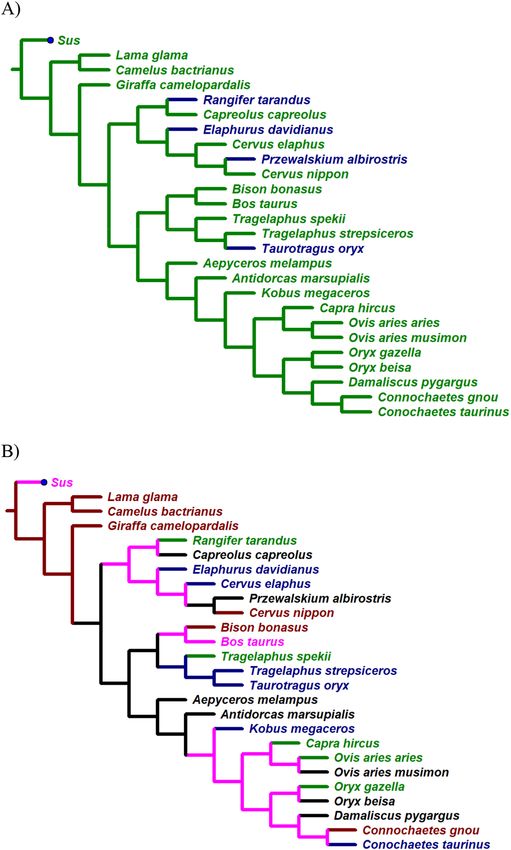
www.nature.com/scientificreports/

                                           Figure 1.  Variable shapes of the “oval window” in the manica flexoria of the pectoral limb (view of the adductor
                                           area). (A) European roe deer, (B) Indochinese sika deer, (C) Guanaco. Photos by M. P.

                                           Figure 2.  Variable shapes of the “oval window” in the manica flexoria of the pectoral limb (view of the adductor
                                           area). (A) Reindeer, (B) Domestic sheep, (C) Beisa oryx. Photos by M. P.

                                           Figure 3.  Variable shapes of the “oval window” in the manica flexoria of the pectoral limb (view of the adductor
                                           area). (A) Western sitatunga, (B) European bison, (C) Giraffe. Photos by M. P.

                                           (3)      Oval elongated shape—length to width OWMF ratio 2.25–2.75:1—in Beisa oryx (Fig. 2C), Blesbok, Euro-
                                                    pean mouflon, European roe deer (Fig. 1A), Impala, Springbok, and White-lipped deer;
                                           (4)      Oval shape very elongated—length to width OWMF ratio more than 2.75:1—in Bactrian camel, Black
                                                    wildebeest, European bison (Fig. 3B), Giraffe (Fig. 3C), Guanaco (Fig. 1C), and Indochinese sika deer
                                                    (Fig. 1B).

          Scientific Reports |   (2022) 12:4362 |                 https://doi.org/10.1038/s41598-022-08303-z                                              4

Vol:.(1234567890)
www.nature.com/scientificreports/
Figure 4. Evolution of knee-clicks (A) and OWMF length–width ratio (B) as reconstructed by the maximum
parsimony approach. For more details see the body of text. Legend to (A): no knee-click in repertoire = green,
knee-click present = blue. Legend to (B): type 1 of OWMF = green, type 2 = blue, type 3 = black, type 4 = brown,
unresolved = pink.
However, these groups are not shared by all species within the same genus (Cervus, Connochaetes, Oryx, Ovis,
Tragelaphus); only in two cases (both camelid species, and Greater kudu, and Common eland) is the particular
shape shared by closely related species (i.e., in the sister-group configuration on the phylogenetic tree—Fig. 4B).
The discrimination between species included in this study is highly significant (Wilks’s Lambda = 0.0000036,
F = 2063.9, p < 0.0001). Specifically, all specimens in 15 of the 20 species analysed were correctly classified using
the classification matrix. Only several individuals were misclassified, specifically: two Impalas—once as an Euro-
pean roe deer and once as a White-lipped deer; two Springboks—once as an Impala and once as a White-lipped
deer; two White-lipped Deer—once as an Impala and once as a Springbok; two European roe deer as Impala;
and one Blue wildebeest as a Greater kudu.
Scientific Reports | (2022) 12:4362 | https://doi.org/10.1038/s41598-022-08303-z 5
Vol.:(0123456789)www.nature.com/scientificreports/
Suidae
Camelidae
superficial and deep
Ar odactyla
digital flexors
Giraffidae
manica flexoria
Ruminan a
Cervidae
musculi adductores
digitorum
Bovidae
Figure 5. Simplified phylogenetic tree of even-toed ungulates (based on Hassanin et al.44) with a specification
of the distribution of several morphological traits of the autopod mentioned in the “Discussion”.
All other statistical comparisons of the OWMF for body size, both digits, sex and age as main effects were
non-significant (p-values > 0.05) indicating the conservative nature of the OWMF parameters. However, for age,
the p-values were around 0.10 in all three inspected species (European roe deer, Domestic goat, and Domestic
sheep), and for sex in the Giraffe and some bovids (Springbok, Black and Blue wildebeests, Blesbok, Giraffe,
Gemsbok, European mouflon, Common eland, Greater kudu) with p-values around 0.08 or around 0.10 in the
case of Impala.
The evolution of knee-clicks (Fig. 4A) was reconstructed successfully for all nodes of the phylogenetic tree.
Interestingly, the evolution of OWMF, as reconstructed (Fig. 4B), was more complex, and OWMF types do not
fit with the distribution of knee-clicks produced by some bovids and cervids.
Discussion
The anatomy of the autopod of the forelimb is diverse in respect of specific phylogenetic clades of even-toed
ungulates (for overview see Fig. 5). Specifically, the superficial and deep digital flexor are always on the fore-
limb of the even-toed ungulates, but musculi adductores digitorum can be found only in ruminant ungulates
(Ruminantia). Manica flexoria is not formed in pigs as representatives of non-ruminant even-toed u ngulates22,35.
36
Constantinescu et al. dealt with the construction of the suspension apparatus of the spinal joint and the digital
flexors on the forelimb of the llama (Lama glama). They confirmed the anatomical similarity of the muscles
around the spinal joint of the llama and other ruminants. However, they found the absence of interflexor mus-
cles (musculi interfloxorii) and the presence of musculi lumbricales, which are more typical of species with more
than two digits37,38.
Manica flexoria arose independently in equids (Equidae)39, despite the lack of development of the musculi
adductores digitorum22,23,40. The tendon of the superficial digital flexor forms a cuff with a strip of connective
tissue that is located closer to the bone30,41. This band of connective tissue is histologically variable and is divided
into membranous and tendinous types. The membranous type may take the form of a "synovial bridge", "fibrous
bridge" or "broad synovial bridge". The tendinous type is then "symmetric X-crossing", "asymmetric X-crossing"
or "oblique crossing"42. The surface digital flexor together with the auxiliary ligament band encircle the tendon of
the deep digital flexor, thereby fixing it in position. Interestingly, manica flexoria is 2.5 cm longer on the horse’s
forelimb than on the pelvic l imb43.
Besides phylogenetic inherence (see above), the observed conservatism of autopodium anatomy could be
caused by functional constraints which optimize transport costs, escape and relaxing possibilities13,14. The same
constraints seem to be valid for detailed parameters of the OWMF which exhibit marked similarities inside
particular species. The similarity of the left and right side of the autopodia is e xpected45,46, whilst the distinctive-
ness between the OWMF of the third and fourth digits indicates some former selection for the symmetry of the
autopodium which increases movement efficiency and e ndurance25.
On the other hand, the species-specific OWMF parameters indicate a differentiation, especially in the cer-
vids and bovids, where the diversity has been higher than in giraffids or camelids (for former diversity see e.g.,
Janis47, for the current diversity see Groves and Grubb48). Since the recognized types based on the length–width
ratio of the OWMF indicate very limited concordance to phylogenetic relationships (see phylogenetic trees in
this study or for example, Hassanin et al.44), this might indicate a differentiation in some close relatives due to
species-specific preferences for specific habitats or for preferred types of m otion21,49–51. More in-depth taxonomic
sampling would be beneficial to clarify the incidence, size, and shape diversity of the OWMF in Artiodactyla.
The following taxa above the genus level (using Groves and Grubb48) are candidates for such further inspection:
Tragulidae; Moschidae; Antilocapridae; Alceini and Muntiacini within Cervidae; and Boselaphini, Neotragini,
Cephalophini and Oreotragini within Bovidae.
Animals of multiple ages were included in this investigation so as to study the possible dependence of the size
and shape of OWMF on the age of the animal. The youngest studied were stillborn domestic cattle and domestic
goats; the oldest was a 22-year-old European bison male. The shape and ratio of the length and width of the oval
window were almost identical for all monitored categories within one species. Thus, it can be assumed that in
Scientific Reports | (2022) 12:4362 | https://doi.org/10.1038/s41598-022-08303-z 6
Vol:.(1234567890)www.nature.com/scientificreports/
older animals with increased cumulative mobility, there is no marked increase or decrease in the OWMF. It was
also confirmed that animals that have never walked (stillborn due to foetal lung atelectasis) had an oval window
developed on both attachment tendons of the adductors. This indicates that the OWMF is a physiological struc-
ture, and its presence is "programmed" in advance during intrauterine development.
The discovery of the non-described (not specified for example in K olda24, Sisson and Grossman35, Najbrt
et al.22, Nickel et al.25, Barone38, König and Liebich23, König et al.52, or in Böhmer et al.53 reporting on extensive
mammal species sampled) oval window in the manica flexoria (OWMF) is surprising. A limited attention to
some detailed configurations of the tendon apparatus in the autopodium, the limited conspicuousness of the
OWMF, and using domestic cattle as the common model organism (e.g., Hedges)54 could be partly responsible
for this oversight. This indicates that there are some gaps in basic comparative anatomical data (e.g. Guillerme
and Cooper55, Conde et al.)56 and that some discoveries are still possible (e.g. K lima57, Crole and S oley58, Shad-
wick et al.59, Frey et al.60).
The appearance and function of the oval window in the OWMF is unclear. The most likely function is to help
balance the pressure inside the ligament cuff and reduce the friction of the areas of the muscle tendons touching/
rubbing against each other. However, this assumption has not yet been physically and technically confirmed and
thus opens up a new field for further research. Another possible function is to increase the extent of digit flexion
in species that have longer and wider OWMFs; manica flexoria with little or no oval window is more rigid and
strong. This hypothesis is confirmed by the autopodies of the Guanaco and Bactrian camel with a large OWMF,
which, compared to unguligrad even-toed ungulates, are semi-digitigrade with a higher range of movements
in the t oes36.
The importance of the OWMF for the self-flexion and extension of the digits of even-toed ungulates has not
yet been elucidated, but there is importance for the veterinary medicine discipline; as example, it is possible to
"pass" through the oval window in the manica flexoria inside the cuff with an arthroscope and to evaluate the
damage and changes of the tendons, for example in septic t endosynovitis61. There is a sufficient distance between
the bone base and the manica flexoria to allow the insertion of the endoscope and its movement in the individual
muscle layers.
An interesting finding was that in domestic cattle, the OWMF does not develop on the forelimb even though
it is developed in the hind limb (Supplementary Fig. S2). Its shape and the ratio of length and width are very
similar to that of the European bison, which, however, has an oval window on both limbs. Perhaps this is due to
the domestication of cattle, which, unlike its "wild" relatives, spends most of its life in a limited area of stables
or pastures, the details surveyed in respect of these studies would be beneficial (e.g., O’Regan and K itchener62,
Keller et al.63, for references related to captivity-induced changes see also Robovský et al.)64.
The shape of the oval window in the manica flexoria among the inspected species was highly variable. The
name "oval window" suggested and used is not entirely suitable for some species from this study. For example,
in the reindeer the window shape is rather triangular with the base located proximally, whilst it is considerably
elongated and spindle-shaped in the guanaco. However, more than three-quarters of the species in the study had
an oval OWMF, thus this "functional" name was retained.
Another interesting finding was that the tendon of the deep digital flexor at the level of the OWMF is a few
millimetres wider and higher than what it is proximal and distal to this site (Supplementary Fig. S1). In the his-
tological assessment of this "swelling" in several selected individuals (Common eland, Bactrian camel, European
bison), areas of cartilaginous tissue surrounded by normal connective tissue were found. The reason for this
phenomenon is unclear, but it is most likely an adaptive response of the tendon to the mechanical demand due
to the higher weight and age of the individual65,66.
The evolution of knee-clicks (Fig. 4A) was reconstructed successfully for all nodes of the phylogenetic tree.
Knee-clicks seems to arise independently four times—specifically three times in cervids and once in bovids (in
blue—Fig. 4A). However, the evolution of OWMF, as reconstructed (Fig. 4B), was more complex and not resolved
for some nodes and clades in cervids and advanced bovids which prevents the description of the evolution of
OWMF in detail. Since none of the absolute or relative (not shown) OWMF parameters fit exclusively with the
distribution of knee-clicks produced by some bovids and cervids during movement, the mechanism responsible
for this sound remain cryptic from the present anatomical perspective.
Therefore, the mechanism responsible for this specific sound requires further investigation and more analyti-
cal approaches. Knee-clicks have been documented in several species by acoustic analysis3,8,9, and Bro-Jørgensen
and Dabelsteen3 identified knee-clicking as the honest signal of body size in the Common eland, using the
comparison of acoustic parameters of knee-clicks and several other phenotype traits of inspected individuals.
Despite the possibility to document knee-clicks readily by acoustic a nalysis3,8,9, it is not easily detectable from
where this sound originates, as reported in our pilot study and as already reviewed by Mohr5,11,67 one century
ago. Mohr11,67 mentioned several methods used by herself or other authors such as the fixing of particular limb
regions by linen, experimental production of knee-clicks by bending of specific parts of limbs in dead individuals
or using the stethoscope in live animals, but no progress has been done since that time. There are some obstacles
in attempts to find the source of the knee-clicks’ sound. Firstly, it is necessary to use live animals or fresh carcass
material before the development of rigor mortis, as documented by M ohr11,67 in the Reindeer, and as also shown
by unsuccessful attempts to obtain knee-clicks post-mortem via different limb positions and pressure involved
on different parts of the limbs in an attempt to mimic movement of the limb during walking within the present
study. Secondly, since an acoustic camera could not identify a single area on the forelimb during sound emission
on a walking eland antelope, it is necessary to use well cooperative/habituated11 representatives of species pro-
ducing this type of sound in vivo, moreover under standardized conditions in order to minimize sources of the
(acoustic) noise under natural or captive conditions. Finally, any generalization obtained from one species might
be limited due to independent origins of knee-clicks in even-toed ungulates, as detected in the present study.
Scientific Reports | (2022) 12:4362 | https://doi.org/10.1038/s41598-022-08303-z 7
Vol.:(0123456789)www.nature.com/scientificreports/
Material and methods
To find the source of the emitted clicking sound on a tame adult eland bull kept at Eland farm (Czech University
of Life Sciences Prague), an acoustic camera (Norsonic AS, Tranby, Norway) provided and operated by Ekola
Ltd. Prague was used. The sound(s) were measured in a barn, where the eland herd is housed, from a distance of
3–4 m, during the walking of the inspected individual, when the clicking sound was clearly emitted. As an addi-
tional trial, the whole forelimb of another adult eland bull was evaluated post-mortem (this bull was slaughtered
due to regular reduction as part of farmed herd management). Before slaughter, this animal was emitting clicking
sounds during walking. The whole skin-on forelimb was removed 1.5 h after post-mortem and stored at 7 °C until
the next day, when the forelimb was transported to the biomechanical lab at the Department of Anatomy and
Biomechanics, (Faculty of Physical Education and Sport Regulations, Charles University Prague). A stethoscope,
sound recorder and palpation were used for sound and vibration detection in endeavours to obtain knee-clicks
post-mortem via different limb positions and pressure involved on different parts of the limbs in an attempt to
mimic movement of the limb during walking.
To conduct a comparative study of thoracic autopodia (manus—from wrist to hoof), 25 species of even-toed
ungulates (Artiodactyla, see Asher and Helgen, and Prothero et al.)68,69 of four families (Camelidae, Giraffidae,
Cervidae and Bovidae), consisting of various ages and sexes were investigated (for detail list see Table 2) (it
contains references 49,70–83).
Altogether 312 autopods of the forelimb originating from 156 individuals were studied (Table 1). The origin
of the animals varied and included zoos in the Czech Republic (Brno Zoo, Dvůr Králové Zoo, Chomutov Zoo,
Jihlava Zoo, Olomouc Zoo, Ostrava Zoo, Pilsen Zoo, Prague Zoo, and Ústí nad Labem Zoo), as well as private
breeders, associations, and school institutions (VFU Brno, CZU Prague, Miskovice u Kutné Hory). Additional
material was also obtained during the harvest of game species in the Republic of South Africa (Game farms near
Bredasdorp and Witsand—Springbok, Black wildebeest and Blesbok; Game ranches close to Modimole—Impala,
Blesbok and Blue wildebeest) and game ranches in Namibia near Windhoek and Kalkfeld (Springbok, Giraffe
and Gemsbok). In this case, these were wild animals kept under minimal husbandry conditions intended for
meat production. As these animals were all from either cadavers (who had died from various causes) or were
collected from carcasses that were part of a standard harvesting for meat management routine, no animal ethics
approvals were required. The sex of the animals used in the study was almost evenly represented with a slight
predominance of males over females (82 males and 74 females). Only two age groups were distinguished in the
study. The first and most numerous were sexually and physically mature individuals (148 individuals), followed
by new-borns and calves (8 individuals; hereafter labelled as non-adult individuals).
Each autopodium underwent a thorough anatomical autopsy focusing on the macroscopic structure of super-
ficial and deep digital flexors and short digital muscles, and their tendons, as well as the topographic relation-
ships of the structures around the manica flexoria and sesamoid bones. First, the skin was removed from the
autopods and then the manica flexoria was dissected from the bone base (Supplementary Fig. S3). The presence
or absence of any different structure from known anatomical parts or distinct species, sexes and age groups
were determined. A yet to be described opening, a so-called "oval window" of the manica flexoria (abbrevi-
ated as OWMF) (Supplementary Fig. S4) was observed in the adduct tendon on the tendon’s surface facing the
bone; its shape and bilateral symmetry were determined. Using a calliper or a metric band, two dimensions of
this hole were obtained, namely its length and width (Supplementary Fig. S5). The term length is defined as the
dimension between the most proximal and the most distal edge of the window, the width the distance between
the axial and abaxial edge. Autopsies were performed at the Institute of Anatomy, Histology and Embryology
(Veterinary University in Brno) and in laboratories designated for that purpose at Czech University of Life Sci-
ences Prague and Jiří Orten Grammar School in Kutná Hora. The material originating from individuals obtained
by controlled harvesting in South Africa and Namibia had to be subjected to on-site autopsy in field conditions
and in local abattoirs.
The obtained measurements (length and width of the OWMF) are specified in millimetres (mm) as
means ± SD (Table 2) but were analysed statistically as primary values in respect of sex (male vs female), age
(non-adult vs adult), body side (left vs right), digit (third vs fourth digit) and species (all species included in this
study). Although the Shapiro-Wilks test recognized some data as not being deviant from normality, the same
nonparametric test variant (Mann–Whitney test) for all species was applied so as to minimise the risk of false
positive results (type I error) due to small sample sizes. Since non-adult individuals were available for only three
species (Domestic goat, European roe deer and Domestic sheep), the effect of age was only evaluated in these
species, whilst for species comparisons, only adult specimens were included to maximize comparability. In sum-
mary, species differences were analysed using Discriminant analysis, and other category differences using the
non-parametric Mann–Whitney test and Sign test. Significance was considered when p ≤ 0.05. Microsoft Excel
under Microsoft 365 and Statistica ver. 13.5.0.17 (copyright TIBCO Software) were used for calculations and
statistical comparisons. Some species were not analysed within all statistical comparisons due to their sample
size being less than three individuals (Table 1).
To compare the evolutionary distribution of knee-clicks and OWMF, the absolute and relative size of the
OWMF (see Table 2) using body weight as a proxy of the body size, were utilised. Sources of body w eights49,70–83
are specified in Table 2. The evolution of knee-clicks and OWMF (specifically, the length–width ratio) was
optimized by NONA (ver. 2.0) and WINCLADA interface (ver. 1.00.0884) using the unweighted maximum-
parsimony approach on a simplified and consensual phylogenetic tree adopted from Pitra et al.85, Hernández
Fernández & V rba86, Hassanin et al.44 and Chen et al.87. The topology of the phylogenetic tree was constrained
for reconstruction. No preference to ACCTRAN nor DELTRAN optimization were given when alternative
reconstructions were of equal cost.
Scientific Reports | (2022) 12:4362 | https://doi.org/10.1038/s41598-022-08303-z 8
Vol:.(1234567890)www.nature.com/scientificreports/
Avg. weight Avg. weight Avg. weight
(kg) (kg) (kg)
Scientific name Common name M F M+F Sources N Sex Age
Aepyceros mela- 70
Impala 56.9 43.8 50.35 15 13M/2F 15A
mpus
Antidorcas 70
Springbok 40.7 35.5 38.1 18 8M/10F 18A
marsupialis
Bison bonasus European bison 718 423 570.5 70
1 1M 1S
Bos taurus 70
Domestic cattle 384 327.5 355.75 6 3M/3F 2J + 2A + 2S
taurus
Camelus bactri- 71,72
Bactrian camel 600 450 525 2 2F 2S
anus bactrianus
Capra hircus 73
Domestic goat 50 30 40 6 3M/3F 2J + 2A + 2S
hircus
Capreolus European roe 70
24.2 23.4 23.8 6 3M/3F 2J + 4A
capreolus deer
Cervus elaphus Red deer 185.1 140.2 162.65 70
3 1M/2F 2A + 1S
Cervus nippon Indochinese sika 74
90 50 70 4 2M/2F 4A
pseudaxis deer
Connochaetes 70
Black wildebeest 166.7 135 150.85 6 3M/3F 6A
gnou
Connochaetes 70
Blue wildebeest 235.3 184.9 210.1 10 5M/5F 10A
taurinus taurinus
Damaliscus 75
Blesbok 70 60 65 8 4M/4F 8A
pygargus phillipsi
Elaphurus Père David’s 70
207.3 149.9 178.6 4 2M/2F 4A
davidianus deer
Giraffa camelo- 70,76–78
Giraffe* 1190.2 814.3 1002.25 16 8M/8F 15A + 1S
pardalis*
Kobus megaceros Nile lechwe 105 75 90 49,75,79
1 1M 1A
Lama glama 70,71
Guanaco 109.5 99 104.25 1 1M 1S
guanicoe
Oryx beisa beisa Beisa oryx 176.4 161.7 169.05 80
4 2M/2F 2A + 2S
Oryx gazella Gemsbok 178 166.4 172.2 70
12 6M/6F 12A
German gray
Ovis aries aries 67.9 44.9 56.4 70
6 3M/3F 2J + 2A + 2S
heath sheep
Ovis aries European 81
42.5 35 38.75 6 2M/4F 6A
musimon mouflon
Przewalskium White-lipped 82
204.2 125 164.6 4 2M/2F 2A + 2S
albirostris deer
Rangifer taran- 70
Reindeer 145 85.8 115.4 3 1M/2F 1A + 2S
dus
Taurotragus oryx Common eland 647.3 415.8 531.55 70
6 3M/3F 6A
Tragelaphus Western sitat- 83
115 55 85 1 1M 1S
spekii gratus unga
Tragelaphus 70
Greater kudu 240.8 159.2 200 7 4M/3F 7A
strepsiceros
Total 156 82M/74F 8J + 129A + 19S
Table 2. List of taxa (ordered alphabetically according to the scientific name) with common names, average
weight of species extracted from the literature, and individuals inspected in this study. *We evaluated a
population equivalent to the Angolan giraffe (= angolensis Lydekker, 1903). avg. average, N sample size, sex: F
female, M male, age: A adult, J neonate or juvenile, S senescent.
Graphics. Photographs used were taken by M. P. Phylogenetic trees were produced using WINCLADA
(v1.00.0884), IrfanView v4.57—64 bit downloaded from https://www.irfanview.com/ and Microsoft 365 interface
downloaded from https://www.microsoft.com/cs-cz/microsoft-365.
Ethical statement. For the study, material was received from animals slaughtered for meat production, or
euthanised due to health reasons or that had died naturally. No single animal was slaughterer or euthanised to
gather material/tissue for this study and all causes of death were unrelated to the musculoskeletal system. All
procedures followed Czech or international laws for manipulation and culling of farmed animals or veterinary
and husbandry laws applied to zoo gardens. No extra permission/ethical clearance or approval by an ethical
committee was necessary since all manipulations with study material and procedures were done post-mortem
and not required by Czech legislation.
Scientific Reports | (2022) 12:4362 | https://doi.org/10.1038/s41598-022-08303-z 9
Vol.:(0123456789)www.nature.com/scientificreports/
Data availability
The data matrix is available in the Supplementary online material, other data subsets used and/or analysed during
this study are available from the corresponding author on request.
Received: 14 July 2021; Accepted: 3 March 2022
References
1. Grubb, P. Order Artiodactyla. In Mammal Species of the World 3rd edn (eds Wilson, D. E. & Reeder, D. M.) 637–722 (The Johns
Hopkins University Press, 2005).
2. Kingdon, J. East African Mammals. An Atlas of Evolution in Africa Vol. 3 (Academic Press, 1982).
3. Bro-Jørgensen, J. & Dabelsteen, T. Knee-clicks and visual traits indicate fighting ability in eland antelopes: Multiple messages and
back-up signals. BMC Biol. 6, 47. https://doi.org/10.1186/1741-7007-6-47 (2008).
4. Bro-Jørgensen, J. & Beeston, J. Multimodal signalling in an antelope: Fluctuating face masks and knee-clicks reveal the social status
of eland bulls. Anim. Behav. 102, 231–239. https://doi.org/10.1016/j.anbehav.2015.01.027 (2015).
5. Mohr, E. Über das “Knacken” bei einigen Paarhufern, besonders beim Renntier. Biol. Zentralbl. 37, 177–188 (1917).
6. Müller-Using, D. & Schloeth, R. Das Verhalten der Hirsche (Cervidae). Handb. Zool. 28, 1–60 (1967).
7. Schaller, G. B. & Hamer, A. Rutting behavior of Père David’s deer, Elaphurus davidianus. Zool. Garten N. F. 48, 1–15 (1975).
8. Pelosse, J. L. Akustische Untersuchungen über das Knacken der Extremitäten beim Rentier (Rangifer tarandus). Bongo 6, 79–84
(1982).
9. Miura, S., Ohtaishi, N., Kaji, K. & Wu, J. A preliminary study of behavior and acoustic repertoire of captive white-lipped deer,
Cervus albirostris, China. J. Mamm. Soc. Japan 13, 105–118. https://doi.org/10.11238/jmammsocjapan1987.13.105 (1988).
10. Toweill, D. E. & Thomas, J. W. North American Elk: Ecology and Management (Smithsonian Institution Press, 2002).
11. Mohr, E. Nochmals über das “Knacken” beim Rentier. Biol. Zentralbl. 39, 251–256 (1919).
12. Wemmer, C. M., Collins, L. R., Beck, B. B. & Wemmer, C. M. The ethogram. In The Biology and Management of an Extinct Species
Père David’s Deer (eds Beck, B. B. & Wemmer, C. M.) 91–125 (Noyes Pubns, 1983).
13. Janis, C. M. & Wilhelm, P. B. Were there mammalian pursuit predators in the Tertiary? Dances with wolf avatars. J. Mammal. Evol.
1, 103–125. https://doi.org/10.1007/BF01041590 (1993).
14. Hermanson, J. W. & MacFadden, B. J. Evolutionary and functional morphology of the knee in fossil and extant horses (Equidae).
J. Vertebr. Paleontol. 16, 349–357. https://doi.org/10.1080/02724634.1996.10011321 (1996).
15. Janis, C. M., Theodor, J. M. & Boisvert, B. Locomotor evolution in camels revisited: A quantitative analysis of pedal anatomy and
the acquisition of the pacing gait. J. Vertebr. Paleontol. 22, 110–121. https://doi.org/10.1671/0272-4634(2002)022[0110:LEICRA]
2.0.CO;2 (2002).
16. Clifford, A. B. The evolution of the unguligrade manus in artiodactyls. J. Vertebr. Paleontol. 30, 1827–1839. https://d oi.o
rg/1 0.1 080/
02724634.2010.521216 (2010).
17. Biancardi, C. M. & Minetti, A. E. Biomechanical determinants of transverse and rotary gallop in cursorial mammals. J. Exp. Biol.
215, 4144–4156. https://doi.org/10.1242/jeb.073031 (2012).
18. Levering, D., Hopkins, S. & Davis, E. Increasing locomotor efficiency among North American ungulates across the Oligocene-
Miocene boundary. Palaeogeogr. Palaeoclimatol. Palaeoecol. 466, 279–286. https://doi.org/10.1016/j.palaeo.2016.11.036 (2017).
19. Zhang, Q., Xu, K. & Ding, X. Investigation of feet functions of large ruminants with a decoupled model of equivalent mechanism.
Biol.Open 6, 407–414. https://doi.org/10.1242/bio.023630 (2017).
20. Doube, M. et al. Limb bone scaling in hopping macropods and quadrupedal artiodactyls. R. Soc. Open Sci. 5, 180152. https://doi.
org/10.1098/rsos.180152 (2018).
21. Short, R. A. & Lawing, A. M. Geography of artiodactyl locomotor morphology as an environmental predictor. Divers. Distrib. 27,
1818–1831 (2021).
22. Najbrt, R. et al. Veterinary Anatomy 1 (SZN, 1980) (in Czech).
23. König, H. E. & Liebich, H. G. Anatomy of Domestic Mammals. Part 1: Musculoskeletal System (H&H, 2003) (in Czech).
24. Kolda, J. Comparative Anatomy of Domestic Animals III-IV Arthrology, Myology Including the Mechanics of Movement (Studentská
organizace čs. veterinárních mediků, 1950) (in Czech).
25. Nickel, R., Schummer, A. & Seiferle, E. The Anatomy of the Domestic Animals, The Locomotor System of the Domestic Mammals
Vol. 1 (Verlag Paul Parey, 1986).
26. Nogueira, G. M., Cattelan, J. W., Pereira, W. A. B., Moraes, P. C. & Duarte, C. A. Ultrasonographic characterization of flexor
tendinous structure muscle and ligaments of the distal limb of crossbred heifers. Arq. Bras. Med. Vet. Zootec. 63, 600–608. https://
doi.org/10.1590/S0102-09352011000300011 (2011).
27. Bertagnoli, A., Raber, M., Morandi, N., Mortellaro, C. M. & Steiner, A. Tenovaginoscopic approach to the common digital flexor
tendon sheath of adult cattle: Technique, normal findings and preliminary results in four clinical cases. Vet. J. 191, 121–127. https://
doi.org/10.1016/j.tvjl.2010.12.009 (2012).
28. Kofler, J. & Edinger, H. K. Diagnostic ultrasound imaging of soft tissues in the bovine distal limb. Vet. Radiol. Ultrasound 36,
246–252. https://doi.org/10.1111/j.1740-8261.1995.tb00255.x (1995).
29. Blaser, M. et al. Arthroscopic approaches to the fetlock joint of adult cattle: A cadaver study. Vet. J. 193, 701–706. https://doi.org/
10.1016/j.tvjl.2012.03.007 (2012).
30. Nixon, A. J. Endoscopy of the digital flexor tendon sheath in horses. Vet. Surg. 19, 266–271. https://doi.org/10.1111/j.1532-950x.
1990.tb01182.x (1990).
31. Denoix, J.-M. & Busoni, V. Ultrasonographic anatomy of the accessory ligament of the superficial digital flexor tendon in horses.
Equine Vet. J. 31, 186–191. https://doi.org/10.1111/j.2042-3306.1999.tb03170.x (1999).
32. Seignour, M., Coudry, V., Norris, R. & Denoix, J.-M. Ultrasonographic examination of the palmar/plantar aspect of the fetlock in
the horse: Techniques and normal images. Equine Vet. Educ. 24, 19–29. https://doi.org/10.1111/j.2042-3292.2011.00192.x (2012).
33. Werpy, N. M., Denoix, J.-M., McIlwraith, C. W. & Frisbie, D. D. Comparison between standard ultrasonography, angle contrast
ultrasonography, and magnetic resonance imaging characteristics of the normal equine proximal suspensory ligament. Vet. Radiol.
Ultrasound 54, 536–547. https://doi.org/10.1111/vru.12051 (2013).
34. El-Shafey, A. & Kassab, A. Computed tomography and cross-sectional anatomy of the metatarsus and digits of the One-humped
Camel (Camelus dromedarius) and Buffalo (Bos bubalis). Anat. Histol. Embryol. 42, 130–137. https://doi.org/10.1111/j.1439-0264.
2012.01174.x (2013).
35. Sisson, S. & Grossman, J. D. The Anatomy of the Domestic Animals 4th edn. (W. B. Saunders Company, 1953).
36. Constantinescu, G. M., Reed, S. K. & Constantinescu, I. A. The suspensory apparatus and digital flexor muscles of the Llama (Lama
glama) 1. Thoracic limb. Int. J. Morphol. 26, 543–550. https://doi.org/10.4067/S0717-95022008000300006 (2008).
37. Ellenberger, W. & Baum, H. Handbuch der vergleichenden Anatomie der Haustiere 18th edn. (Springer, 1977).
38. Barone, R. Anatomie Comparée des Mammiferes Domestiques Tome 2, Arthrologie et myologie (Vigot, 2000).
Scientific Reports | (2022) 12:4362 | https://doi.org/10.1038/s41598-022-08303-z 10
Vol:.(1234567890)www.nature.com/scientificreports/
39. Grassé, P.-P. Traité de Zoologie, Anatomie, Systématique, Biologie XVII, Mammifères: Les Orders: Anatomie, Ethologie, Systématique
(Masson and Co., 1955).
40. Denoix, J.-M. Essentials of Clinical Anatomy of the Equine Locomotor System (CRC Press, 2019).
41. Denoix, J.-M. Functional anatomy of tendons and ligaments in the distal limbs (manus and pes). Vet. Clin. N. Am. Equine Pract.
10, 273–322. https://doi.org/10.1016/s0749-0739(17)30358-9 (1994).
42. Jordana, M. et al. Anatomical description of the presence and variability of the digital manica flexoria in the equine digital flexor
tendon sheath. Anat. Histol. Embryol. 46, 9–16. https://doi.org/10.1111/ahe.12224 (2017).
43. Findely, J. A., Ricci, E. E. & Singer, E. E. An anatomical and histological study of the equine proximal manica flexoria. Vet. Comp.
Orthop. Traumatol. 30, 91–98. https://doi.org/10.3415/VCOT-16-01-0016 (2017).
44. Hassanin, et al. Pattern and timing of diversification of Cetartiodactyla (Mammalia, Laurasiatheria), as revealed by a comprehensive
analysis of mitochondrial genomes. C. R. Biol. 335, 32–50. https://doi.org/10.1016/j.crvi.2011.11.002 (2012).
45. Baruzzi, C., Nawroth, C., McElligott, A. G. & Baciadonna, L. Motor asymmetry in goats during a stepping task. Laterality 23,
599–609. https://doi.org/10.1080/1357650X.2017.1417993 (2018).
46. Leliveld, L. M. C. From science to practice: A review of laterality research on ungulate livestock. Symmetry 11, 1157. https://doi.
org/10.3390/sym11091157 (2019).
47. Janis, C. Evolution of horns in ungulates: Ecology and paleoecology. Biol. Rev. 57, 261–318. https://doi.org/10.1111/j.1469-185X.
1982.tb00370.x (1982).
48. Groves, C. & Grubb, P. Ungulate Taxonomy (Johns Hopkins University Press, 2011).
49. Spinage, C. A. The Natural History of Antelopes (Facts On File Publications, 1986).
50. Putman, R. The Natural History of Deer (Comstock Publishing Associates, 1988).
51. Geist, V. Deer of the World: Their Evolution, Behaviour, and Ecology (Stackpole Books, 1998).
52. König, H. E. et al. Sehnen und Bänder an der Rinderzehe. Wien. Tierärztl. Monat. 100, 55–60 (2013).
53. Böhmer, C., Theil, J.-C., Fabre, A.-C. & Herrel, A. Atlas of Terrestrial Mammal Limbs (CRC Press, 2020).
54. Hedges, S. B. The origin and evolution of model organisms. Nat. Rev. Genet. 3, 838–849. https://doi.org/10.1038/nrg929 (2002).
55. Guillerme, T. & Cooper, N. Assessment of available anatomical characters for linking living mammals to fossil taxa in phylogenetic
analyses. Biol. Lett. 12, 20151003. https://doi.org/10.1098/rsbl.2015.1003 (2016).
56. Conde, D. A. et al. Data gaps and opportunities for comparative and conservation biology. Proc. Natl. Acad. Sci. USA 116, 9658–
9664. https://doi.org/10.1073/pnas.1816367116 (2019).
57. Klima, M. Development of the cetacean nasal skull. Adv. Anat. Embryol. Cell Biol. 149, 1–143. https://doi.org/10.1007/978-3-642-
58612-5 (1999).
58. Crole, M. R. & Soley, J. T. Evidence of a true pharyngeal tonsil in birds: A novel lymphoid organ in Dromaius novaehollandiae and
Struthio camelus (Palaeognathae). Front. Zool. 9, 21. https://doi.org/10.1186/1742-9994-9-21 (2012).
59. Shadwick, R. E., Goldbogen, J. A., Potvin, J., Pyenson, N. D. & Vogl, A. W. Novel muscle and connective tissue design enables high
extensibility and controls engulfment volume in lunge-feeding rorqual whales. J. Exp. Biol. 216, 2691–2701. https://doi.org/10.
1242/jeb.081752 (2013).
60. Frey, R., Reby, D., Fritsch, G. & Charlton, B. D. The remarkable vocal anatomy of the koala (Phascolarctos cinereus): Insights into
low-frequency sound production in a marsupial species. J. Anat. 232, 575–595. https://doi.org/10.1111/joa.12770 (2018).
61. Anderson, D. E. & St. Jean, G. Diagnosis and management of tendon disorders in cattle. Vet. Clin. North Am. Food Anim. Pract.
12, 85–116. https://doi.org/10.1016/s0749-0720(15)30438-2 (1996).
62. O’Regan, H. J. & Kitchener, A. C. The effects of captivity on the morphology of captive, domesticated and feral mammals. Mam.
Rev. 35, 215–230. https://doi.org/10.1111/j.1365-2907.2005.00070.x (2005).
63. Keller, A., Clauss, M., Muggli, E. & Nuss, K. Even-toed but uneven in length: The digits of artiodactyls. Zoology 112, 270–278.
https://doi.org/10.1016/j.zool.2008.11.001 (2009).
64. Robovský, J., Melichar, L. & Gippoliti, S. Zoos and conservation in the anthropocene: Opportunities and problems. In Problem-
atic Wildlife II: New Conservation and Management Challenge in the Human-Wildlife Interactions (eds Angelici, F. A. & Rossi, L.)
451–484 (Springer, 2020).
65. Tichý, F., Horký, D., Kociánová, I. & Gorošová, A. Histology: Cytology and General Histology (VFU Brno, 2000).
66. Montero, J. A., Lorda-Diez, C. I. & Hurle, J. M. Regenerative medicine and connective tissues: Cartilage versus tendon. J. Tissue
Eng. Regen. Med. 6, 337–347. https://doi.org/10.1002/term.436 (2012).
67. Mohr, E. “Knacken” beim toten Rentier. Biol. Zentralbl. 46, 231–232 (1926).
68. Asher, R. J. & Helgen, K. M. Nomenclature and placental mammal phylogeny. BMC Evol. Biol. 10, 102. https://doi.org/10.1186/
1471-2148-10-102 (2010).
69. Prothero, D. R. et al. On the unnecessary and misleading taxon “Cetartiodactyla”. J. Mammal. Evol. https://d oi.o
rg/1 0.1 007/s 10914-
021-09572-7 (2021).
70. Peréz-Barbería, F. J. & Gordon, I. J. Differences in body mass and oral morphology between the sexes in the Artiodactyla: Evolu-
tionary relationships with sexual segregation. Evol. Ecol. Res. 2, 667–684 (2000).
71. Franklin, W. L. Family Camelidae (camels). In Handbook of the Mammals of the World Vol. 2 (eds Wilson, D. E. & Mittermeier, R.
A.) 206–246 (Lynx Edictions, 2011).
72. Lensch, J. The two-humped camel (Camelus bactrianus). World Animal Review: FAO Editorial Group http://w ww.f ao.o rg/3/x 1700t/
x1700t05.htm#TopOfPage (1999).
73. Coblentz, B. Capra hircus. Global Invasive Species Database http://www.iucngisd.org/gisd/species.php?sc=40 (2021).
74. Banwell, B. The Sika (The Halcyon Press, 2009).
75. Groves, C. P. & Leslie, D. M. Family Bovidae (hollow-horned ruminants). In Handbook of the Mammals of the World Vol. 2 (eds
Wilson, D. E. & Mittermeier, R. A.) 447–779 (Lynx Edictions, 2011).
76. Ciofolo, C. & Le Pendu, Y. Giraffa camelopardalis Giraffe. In Mammals of Africa: Volume VI: Pigs, Hippopotamuses, Chevrotain,
Giraffes, Deer and Bovids (eds Kingdon, J. & Hoffmann, M.) 98–109 (Bloomsbury Publishing, 2013).
77. Gloneková, M. et al. The weight of Rothschild giraffe: Is it really well known? Zoo Biol. 35, 423–431. https://doi.org/10.1002/zoo.
21308 (2016).
78. Skinner, J. D. & Mitchell, G. Family Giraffidae (Giraffe and Okapi). In Handbook of the Mammals of the World Vol. 2 (eds Wilson,
D. E. & Mittermeier, R. A.) 788–802 (Lynx Edictions, 2011).
79. Castelló, J. R. Bovids of the World. Antellopes, Gazelles, Cattle, Goats, Sheep, and Relatives (Princeton University Press, 2016).
80. Ledger, H. Weights of some East African mammals (2). Afr. J. Ecol. 2, 159. https://doi.org/10.1111/j.1365-2028.1964.tb00205.x
(1964).
81. Röhrs, M. Ovis ammon musimon (Pallas, 1811)—Mufflon. In Handbuch der Säugetiere Europas (eds Niethammer, J. & Krapp, F.)
435–449 (Aula-Verlag, 1986).
82. Leslie, D. M. Jr. Przewalskium albirostre (Artiodactyla: Cervidae). Mamm. Species 42, 7–18. https://doi.org/10.1644/849.1 (2010).
83. Furstenburg, D. Focus on the sitatunga (Tragelaphus spekii). S. A. Hunter 01024, 40–43 (2009).
84. Nixon, K. C. Winclada (Beta) Ver. 0.9.9 (Published by the Author, 1999).
85. Pitra, C., Fickel, J., Meijaard, E. & Groves, C. P. Evolution and phylogeny of old world deer. Mol. Phylogenet. Evol. 33, 880–895.
https://doi.org/10.1016/j.ympev.2004.07.013 (2004).
Scientific Reports | (2022) 12:4362 | https://doi.org/10.1038/s41598-022-08303-z 11
Vol.:(0123456789)You can also read