Masseter muscle parameters can function as an alternative for skeletal muscle mass assessments on cross-sectional imaging at lumbar or cervical ...
←
→
Page content transcription
If your browser does not render page correctly, please read the page content below
Original Article

Masseter muscle parameters can function as an alternative for
skeletal muscle mass assessments on cross-sectional imaging at
lumbar or cervical vertebral levels
Hugo C. van Heusden1, Najiba Chargi1, Jan Willem Dankbaar2, Ernst J. Smid3, Remco de Bree1^
1
    Department of Head and Neck Surgical Oncology, University Medical Center Utrecht and Utrecht University, Utrecht, The Netherlands;
2
    Department of Radiology and Nuclear Medicine, University Medical Center Utrecht and Utrecht University, Utrecht, The Netherlands;
3
Department of Radiation Oncology, University Medical Center Utrecht and Utrecht University, Utrecht, The Netherlands

Contributions: (I) Conception and design: R de Bree, HC van Heusden, JW Dankbaar; (II) Administrative support: HC van Heusden; (III) Provision
of study materials or patients: EJ Smid, N Chargi, HC van Heusden; (IV) Collection and assembly of data: HC van Heusden; (V) Data analysis and
interpretation: HC van Heusden, N Chargi, R de Bree; (VI) Manuscript writing: All authors; (VII) Final approval of manuscript: All authors.

Correspondence to: Remco de Bree, MD, PhD. Department of Head and Neck Surgical Oncology, University Medical Center Utrecht, House Postal
Number Q.05.4.300, PO BOX 85500, 3508 GA, Utrecht, The Netherlands. Email: R.deBree@umcutrecht.nl.

                  Background: Patients with head and neck cancer are at increased risk of developing low skeletal muscle
                  mass (SMM), which is associated with adverse treatment outcomes and prognosis. Low SMM is most
                  commonly assessed by the skeletal muscle cross sectional area (CSA) at the third lumbar vertebra (L3) or
                  more recently the third cervical vertebra (C3). L3 is not routinely imaged and C3 may be impacted by disease
                  or treatment. As an alternative we analyzed masseter muscle characteristics and their relationship with L3
                  and C3 skeletal muscle CSA and overall survival (OS).
                  Methods: In this single-center retrospective study, 99 patients with head and neck cancer who underwent
                  whole body FDG-PET/CT-scans were reviewed. Of these patients, L3 CSA, C3 CSA, masseter CSA,
                  masseter thickness, masseter volume, masseter Hounsfield Unit values, lumbar skeletal muscle index (LSMI),
                  cervical skeletal muscle index (CSMI), and masseter skeletal muscle index (MSMI) were recorded and
                  correlated with each other and with OS.
                  Results: We included 72 male and 27 female patients. The masseter muscle parameters differed
                  significantly between sexes. The Spearman correlation coefficients for C3 CSA–Masseter volume and L3
                  CSA–Masseter volume were 0.639 and 0.531 (P
16                                                                             van Heusden et al. Masseter parameters indicate total SMM

Introduction                                                                 The purpose of this study was firstly to investigate
                                                                          whether masseter muscle quantity measures correlate
Head and neck squamous cell carcinoma (HNSCC) is
                                                                          with the CSA at C3 and L3. Secondly, the study sought to
the seventh most common type of cancer worldwide with
                                                                          investigate the association between these masseter muscle
890,000 new cases and 450,000 deaths in 2018 (1). About
                                                                          parameters and OS.
two thirds of patients present with advanced stage disease.
HNSCC at this stage is associated with a poor 5-year
overall survival (OS) of less than 50%. There is a need for               Methods
accurate prognostic factors to tailor treatment for HNSCC
                                                                          Ethical considerations
patients, and sarcopenia is emerging as a novel candidate in
HNSCC (2-4).                                                              The study was conducted in accordance with the
   Sarcopenia is defined as the loss of skeletal muscle mass              Declaration of Helsinki (as revised in 2013). The study was
(SMM) and muscle function (5), although measurements of                   approved by the Medical Ethical Research Committee of
only SMM are often used in literature. Sarcopenia is often                the University Medical Center Utrecht (approval ID 17-
the result of cancer cachexia (6,7).                                      365/C) and individual consent for this retrospective analysis
   Patients with HNSCC are at an increased risk for                       was waived.
cancer related cachexia and sarcopenia. This is partly due
to dysphagia caused by tumor localization or its treatment
                                                                          Patient and study design
and side effects thereof. Moreover, patients with HNSCC
might present with underlying malnutrition caused by poor                 We reviewed patients with newly diagnosed, pathologically
diet, tobacco use or alcohol abuse (8,9). Low SMM cancer                  proven HNSCC who underwent a whole body FDG-PET/
patients treated with surgery are at risk for complications               CT-scan between 2010 and 2018 at the University Medical
and decreased survival (10). In HNSCC, low SMM has been                   Center Utrecht, the Netherlands (UMCU). Indications
associated with and increased risk of surgical complications              to perform a whole body FDG-PET/CT-scan in our
and cisplatin dose limiting toxicity and with decreased survival          institute were clinical suspicion of advanced (III/IV) stage
(11-13). Low SMM can be considered as an emerging                         at presentation, carcinoma of unknown primary tumor,
biomarker for the clinical setting in HNSCC patients (14).                recurrent disease and second primary tumor. Patients with
   While the gold standard for total SMM assessment is full               previous HNSCC or second primary tumor were excluded.
body imaging, earlier research has shown that the muscle                  Patient scans who were incomplete, of insufficient quality or
cross-sectional area (CSA) measured on a single abdominal                 incompatible with current imaging software were excluded
cross-sectional slice at the level of the third lumbar                    from further analysis.
vertebra (L3) on computed tomography (CT) imaging can                        Patient factors with known or expected relation to HNC
provide accurate estimates of patient’s total SMM (15).                   outcome measures or development of sarcopenia were
Unfortunately, patients treated for head and neck cancers do              collected: age at diagnosis, gender, histological diagnosis,
not usually have imaging performed at this level. Therefore,              comorbidities scored using the Charlson Comorbidity
a method was developed to assess SMM on a single CT                       Index (CCI) and the ACE-27 score, tumor site and tumor
slide at the level of the third cervical vertebra (C3) in head            staging according to the 7th edition of the UICC TNM
and neck cancer patients (16). However, CSA assessment at                 classification system, human papillomavirus (HPV) status
this level may be impaired by extension of primary tumor                  for oropharyngeal carcinomas, weight loss 6 months before
and/or lymph nodes or previous treatment. Moreover,                       diagnosis and treatment regimens.
accurate assessment is time consuming (17,18). There
is a need for a reliable index muscle that is consistently
                                                                          Radiological assessment
present on routine imaging, is rarely impacted by disease
or treatment and is quick and easy to characterize using                  Segmentation of muscle tissue at the level of C3 and L3
commonly used imaging software. For this purpose, we                      was manually performed using the commercially available
propose the masseter muscle. The masseter muscle has been                 software package SliceO-matic (Tomovision, Canada). For
shown to be adequate in determining SMM and predicting                    analysis of the CSA at the level of C3, a standard method
mortality in other fields of medicine (19-21).                            for slide selection was used, where the first slide to show

© Quantitative Imaging in Medicine and Surgery. All rights reserved.   Quant Imaging Med Surg 2022;12(1):15-27 | https://dx.doi.org/10.21037/qims-21-43
Quantitative Imaging in Medicine and Surgery, Vol 12, No 1 January 2022 17
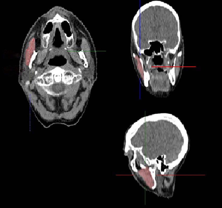
Ar: 267.59 mm2 Ar: 236.75 mm2
Av: 39.1 HU Av: 51.8 HU 11.4 mm
SD: 36.4 SD: 28.2
33.4 mm
Figure 1 Example of masseter delineation using Intellispace.
the entire vertebral arc and the transverse and spinous Figure 2 A 3D example of the measuring of masseter volume using
process when scrolling in a cephalad to caudad direction the TumorTracking feature in Intellispace.
was selected. Skeletal muscle tissue was identified using
Houndsfield Unit (HU) range settings from −29 to 150 HU
and the outer contours of the sternocleidomastoid and calculated after segmenting the entire muscle (Figure 2).
paravertebral muscles were traced manually. The CSA at the MT of the masseter was determined using the
level of C3 was determined as the sum of delineated areas measuring-tool included in Intellispace (mm). HUROI was
of the paravertebral muscles and both sternocleidomastoid determined in a 1-centimeter diameter circle on the same
muscles within a HU range of −29 to 150 HU in cm2. level as MCSA.
For analysis of the CSA at level L3 the muscle groups Since the state of a patient’s teeth may impact masseter
analyzed were the psoas, paravertebral and the anterior function and size each patient was examined for the
abdominal wall. presence of dental elements (22). Dental status was scored
For assessment of the masseter muscle, Intellispace as follows: [0] no missing dentition, [1] one or more missing
(version 14, Phillips, Netherlands) was chosen for its ability teeth, [2] total absence of dentition.
to measure the volume of a selected structure (e.g., the Presence of scattering cause by (dental) implants was
masseter muscle) using the TumorTracking feature which scored as follows: [0] no scattering present, [1] slight
allows for rapid tissue volume assessment. The following scattering present, [2] significant scattering present.
muscle parameters were chosen and measured: masseter Measurements were performed bilaterally for each patient
cross-sectional area (MCSA), masseter muscle volume (MV), and an average was calculated and used for further analysis.
masseter muscle maximum thickness (MT). Furthermore, Earlier research has shown that there is excellent
measurements of muscle quality defined by the Hounsfield agreeability between image scoring software programs used
unit (HU) and expressed as the average HU of all measured for measuring CSA (23). Therefore, we found it acceptable to
tissue (HU tot) and in a region of interest (HU ROI) were use the two programs independently and compare the data.
collected. MCSA was measured at the level of the dens of
the second cervical vertebra (Figure 1).
Body composition measurements
Coronal tilt alignment was made according to a tangent
running through the dens and hard palate. MCSA was Weight and height were recorded during a patient’s first
measured by outlining the outer surface of masseter after consultation at our out-patient clinic and used to calculate
which IntelliSpace automatically calculated the surface body mass index (BMI) and body surface area (BSA) using
area (mm 2), a method independent of the HU value of the Mosteller formula (24). Lumbar skeletal muscle index
the defined area. MV (cm3) and HUtot were automatically (LSMI), cervical skeletal muscle index (CSMI) and masseter
© Quantitative Imaging in Medicine and Surgery. All rights reserved. Quant Imaging Med Surg 2022;12(1):15-27 | https://dx.doi.org/10.21037/qims-21-4318 van Heusden et al. Masseter parameters indicate total SMM
skeletal muscle index (MSMI) were calculated by dividing Results
the corresponding patient’s CSA values by patient’s squared
Search and inclusion
height. There is, to our knowledge, no scientific consensus
on a cut-off point for MSMI. We therefore designated In total 139 patients who had undergone a CT-scan were
patients present in the lowest quartile of MCSA for their screened for study viability. Of these patients 15.2% (n=21)
specific gender as “low MSMI”. had (partially) missing imaging and were subsequently
excluded. Furthermore, in 3.6% (n=5) of included patients
the available imaging was of insufficient quality for analysis
Overall survival
either due to low resolution or poor image quality. In total,
The status of the patient (alive/deceased) was acquired from 113 whole body FDG-PET/CT-scans were included for
the UMCU electronic patient data system on date of last further image analysis.
follow-up. OS was defined as the time between the date
of histologic diagnosis and death, or date of last follow-
Patient, tumor and treatment characteristics
up. UMCU patient system is linked to the provincial
government register and is updated continuously for patients In total 99 patients were included, with a median age of
living in the Utrecht province. Patients were considered 61.69 (IQR, 56.0–68.40) years. Of the included patients,
alive if no date of death was available on date of last follow- 81 (71.7) were male. A minority of patients had no history
up or if there was no physician note reporting on their death. of alcohol consumption (n=32, 28.3%) or smoking (n=13,
Cause of date was determined by physician’s notes. 13.1%). Forty patients (40.4%) were categorized as having
normal weight based on BMI score. Most patients presented
tumorous disease localized in the oropharynx (n=66, 66.7%)
Statistical analysis
of which 16 (16.2%) were HPV-positive. Most patients
SPSS 26 for Windows (IBM, Armonk, NY, USA) was presented with a clinical stage IV disease (n=64, 64.6%) and
used for analysis. Descriptive statistics were calculated patients were most commonly treated with a combination
with the continuous variables presented as mean (standard of radiotherapy and systemic therapy (n=53, 46.9%).
deviation) or median (interquartile range). Discrete Twenty-six patients were designated as “Low MSMI”
variables were displayed as counts (percentages). Normality and seventy-three as “Normal MSMI”. There was a
was investigated by using the Kolmogorov-Smirnov test. statistically significant difference between these groups for
Characteristics and muscle measurements were analyzed LSMI, C3 CSA, CSMI and BMI (P=0.015, 0.019 and 0.014,
using independent-samples t-test for normally distributed respectively).
variables, independent-samples test for skewed variables See Tables 1,2 for patient, tumor and treatment
and Fisher’s exact test or Pearson’s chi-squared test for characteristics and the comparison of these characteristics
categorical variables. Spearman correlation coefficients between the low MSMI and normal MSMI groups.
were calculated to establish the relationship between
L3 measurements, C3 measurements and masseter
Body composition measurement
measurements. A correlation coefficient of (−)0.8 to (−)1
was interpreted as a very strong correlation, (−)0.6–0.8 as Table 3 shows a significant difference based on sex for HUtot
strong, (−)0.4 to (−)0.6 as moderate, and (−)0.2 to (−)0.4 and HUROI (both P=0.049), BSA, L3 CSA, C3 CSA, MCSA,
as a low correlation (25). Radiological measurements and MV, MT, L3 SMI and MSMI (all PQuantitative Imaging in Medicine and Surgery, Vol 12, No 1 January 2022 19
Table 1 Baseline characteristics of included patients and differences between low and normal masseter muscle index
All patients (n=99), Normal MSMI (n=73), Low MSMI (n=26),
Characteristics P
N (%) or mean (± SD) N (%) or mean (± SD) N (%) or mean (± SD)
Median age in years (IQR) 61.77 (55.95–67.42) 61.85 (54.84–68.40) 62.26 (57.18–65.14) NS
Male sex 72 (72.7) 53 (72.6) 19 (73.1) NS
Deceased 48 (48.5) 30 (41.1) 18 (69.2) NS
Alcohol intake NS
Never 26 (26.4) 18 (24.7) 8 (30.8)
Light consumption (≤1 U/day) 24 (24.2) 15 (20.5) 9 (34.6)
Moderate consumption (>1 and 4 U/day) 21 (21.2) 17 (23.3) 4 (15.4)
Smoking status NS
Never 13 (13.1) 13 (17.8) 0 (0)
Currently smoking 53 (53.5) 34 (46.6) 18 (69.2)
Former smoker 30 (33.3) 26 (35.6) 3 (11.5)
ACE-27 categories NS
None 18 (18.2) 13 (17.8) 5 (19.2)
Mild 33 (33.3) 25 (34.3) 8 (30.8)
Moderate 33 (31.3) 24 (31.5) 8 (30.8)
Severe 17 (17.2) 13 (16.5) 5 (19.2)
Charlson Comorbidity Index score NS
No risk [0] 2 (2.0) 2 (2.7) 0 (0.0)
Low risk [1–2] 42 (42.4) 30 (41.1) 12 (46.2)
Moderate risk [3–4] 36 (36.4) 26 (35.6) 10 (38.5)
High risk (≥5) 19 (19.2) 15 (20.5) 4 (15.4)
Body mass index 0.014
30 (obese) 9 (9.1) 9 (12.3) 0 (0.0)
Body surface area 3.71 (±0.90) 3.82 (±0.93) 3.39 (±0.84) 0.043
C3 CSA 38.46 (±8.53) 39.74 (±7.83) 34.89 (±8.35) 0.019
L3 CSA 140.50 (±30.77) 143.84 (±30.45) 131.11 (±30.30) NS
LSMI 45.18 (±8.21) 46.48 (±7.90) 41.51(±8.17) 0.015
CSMI 12.40 (±2.34) 12.86 (±2.13) 11.09 (±2.45) 0.001
Comparison of patient characteristics based on MSMI classification. P values >0.05 are shown as non-significant (NS). MSMI, masseter
skeletal muscle index; ACE-27, adult co-morbidity evaluation 27; C3 CSA, cervical muscle cross sectional area; L3 CSA, lumbar muscle
cross sectional area; LSMI, lumbar skeletal muscle index; CSMI, cervical skeletal muscle index.
© Quantitative Imaging in Medicine and Surgery. All rights reserved. Quant Imaging Med Surg 2022;12(1):15-27 | https://dx.doi.org/10.21037/qims-21-4320 van Heusden et al. Masseter parameters indicate total SMM
Table 2 Tumor and treatment characteristics of included patients based on low and normal masseter muscle index
All patients (n=99), Normal MSMI (n=73), Low MSMI (n=26),
Tumor characteristics P
N (%) or mean (±SD) N (%) or mean (±SD) N (%) or mean (±SD)
Localisation NS
Oral cavity 6 (6.1) 3 (4.1) 3 (11.5)
Oropharynx 66 (66.7) 50 (68.5) 16 (61.5)
Nasofarynx 3 (3.0) 2 (2.7) 1 (3.8)
Hypopharynx 17 (17.2) 12 (16.4) 5 (19.2)
Larynx 6 (6.1) 5 (6.8) 1 (3.8)
Lymph node 1 (1.0) 1 (1.4) 1 (3.8)
HPV-status NS
Negative 60 (60.6) 43 (58.9) 17 (65.4)
Positive (all oropharynx) 16 (16.2) 15 (20.5) 1 (3.8)
Not recorded 23 (23.2) 15 (20.5) 8 (30.8)
T-staging NS
T0 1 (0.9) 1 (1.4) 0 (0.0)
T1 18 (18.2) 14 (19.2) 4 (15.4)
T2 32 (32.3) 25 (34.2) 7 (26.9)
T3 22 (22.2) 15 (20.5) 7 (26.9)
T4a,b 26 (26.2) 18 (24.7) 8 (30.8)
N-staging NS
N0 36 (36.4) 27 (37.0) 9 (34.6)
N1 17 (17.2) 12 (16.4) 5 (19.2)
N2a,b,c 45 (45.5) 33 (45.1) 12 (46.2)
N3 1 (1.0) 1 (1.4) 0 (0.0)
M-staging
M0 92 (92.9) 69 (94.5) 23 (88.5) NS
M1 2 (2.0) 2 (2.7) 0 (0.0)
Mx 5 (5.1) 2 (2.7) 3 (11.5)
Clinical staging NS
Stage I 3 (3.0) 2 (2.7) 1 (3.48
Stage II 14 (14.1) 15 (15.1) 3 (11.5)
Stage III 18 (18.2) 14 (19.2) 4 (15.4)
Stage IV 64 (64.6) 46 (63.0) 18 (69.2)
Treatment characteristics NS
Treatment modality
Surgery with or without (chemo)radiotherapy 26 (26.2) 21 (28.8) 5 (19.2)
Radiotherapy 25 (25.3) 18 (24.7) 7 (26.9)
Radiotherapy with concurrent cisplatin, 48 (48.5) 34 (46.6) 14 (53.2)
carboplatin or cetuximab
Comparison of tumor and treatment characteristics based on MSMI classification. Statistically significant relationships are highlighted in
bold. P values >0.05 are shown as non-significant (NS). MSMI, masseter skeletal muscle index; HPV, human papillomavirus.
© Quantitative Imaging in Medicine and Surgery. All rights reserved. Quant Imaging Med Surg 2022;12(1):15-27 | https://dx.doi.org/10.21037/qims-21-43Quantitative Imaging in Medicine and Surgery, Vol 12, No 1 January 2022 21 Table 3 Body composition measurements per sex Characteristic Total (n=99) Male (n=72) Female (n=27) P BMI at diagnosis* 24.38 (21.52–26.72) [14.9–40.1] 24.65 (22.08–26.86) [14.90–38.40] 22.05 (18.65–26.18) [15.8–40.1] NS BSA at diagnosis 3.71 (0.90) [2.01–6.54] 3.94 (0.82) [2.56–6.54] 3.10 (0.82) [2.01–5.77)
22                                                                                         van Heusden et al. Masseter parameters indicate total SMM

 Table 5 Effect of scattering on masseter left-right deviation
 Measurement                  Scatter score 0 (n=32)                      Scatter score 1 (n=39)                   Scatter score 2 (n=28)                  P
               2
 MCSA (mm )            37.78 (17.07–37.78) [0.78–117.42]          37.32 (17.76–65.54) [0.00–158.55]         29.91 (15.00–54.79) [3.82–184.62]             NS

 MV (cm3)                  1.45 (0.83–2.42) [0.02–5.31]                 0.56 (0.24–1.73) [0.02–5.41]            1.51 (0.83–2.30) [0.05–4.25]             0.011

 MT (mm)                   0.85 (0.50–2.03) [0.00–8.00]                 1.20 (0.50–2.00) [0.00–6.10]            1.00 (0.43–1.88) [0.10–3.20]              NS

 HUtot                     2.85 (1.10–4.25) [0.0–25.50]                5.40 (2.40–8.80) [0.30–374.00]          8.85 (4.33–16.48) [0.50–26.60]            0.001

 HUROI                   8.65 (4.28–15.75) [0.10–38.98]                7.30 (4.40–17.00) [1.00–58.60]        13.00 (3.53–20.38) [0.30–548.90]             NS
 Effect of scattering on deviations in masseter assessment. Scatter score is defined as follows: 0 = no scattering present, 1 = slight
 scattering present, 2 = significant scattering present. Statistically significant relationships are highlighted in bold. P values >0.05 are
 shown as non-significant (NS). All variables are shown as median (IQR) [range]. MCSA, masseter cross sectional area; MV, masseter
 volume; MT, masseter maximum thickness; HUtot, the total HU-value of the measured tissue; HUROI, the HU value of a 1 cm diameter circle
 in the measured tissue.

 Table 6 Effect of dental status on masseter measurements
 Measurement                               Dental score, 0 (n=77)                Dental score, 1 (n=16)             Dental score, 2 (n=20)                P
                   2
 MCSA* (mm )                                    403.71 (88.68)                       380.04 (71.90)                      375.30 (73.73)                  NS
           3
 MV* (cm )                                       19.17 (5.90)                         16.68 (4.90)                        15.57 (2.84)                  0.017

 MT (mm)                                    12.75 (10.98–15.45)                   13.05 (10.45–15.13)                 12.10 (10.41–13.78)                NS

 HUtot                                     115.60 (98.15–133.15)                 108.55 (96.3–116.35)                95.85 (83.78–112.08)               0.010

 HUROI                                      56.85 (48.85–65.83)                   56.05 (47.73–61.34)                 53.03 (47.33–60.58)                NS
 Effect of dental status on masseter parameters. Dental score is defined as follows: Dental status was scored as follows: 0= no missing
 dentition, 1= one or more missing teeth, 2= total absence of dentition. Variables noted by an asterisk are shown as mean (SD), unnoted
 variables are shown as median (IQR). Statistically significant relationships are highlighted in bold. P values >0.05 are shown as
 non-significant (NS). MCSA, masseter cross sectional area; MV, masseter volume; MT, masseter maximum thickness; HUtot, the total
 HU-value of the measured tissue; HUROI, the HU value of a 1 cm diameter circle in the measured tissue.

were strongly correlated or dependent on each other (e.g.,                            Discussion
MSMI, low MSMI and MCSA) the variable with the lowest
                                                                                      Patients with head and neck cancers are at an increased
P value was included in the multivariate analysis. As to not
                                                                                      risk of sarcopenia compared to patients with other types
exceed the >10 events per variable rule four variables were
                                                                                      of cancer (8,9,27). Previous reports have established that
included (26). This left MSMI-classification, C3 CSA, LSMI
and CCI as included variables. Low MSMI and CCI score                                 measuring muscle mass at level L3 on CT-scans is a reliable
remained as the only independent predictors of OS (HR                                 method for assessing total body SMM. Unfortunately,
2.227, P=0.009 and HR 1.338, P
Quantitative Imaging in Medicine and Surgery, Vol 12, No 1 January 2022 23 Table 7 Spearman correlation coefficients of the different skeletal mass measurements Relation Correlation coefficient P LSMI-MSMI 0.278
24                                                                                                 van Heusden et al. Masseter parameters indicate total SMM

 Table 9 Multivariable Cox regression analysis of the effect of risk factors and radiological muscle measurements on survival
 Risk factor                                                             HR                                                 95% CI                                            P

 Charlson Comorbidity Index                                            1.338                                              1.158–1.532                                       0.05
 are shown as non-significant (NS). MCSA, masseter cross-sectional area, MSMI, masseter skeletal muscle index, LSMI, lumbar skeletal
 muscle index, C3 CSA, cervical muscle cross sectional area.

                            1.0                                   MSMI                                                    1.0
                                                               Normal MSMI
                                                               Low MSMI
                                                               Normal MSMl-censored
                            0.8                                Low MSMl-censored                                          0.8

                                                                                                   Survival probability
     Survival probability

                                                               Log rank P=0.015

                            0.6                                                                                           0.6

                            0.4                                                                                           0.4
                                                                                                                                       LSMI
                                                                                                                                       Normal MSMI
                                                                                                                                       Low MSMI
                                                                                                                          0.2          Normal MSMl
                            0.2                                                                                                        Low MSMl

                                                                                                                                       Log rank P=0.188
                                                                                                                          0.0
                            0.0
                              0.00   20.00   40.00     60.00        80.00         100.00                                        0.00       20.00          40.00     60.00    80.00   100.00
                                             Time in months                                                                                                Time in months

Figure 3 Kaplan-Meier curve displaying differences in survival                                Figure 4 Kaplan-Meier curve displaying differences in survival
probability based on MSMI classification. MSMI, masseter skeletal                             probability based on LSMI classification using the pre-defined cut-
muscle index.                                                                                 off of 43.2 cm2. LSMI, lumbar skeletal muscle index.

associations for most masseter parameters with muscle mass                                    as viable options when significant scattering is present. Our
on level L3 and C3, with MV being the strongest followed                                      included patient group had 3 (4.1%) patients with tumors in
by MCSA. Low MSMI was shown to be an independent                                              the oral cavity. These tumors were always unilateral with no
predictor for OS in multivariate analysis.                                                    significant ingrowth into the masseter muscle. Consequently,
   We found that the scatter-score had a significant impact on                                they did not significantly impact the masseter muscles. If
MV and Masseter HU measurements. It stands to reason that                                     significant impairment would be present, one solution could
scattering results in unreliable masseter HU-measurements,                                    be that in the rare cases where the muscle is unilaterally
as scattering generally causes a larger spread of pixel values                                significantly affected, a contralateral masseter measurement is
shown on imaging. The method we used to determine MV                                          counted twice.
used the TumorTracking feature included in IntelliSpace                                          Our findings are consistent with other studies which
which utilizes the pixel values recorded and inputs them into                                 determine that MCSA predicts mortality in patients
an algorithm to determine whether certain areas are related                                   suffering from blunt trauma, traumatic brain injury or
to each other. It follows that a larger spread in pixel-values                                survival after carotid endarterectomy (19-21). However,
decreases the reliability of the algorithm. Manual adjustment                                 differences between our study and earlier scientific reports
of the measured area was often required to fully include all                                  should be noted. Oksala et al., Wallace et al. and Hu et al.
masseter muscle tissue, although this too becomes unreliable                                  all used the MCSA measured at 2 cm below the arcus
when significant scattering is present. However, we found                                     zygomaticus. In our study we chose the first slice showing
no significant relationship between scatter-score and MT,                                     the dens of the C2 vertebra as our landmark as this was
HUROI and MCSA (and subsequently MSMI) leaving these                                          easily identifiable when scrolling in cephalad-to-caudad

© Quantitative Imaging in Medicine and Surgery. All rights reserved.                       Quant Imaging Med Surg 2022;12(1):15-27 | https://dx.doi.org/10.21037/qims-21-43
Quantitative Imaging in Medicine and Surgery, Vol 12, No 1 January 2022 25
fashion. Secondly, whereas Wallace et al. and Hu et al. did and CCI. Patients classified as having low MSMI had
not correct for head tilt, Oksala et al. adjusted their CT- significantly decreased overall survival. In patients without
scans for both sagittal and coronal head tilt. Based on a cross-sectional imaging at level L3 or C3 or with impaired
consensus discussion we chose to only adjust for coronal C3 measurements, masseter muscle parameters could serve
head tilt. Using our center’s patient positioning protocol, as a (swifter assessable) alternative for SMM assessed by
we expected very little to no sagittal tilt in our imaging. cross-sectional muscle area measurements at these vertebral
We corrected the observed MCSA by dividing by levels. We recommend further studies to determine
squared body height to determine a masseter muscle mass factors influencing masseter parameters as to formulate an
index (MSMI). The masseter muscle characteristics are improved method to correct for individual patient factors,
dependent on various factors such as dental status and e.g., dental status, previous dental disease, previous cancer
craniofacial structure (22,28). MCSA was adjusted by body treatment and facial morphologic features. Subsequently
height, as it has been established that muscle mass corrected this research should correlate masseter parameters with
by body height is an accurate adjustment method for other muscle strength and physical performance.
CSA measurements (29). We found a significant difference
in muscle mass and body composition indicators for groups
Acknowledgments
based on MSMI. Furthermore, we found a significant
difference in OS between patients classified as normal or Funding: None.
low MSMI classification (P=0.015). In multivariate analysis
low MSMI classification significantly predicted OS.
Footnote
Another limitation of our retrospective design is that
patient frailty and sarcopenia as defined by the European Conflicts of Interest: All authors have completed the ICMJE
Working Group on Sarcopenia in Older People (EWGSOP) uniform disclosure form (available at https://dx.doi.
could not be assessed. Sarcopenia is diagnosed by evaluating org/10.21037/qims-21-43). The authors have no conflicts
muscle mass, muscle quality, muscle strength and physical of interest to declare.
performance (6). Further prospective studies are needed
that correlate masseter findings with muscle strength (e.g., Ethical Statement: The authors are accountable for all
by grip strength) and physical performance (e.g., by the aspects of the work in ensuring that questions related
Short Physical Performance Battery and the Timed Up and to the accuracy or integrity of any part of the work are
Go-test). appropriately investigated and resolved. The study was
Finally, whole-body PET-CT-scans are only performed conducted in accordance with the Declaration of Helsinki (as
in patients with clinical suspicion of advanced (III/IV) revised in 2013). The study was approved by the Medical
stage at presentation. We reason that this does not cause Ethical Research Committee of the University Medical
any significant bias in our study as Swartz et al. found no Center Utrecht (approval ID 17-365/C) and individual
significant difference in C3 or L3 CSA between patients consent for this retrospective analysis was waived.
with traumatic injury and head and neck cancer allowing
for extrapolation to both healthy patients and patients with Open Access Statement: This is an Open Access article
malignant disease (14). distributed in accordance with the Creative Commons
Attribution-NonCommercial-NoDerivs 4.0 International
License (CC BY-NC-ND 4.0), which permits the non-
Conclusions
commercial replication and distribution of the article with
We conclude that several masseter muscle parameters, the strict proviso that no changes or edits are made and the
namely MV, MCSA and MT, are significantly correlated original work is properly cited (including links to both the
(varying from moderate to strong) with cross-sectional formal publication through the relevant DOI and the license).
muscle area at cervical and lumbar level. Additionally, See: https://creativecommons.org/licenses/by-nc-nd/4.0/.
MSMI, defined as MCSA divided by the squared patient’s
length in meters, proved to be an independent predictor
References
for overall survival (HR 2.227), with other covariates for
survival being: Low MSI-classification, C3 CSA, L3 CSA 1. Chow LQM. Head and Neck Cancer. N Engl J Med
© Quantitative Imaging in Medicine and Surgery. All rights reserved. Quant Imaging Med Surg 2022;12(1):15-27 | https://dx.doi.org/10.21037/qims-21-4326 van Heusden et al. Masseter parameters indicate total SMM
2020;382:60-72. R. Low skeletal muscle mass is a strong predictive factor
2. Leemans CR, Snijders PJF, Brakenhoff RH. The for surgical complications and a prognostic factor in oral
molecular landscape of head and neck cancer. Nat Rev cancer patients undergoing mandibular reconstruction
Cancer 2018;18:269-82. with a free fibula flap. Oral Oncol 2020;101:104530.
3. Elicin O, Mahmut Ozsahin E. Head and neck cancer. Med 14. Economopoulou P, de Bree R, Kotsantis I, Psyrri A.
Radiol 2018;91-126. Diagnostic Tumor Markers in Head and Neck Squamous
4. Wong A, Zhu D, Kraus D, Tham T. Radiologically Cell Carcinoma (HNSCC) in the Clinical Setting. Front
Defined Sarcopenia Affects Survival in Head and Neck Oncol 2019;9:827.
Cancer: A Meta-Analysis. Laryngoscope 2021;131:333-41. 15. Shen W, Punyanitya M, Wang Z, Gallagher D, St-Onge
5. Cruz-Jentoft AJ, Bahat G, Bauer J, Boirie Y, Bruyère O, MP, Albu J, Heymsfield SB, Heshka S. Total body skeletal
Cederholm T, Cooper C, Landi F, Rolland Y, Sayer AA, muscle and adipose tissue volumes: estimation from a
Schneider SM, Sieber CC, Topinkova E, Vandewoude M, single abdominal cross-sectional image. J Appl Physiol
Visser M, Zamboni M; Writing Group for the European (1985) 2004;97:2333-8.
Working Group on Sarcopenia in Older People 2 16. Swartz JE, Pothen AJ, Wegner I, Smid EJ, Swart KM, de
(EWGSOP2), and the Extended Group for EWGSOP2. Bree R, Leenen LP, Grolman W. Feasibility of using head
Sarcopenia: revised European consensus on definition and and neck CT imaging to assess skeletal muscle mass in
diagnosis. Age Ageing 2019;48:16-31. head and neck cancer patients. Oral Oncol 2016;62:28-33.
6. Cruz-Jentoft AJ, Sayer AA. Sarcopenia. Lancet 17. Ufuk F, Herek D, Yüksel D. Diagnosis of Sarcopenia
2019;393:2636-46. in Head and Neck Computed Tomography: Cervical
7. Fearon K, Strasser F, Anker SD, Bosaeus I, Bruera E, Muscle Mass as a Strong Indicator of Sarcopenia. Clin Exp
Fainsinger RL, Jatoi A, Loprinzi C, MacDonald N, Otorhinolaryngol 2019;12:317-24.
Mantovani G, Davis M, Muscaritoli M, Ottery F, Radbruch 18. Bozkurt G, Elhassan HA, Mahmutoğlu AS, Çelebi İ,
L, Ravasco P, Walsh D, Wilcock A, Kaasa S, Baracos Mcleod RWJ, Soytaş P, Erol ZN, Sözen E. Neck Muscle
VE. Definition and classification of cancer cachexia: an Mass Index as a Predictor of Post-Laryngectomy Wound
international consensus. Lancet Oncol 2011;12:489-95. Complications. Ann Otol Rhinol Laryngol 2018;127:841-7.
8. Chasen MR, Bhargava R. A descriptive review of the 19. Hu P, Uhlich R, White J, Kerby J, Bosarge P. Sarcopenia
factors contributing to nutritional compromise in Measured Using Masseter Area Predicts Early Mortality
patients with head and neck cancer. Support Care Cancer following Severe Traumatic Brain Injury. J Neurotrauma
2009;17:1345-51. 2018;35:2400-6.
9. Almada-Correia I, Neves PM, Mäkitie A, Ravasco P. 20. Oksala NKJ, Lindström I, Khan N, Pihlajaniemi VJ,
Body Composition Evaluation in Head and Neck Cancer Lyytikäinen LP, Pienimäki JP, Hernesniemi J. Pre-
Patients: A Review. Front Oncol 2019;9:1112. Operative Masseter Area is an Independent Predictor of
10. Weerink LBM, van der Hoorn A, van Leeuwen BL, de Long-Term Survival after Carotid Endarterectomy. Eur J
Bock GH. Low skeletal muscle mass and postoperative Vasc Endovasc Surg 2019;57:331-8.
morbidity in surgical oncology: a systematic review 21. Wallace JD, Calvo RY, Lewis PR, Brill JB, Shackford SR,
and meta-analysis. J Cachexia Sarcopenia Muscle Sise MJ, Sise CB, Bansal V. Sarcopenia as a predictor of
2020;11:636-49. mortality in elderly blunt trauma patients: Comparing
11. Wendrich AW, Swartz JE, Bril SI, Wegner I, de Graeff the masseter to the psoas using computed tomography. J
A, Smid EJ, de Bree R, Pothen AJ. Low skeletal muscle Trauma Acute Care Surg 2017;82:65-72.
mass is a predictive factor for chemotherapy dose-limiting 22. Newton JP, Yemm R, Abel RW, Menhinick S. Changes
toxicity in patients with locally advanced head and neck in human jaw muscles with age and dental state.
cancer. Oral Oncol 2017;71:26-33. Gerodontology 1993;10:16-22.
12. Rier HN, Jager A, Sleijfer S, Maier AB, Levin MD. The 23. van Vugt JL, Levolger S, Gharbharan A, Koek M, Niessen
Prevalence and Prognostic Value of Low Muscle Mass in WJ, Burger JW, Willemsen SP, de Bruin RW, IJzermans
Cancer Patients: A Review of the Literature. Oncologist JN. A comparative study of software programmes for cross-
2016;21:1396-409. sectional skeletal muscle and adipose tissue measurements
13. Ansari E, Chargi N, van Gemert JTM, van Es RJJ, on abdominal computed tomography scans of rectal cancer
Dieleman FJ, Rosenberg AJWP, Van Cann EM, de Bree patients. J Cachexia Sarcopenia Muscle 2017;8:285-97.
© Quantitative Imaging in Medicine and Surgery. All rights reserved. Quant Imaging Med Surg 2022;12(1):15-27 | https://dx.doi.org/10.21037/qims-21-43Quantitative Imaging in Medicine and Surgery, Vol 12, No 1 January 2022 27
24. Verbraecken J, Van de Heyning P, De Backer W, Van P, Vasson MP, Chelle F, Maget B, Antoun S, Bachmann
Gaal L. Body surface area in normal-weight, overweight, P. Prevalence, risk factors and clinical implications of
and obese adults. A comparison study. Metabolism malnutrition in French Comprehensive Cancer Centres.
2006;55:515-24. Br J Cancer 2010;102:966-71.
25. Akoglu H. User's guide to correlation coefficients. Turk J 28. Grünheid T, Langenbach GE, Korfage JA, Zentner A,
Emerg Med 2018;18:91-3. van Eijden TM. The adaptive response of jaw muscles to
26. Rowe AK, Kleinbaum DG, Koplan JP. Practical varying functional demands. Eur J Orthod 2009;31:596-612.
methods for public health practitioners. Am J Prev Med 29. Han DS, Chang KV, Li CM, Lin YH, Kao TW, Tsai KS,
2004;26:252-3. Wang TG, Yang WS. Skeletal muscle mass adjusted by
27. Pressoir M, Desné S, Berchery D, Rossignol G, Poiree height correlated better with muscular functions than that
B, Meslier M, Traversier S, Vittot M, Simon M, Gekiere adjusted by body weight in defining sarcopenia. Sci Rep
JP, Meuric J, Serot F, Falewee MN, Rodrigues I, Senesse 2016;6:19457.
Cite this article as: van Heusden HC, Chargi N, Dankbaar JW,
Smid EJ, de Bree R. Masseter muscle parameters can function
as an alternative for skeletal muscle mass assessments on cross-
sectional imaging at lumbar or cervical vertebral levels. Quant
Imaging Med Surg 2022;12(1):15-27. doi: 10.21037/qims-21-43
© Quantitative Imaging in Medicine and Surgery. All rights reserved. Quant Imaging Med Surg 2022;12(1):15-27 | https://dx.doi.org/10.21037/qims-21-43You can also read