Intracranial arterial stenosis in Caucasian versus Chinese patients with TIA and minor stroke: two contemporaneous cohorts and a systematic review

Page created by Kyle Tucker
 
CONTINUE READING
Intracranial arterial stenosis in Caucasian versus Chinese patients with TIA and minor stroke: two contemporaneous cohorts and a systematic review
Cerebrovascular disease

                                                                                                                                                                J Neurol Neurosurg Psychiatry: first published as 10.1136/jnnp-2020-325630 on 30 March 2021. Downloaded from http://jnnp.bmj.com/ on December 6, 2021 by guest. Protected by
                                      Original research

                                      Intracranial arterial stenosis in Caucasian versus
                                      Chinese patients with TIA and minor stroke: two
                                      contemporaneous cohorts and a systematic review
                                      Xinyi Leng ‍ ‍,1,2 Robert Hurford,1 Xueyan Feng,2 Ka Lung Chan,2 Frank J Wolters,1
                                      Linxin Li ‍ ‍,1 Yannie OY Soo ‍ ‍,2 Ka Sing Lawrence Wong,2 Vincent CT Mok,2
                                      Thomas W Leung ‍ ‍,2 Peter M Rothwell1

►► Additional material is             ABSTRACT                                                           risk of recurrent stroke within 90 days than those
published online only. To view,       Background Intracranial arterial stenosis (ICAS) is an             without ICAS (12.5% vs 5.4%, p
Cerebrovascular disease

                                                                                                                                                              J Neurol Neurosurg Psychiatry: first published as 10.1136/jnnp-2020-325630 on 30 March 2021. Downloaded from http://jnnp.bmj.com/ on December 6, 2021 by guest. Protected by
patients admitted within 24 hours of symptom onset enrolled in            Statistical analyses
the Chinese University of Hong Kong Stroke Registry (CUHK-­               We compared patients’ characteristics between the two cohorts,
SR) from January 2011 to December 2015. TIA was defined as                and characteristics of those with and without ICAS in each
a transient episode of neurological dysfunction caused by focal           cohort. We compared ICAS prevalence between the two cohorts
brain or retinal ischaemia that completely resolved within 24             in different age categories (
Cerebrovascular disease

                                                                                                                                                                  J Neurol Neurosurg Psychiatry: first published as 10.1136/jnnp-2020-325630 on 30 March 2021. Downloaded from http://jnnp.bmj.com/ on December 6, 2021 by guest. Protected by
                                                                                            heart disease, and more of them had AF (table 1). Additionally,
 Table 1        Baseline characteristics of patients in OXVASC and CUHK-­SR
                                                                                            more male patients in OXVASC had ever smoked than male
                                               OXVASC            CUHK-­SR                   patients in CUHK-­SR (64.7% vs 39.5%; p
Cerebrovascular disease

                                                                                                                                                                   J Neurol Neurosurg Psychiatry: first published as 10.1136/jnnp-2020-325630 on 30 March 2021. Downloaded from http://jnnp.bmj.com/ on December 6, 2021 by guest. Protected by
                                                                                                                                                                                                                                                     copyright.
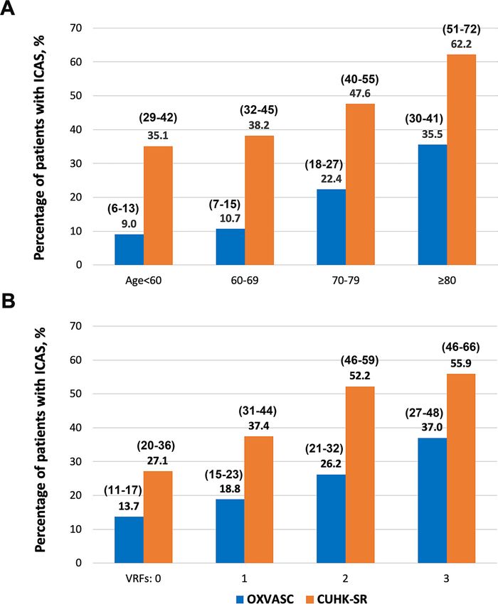
Figure 1 Prevalence of ICAS in minor stroke and TIA patients in Caucasians (OXVASC) and Chinese (CUHK-­SR) in subgroups. (A) Prevalence of ICAS
in OXVASC and CUHK-­SR in different age groups; p
Cerebrovascular disease

                                                                                                                                                                                      J Neurol Neurosurg Psychiatry: first published as 10.1136/jnnp-2020-325630 on 30 March 2021. Downloaded from http://jnnp.bmj.com/ on December 6, 2021 by guest. Protected by
 Table 2        Baseline characteristics of patients with or without ICAS in OXVASC and CUHK-­SR
                                                                 OXVASC (n=1287)                                                CUHK-­SR (n=691)
                                                                 Any ICAS              No ICAS                                 Any ICAS              No ICAS
 Characteristics                                                 (n=257)               (n=1030)              P value           (n=297)               (n=394)            P value
 Age, years                                                       75 (12.2)             68 (13.8)
Cerebrovascular disease

                                                                                                                                                                                                                                                                                                                                                                                                                                                                                                         J Neurol Neurosurg Psychiatry: first published as 10.1136/jnnp-2020-325630 on 30 March 2021. Downloaded from http://jnnp.bmj.com/ on December 6, 2021 by guest. Protected by
                                                                                                                                                                                                                                                                                                                                                                                                                     justified in elderly Caucasian patients presenting with TIA/stroke

                                                                                               P value
                                                                                                                                                                                                                                                                                                                                                                                                                     with multiple vascular risk factors, for better understanding of

                                                                                                              0.003

                                                                                                                                           0.810
                                                                                                                                                                 0.894
                                                                                                                                                                                       0.047
                                                                                                                                                                                                                 0.005
                                                                                                                                                                                                                                       0.069
                                                                                                                                                                                                                                                                                                                                                                                                                     the stroke aetiology.
                                                                                                                                                                                                                                                                                                                                                                                                                        The current study indicated more involvement of the anterior
                                                                                                                                                                                                                                                                                                                                                                                                                     circulation by ICAS in Chinese stroke/TIA patients, with MCA

                                                                                    Multivariable-­adjusted
                                                                                                                                                                                                                                                                                                                                                                                                                     as the most common lesion location, consistent with previous
                                                                                                                                                                                                                                                                                                                                                                                                                     Chinese studies.33–35 For Caucasian patients, our study indicated

                                                                                                              1.23 (1.08 to 1.41)

                                                                                                                                           0.96 (0.70 to 1.33)
                                                                                                                                                                 1.02 (0.74 to 1.40)
                                                                                                                                                                                       1.43 (1.00 to 2.03)
                                                                                                                                                                                                                 1.67 (1.17 to 2.38)
                                                                                                                                                                                                                                       1.34 (0.98 to 1.85)
                                                                                                                                                                                                                                                                                                                                                                                                                     same involvement rates of the anterior and posterior circulations
                                                                                    OR (95% CI)                                                                                                                                                                                                                                                                                                                      by ICAS, with PCA, VA-­V4, MCA and ICA similarly affected.
                                                                                                                                                                                                                                                                                                                                                                                                                     Distribution of ICAS lesions in anterior vs posterior circulations
                                                                                                                                                                                                                                                                                                                                                                                                                     in European Caucasians varied in previous studies. A study in
                                                                                                                                                                                                                                                                                                                                                                                                                     Netherlands (n=786) reported higher ICAS prevalence in poste-
                                                                                               P value
Cerebrovascular disease

                                                                                                                                                                                                J Neurol Neurosurg Psychiatry: first published as 10.1136/jnnp-2020-325630 on 30 March 2021. Downloaded from http://jnnp.bmj.com/ on December 6, 2021 by guest. Protected by
 Table 4      Distribution of ICAS lesions in the two cohorts
                                                                            OXVASC (n=1287)                                             CUHK-­SR (n=691)
                                                                            No of patients with the                                     No of patients with the        No of arteries
                                                                            arteries affected/no of         No of arteries              arteries affected/             affected/total
                                                                            patients with the artery        affected/total no of        no of patients with the        no of arteries
                                                                           imaged (%)                      arteries imaged (%)         artery imaged (%)              imaged (%)
 Any ICAS in anterior circulation                                           155/1287 (12.0)*                                            248/691 (35.9)*
 Any ICAS in posterior circulation                                          155/1287 (12.0)†                                            121/689 (17.6)†
 ICAS in both anterior and posterior circulations                             53/1287 (4.1)‡                                             72/689 (10.4)‡
 ICAS in individual arteries
  Intracranial ICA                                                           68/1287 (5.3)                 90/2574 (3.5)                83/691 (12.0)                 107/1382 (7.7)
  ACA (A1+A2)                                                                30/1287 (2.3)                 31/2574 (1.2)                68/691 (9.8)                   75/1382 (5.4)
  MCA (M1+M2)                                                                85/1287 (6.6)                 93/2574 (3.6)               184/691 (26.6)                 228/1382 (16.5)
  PCA (P1+P2)                                                                83/1287 (6.4)                 98/2574 (3.8)               102/691 (14.8)                 116/1382 (8.4)
  VA (V4)                                                                    82/1287 (6.4)                 97/2574 (3.8)                15/334 (4.5)                   19/668 (2.8)
  BA                                                                         13/1287 (1.0)                 13/1287 (1.0)                23/689 (3.3)                   23/689 (3.3)
 *P
Cerebrovascular disease

                                                                                                                                                                                                   J Neurol Neurosurg Psychiatry: first published as 10.1136/jnnp-2020-325630 on 30 March 2021. Downloaded from http://jnnp.bmj.com/ on December 6, 2021 by guest. Protected by
13 Meseguer E, Lavallée PC, Mazighi M, et al. Yield of systematic transcranial Doppler in         26 NINDS Stroke Genetics Network (SiGN), International Stroke Genetics Consortium
   patients with transient ischemic attack. Ann Neurol 2010;68:9–17.                                 (ISGC). Loci associated with ischaemic stroke and its subtypes (sign): a genome-­wide
14 von Sarnowski B, Schminke U, Tatlisumak T, et al. Prevalence of stenoses and                      association study. Lancet Neurol 2016;15:174–84.
   occlusions of brain-­supplying arteries in young stroke patients. Neurology                    27 Malik R, Chauhan G, Traylor M, et al. Multiancestry genome-­wide association study
   2013;80:1287–94.                                                                                  of 520,000 subjects identifies 32 loci associated with stroke and stroke subtypes. Nat
15 Weimar C, Goertler M, Harms L, et al. Distribution and outcome of symptomatic stenoses            Genet 2018;50:524–37.
   and occlusions in patients with acute cerebral ischemia. Arch Neurol 2006;63:1287–91.          28 Miyawaki S, Imai H, Shimizu M, et al. Genetic variant RNF213 c.14576G>A in various
16 Kim B-­S, Chung P-­W, Park K-­Y, et al. Burden of intracranial atherosclerosis is                 phenotypes of intracranial major artery stenosis/occlusion. Stroke 2013;44:2894–7.
   associated with long-­term vascular outcome in patients with ischemic stroke. Stroke           29 Liu W, Morito D, Takashima S, et al. Identification of Rnf213 as a susceptibility gene
   2017;48:2819–26.                                                                                  for moyamoya disease and its possible role in vascular development. PLoS One
17 Kim BS, Jung HS, Bang OY, et al. Elevated serum lipoprotein(a) as a potential predictor           2011;6:e22542.
   for combined intracranial and extracranial artery stenosis in patients with ischemic           30 Liu W, Hitomi T, Kobayashi H, et al. Distribution of moyamoya disease susceptibility
   stroke. Atherosclerosis 2010;212:682–8.                                                           polymorphism p.R4810K in RNF213 in East and Southeast Asian populations. Neurol
18 Park H-­K, Kim BJ, Han M-­K, et al. One-­Year outcomes after minor stroke or high-­               Med Chir 2012;52:299–303.
   risk transient ischemic attack: Korean multicenter stroke Registry analysis. Stroke            31 Okazaki S, Morimoto T, Kamatani Y, et al. Moyamoya disease susceptibility variant
   2017;48:2991–8.                                                                                   RNF213 p.R4810K increases the risk of ischemic stroke attributable to large-­artery
19 Amarenco P, Lavallée PC, Labreuche J, et al. One-­Year risk of stroke after transient             atherosclerosis. Circulation 2019;139:295–8.
   ischemic attack or minor stroke. N Engl J Med 2016;374:1533–42.                                32 Pu Y, Liu L, Wang Y, et al. Geographic and sex difference in the distribution of
20 Uchiyama S, Hoshino T, Sissani L, et al. Japanese versus non-­Japanese patients with              intracranial atherosclerosis in China. Stroke 2013;44:2109–14.
   transient ischemic attack or minor stroke: subanalysis of TIA ​registry.​org. J Stroke         33 Lee T-­H, Hsu W-­C, Chen C-­J, et al. Etiologic study of young ischemic stroke in Taiwan.
   Cerebrovasc Dis 2019;28:2232–41.                                                                  Stroke 2002;33:1950–5.
21 Uehara T, Tabuchi M, Hayashi T, et al. Asymptomatic occlusive lesions of carotid and           34 Niu J-­W, Gao S, Cui L-­Y, et al. Intracranial atherosclerosis in Chinese young adult
   intracranial arteries in Japanese patients with ischemic heart disease: evaluation by             stroke patients. J Stroke Cerebrovasc Dis 2014;23:1519–23.
   brain magnetic resonance angiography. Stroke 1996;27:393–7.                                    35 Hua Y, Jia L, Xing Y, et al. Distribution pattern of atherosclerotic stenosis in Chinese
22 Bae H-­J, Yoon B-­W, Kang D-­W, et al. Correlation of coronary and cerebral                       patients with stroke: a multicenter registry study. Aging Dis 2019;10:62–70.
   atherosclerosis: difference between extracranial and intracranial arteries. Cerebrovasc        36 Homburg PJ, Plas GJJ, Rozie S, et al. Prevalence and calcification of intracranial arterial
   Dis 2006;21:112–9.                                                                                stenotic lesions as assessed with multidetector computed tomography angiography.
23 Seo W-­K, Yong HS, Koh S-B­ , et al. Correlation of coronary artery atherosclerosis with          Stroke 2011;42:1244–50.
   atherosclerosis of the intracranial cerebral artery and the extracranial carotid artery.       37 Ovesen C, Abild A, Christensen AF, et al. Prevalence and long-­term clinical significance
   Eur Neurol 2008;59:292–8.                                                                         of intracranial atherosclerosis after ischaemic stroke or transient ischaemic attack: a
24 Leng XY, Chen XY, Chook P, et al. Correlation of large artery intracranial occlusive              cohort study. BMJ Open 2013;3:e003724.
   disease with carotid intima-­media thickness and presence of carotid plaque. Stroke            38 Turan TN, Makki AA, Tsappidi S, et al. Risk factors associated with severity and
   2013;44:68–72.                                                                                    location of intracranial arterial stenosis. Stroke 2010;41:1636–40.
25 Kim JS, Kim Y-­J, Ahn S-­H, et al. Location of cerebral atherosclerosis: why is there a        39 Feldmann E. Diagnosis and quantitation of intracranial stenosis. J Neuroimaging
   difference between East and West? Int J Stroke 2018;13:35–46.                                     2009;19 Suppl 1:22S–4.
                                                                                                  40 Feldmann E, Wilterdink JL, Kosinski A, et al. The stroke outcomes and neuroimaging of
                                                                                                     intracranial atherosclerosis (SONIA) trial. Neurology 2007;68:2099–106.

                                                                                                                                                                                                                                                                                     copyright.

8                                                                                             Leng X, et al. J Neurol Neurosurg Psychiatry 2021;0:1–8. doi:10.1136/jnnp-2020-325630
You can also read