In Vitro Models of the Canine Digestive Tract as an Alternative to In Vivo Assays: Advances and Current Challenges
←
→
Page content transcription
If your browser does not render page correctly, please read the page content below
ALTEX, accepted manuscript
                                                                                            published January 13, 2022
                                                                                             doi:10.14573/altex.2109011

Review article
In Vitro Models of the Canine Digestive Tract as an
Alternative to In Vivo Assays: Advances and Current
Challenges*
Charlotte Deschamps1,2, Sylvain Denis1, Delphine Humbert3, Jürgen Zentek4, Nathalie Priymenko5,
Emmanuelle Apper2 and Stéphanie Blanquet-Diot1
1
Université Clermont Auvergne, UMR 454 MEDIS UCA-INRAE, Clermont-Ferrand, France; 2Lallemand Animal Nutrition, Blagnac, France;
3
Dômes Pharma, Pont-du-Château, France; 4Institute of Animal Nutrition, Freie Universität Berlin, Berlin, Germany; 5Toxalim (Research
Center in Food Toxicology), University of Toulouse, INRAE, ENVT, INP-Purpan, UPS, Toulouse, France

Abstract
Dogs occupy a full place in the family and their well-being is of paramount importance to their owners. Digestion, a
complex process involving physicochemical, mechanical, and microbial parameters, plays a central in maintaining
animals healthy. As in vivo studies in dogs are more and more restricted by ethical, regulatory, societal, and cost
reasons, an alternative option resides in the use of in vitro models simulating the different parts of the canine
gastrointestinal tract. This review paper first introduces digestion and gut microbiota as key factors in dog nutrition
and health, under both healthy and diseased conditions (obesity and inflammatory bowel disease), by highlighting
when relevant similarities or differences between human and canine digestion process. Then, we provide for the
first time an in-depth description of currently available models of the canine digestive tract, discuss technical and
scientific challenges that need to be addressed and introduce potential applications of in vitro gut models in food
and veterinary fields. Even if the development of some in vitro models remains limited by a lack of in vivo data in
dogs necessary for a relevant configuration and validation, translation of long-term expertise on human in vitro gut
models to dog’s ecosystem opens avenues for canine in vitro gut model development and their adaptation to specific
digestive conditions associated to various ages, sizes, breeds and/or diets, under both physiological and diseased
states.

1    Introduction

1.1. Dogs in familial and economical contexts
Canis lupus familiaris, also known as domesticated dogs, belongs to Canidae family. Dog is maybe the first animal
domesticated by human around 12.000 years ago (Axelsson et al., 2013). Dogs were initially strict carnivores, but during the
agricultural revolution, they have probably acquired the ability to digest starch and became facultative carnivores (Axelsson et
al., 2013). Nowadays, canine species include approximatively 400 breeds with high morphological and size variabilities and
weight ranging from 1 kg for a Chihuahua to 100 kg for a Saint-Bernard (Grandjean and Haymann, 2010). Pets, especially
dogs, occupy a full place in the family and their health and well-being are therefore of paramount importance to their owners.
          Dogs are estimated to be more than 500 million worldwide, which represents a huge market for petfood and animal
health industry. In 2018, global petfood market size reached $91.1 billion, representing 31% increase within 5 years, with the
need of constantly innovate (e.g., food, probiotics and prebiotics) (Phillips-Donaldson, 2019). A vast array of foods, snacks
and nutritional supplements have been recently developed to support well-being, health, improve aging or prevent diseases.
According to increased interest of owners to maintain dog’s health, petfood was adapted to fit each dog’s lifestyle, for example
for puppies or bitches, sedentary or active, maintenance diet or hypocaloric diet. Different types of canine food are available
and can be classified in three categories: dry food, canned food, and alternative food (biologically appropriate raw food, namely
BARF, homemade food, and feedstuffs). At the interface between petfood and veterinary compounds, pets’ nutritional
supplements represent a specific expanding market with huge range of products. As an example, micronutrients like selenium,
taurine or polyphenols could be added for old dogs, calcium, phosphorus, omega-3 fatty-acids and vitamin E for lactating
bitches, or L-carnitine for athletic dogs. Particular nutritional purposes are also developed to decrease food sensitivities,
digestive or articular care and could be supplemented by prebiotics or probiotics.

*

Received September 1, 2021; Accepted January 11, 2022;
Epub January 13, 2022; © The Authors, 2022.

ALTEX 39(#), ###-###. doi:10.14573/altex.2109011

Correspondence: Stéphanie Blanquet-Diot, PhD
UMR 454 MEDIS
University Clermont Auvergne, INRAe
28 place Henri Dunant
63000 Clermont-Ferrand, France
(mailto:stephanie.blanquet@uca.fr)

                                                                                                                                  1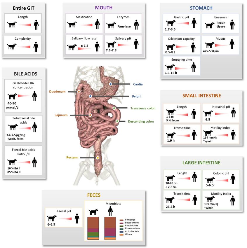
ALTEX, accepted manuscript
                                                                                           published January 13, 2022
                                                                                            doi:10.14573/altex.2109011

          In 2019, pet medicine represents a $17.5 billion market with the sales of veterinary compounds like vaccines,
antiparasitic treatments or antibiotics1. As for human, a large range of products has been developed to improve dog’s health
and hygiene. Drugs are present on different forms: cutaneous applications, injection paths or oral formulation. For oral use,
there are liquid forms (e.g., solution or suspension), semi-solid forms (oily or aqueous formulations) and solid forms (e.g.,
powder, capsules, conventional or sustained-release tablets). Specific treatments have been also developed to avoid lipid
absorption in obesity or decrease pain in inflammatory bowel disease (IBD). Of note, sensibility to specific diseases depends
on dog’s size and breed, with for instance overweight and dental trouble in small dogs and digestive sensibility in larger animals.

1.2. Regulatory context
Strong expansion of veterinary medicine and petfood or oral supplements with health claims is associated with an increasing
specific regulatory context. In Europe, the European petfood industry federation (Fediaf) represents 16 national petfood
industries and aims at proposing a framework for production of safe, nutritious, and palatable petfood. Heads of Medicine
Agencies or national Agency must deliver an authorization for veterinary drug marketing. These agencies evaluate quality and
safety of medicinal product for animals, consumers, users, and environment as well as effectiveness of medicinal product. To
assess digestibility of petfood or bioaccessibility of active compounds (including drugs) in the canine gastrointestinal tract
(GIT), in vivo studies remain the golden standard. In vivo experiments in dogs can also be performed as a model of human gut
due to similarities in digestive physiology (Lui et al., 1986; Akimoto et al., 2000). Worldwide, experimentations are still
conducted on 192.1 million animals with 207 724 experimentations involving dogs in 2015 as reported by the European
Commission in 2017. However, in vivo assays are more and more restricted by regulation, ethical and societal constraints, and
high associated costs. The European and North America “3Rs” rules (adapted from Russel and Burch, 1959) widely encourage
a strong reduction in the number of animals used in research and prone the development of alternative in vitro approaches.
Among in vitro alternatives, models simulating the canine digestive environment (intestinal cell culture, organoids, or in vitro
gut models) can help to answer many scientific questions associated to food and drug behavior during canine digestion.
Providing alternative methods before performing in vivo trials can be of interest to match with owners, regulatory, and health
expectations and go deeper into mechanistic aspects. However, this requires a comprehensive understanding of dog digestive
processes.

1.3. Digestion and gut microbiota as key parameters in dog nutrition and health
Digestion is an essential process at the center of dog health study and of high interest for petfood and veterinary industries.
Canine digestion involves physicochemical (e.g., pH, digestive secretions, transit time), mechanical and microbial parameters.
The gut microbiota and its involvement in dog nutrition and health is becoming a more and more important topic. All these
digestive components affect food digestibility, nutrient absorption, and energy release, but also drug metabolism and
absorption, and survival of probiotic microorganisms. Thus, developing new food or veterinary products needs to consider all
these multi-faceted aspects of canine digestion in order to answer important questions, such as: how physicochemical
parameters modulate food digestibility?; what is the importance of gut microbiota in canine digestion and drug metabolism?;
where drugs are released and absorbed?; how drug bioaccessibility is impacted by food matrix, galenic form, physicochemical
parameters or microbiota?; how probiotic strains survive along the GIT? Yet, recommendations for petfood and drug intakes
are only based on dog body weight or metabolic weight. Future development of new products needs therefore to take into
account not only dog’s weight but also all the variations in digestion process associated to different canine sizes and breeds to
move towards personalized nutrition and veterinary medicine.

2    Methods of literature research and aim of the review

Our literature search was performed using PubMed2 and Google Scholar3 using the keywords “dog” OR “canine” AND
“digestion”, “pH”, “enzyme”, “digestive secretions”, “absorption”, “microbiota”, “bile acids”, “transit time”, “short chain fatty
acids”, “fermentation”, “gas”, “mucus”, “in vitro”, “model” in all available years. The online database search was last
performed in August 2021 on titles, abstracts and key words including original articles, reviews, thesis, and books. Relevant
studies were identified after consultation of the main text, figures and supplementary materials and we extracted information
regarding involved dogs (i.e., number of dogs, age, weight, breed, sex, reproduction state, living environment), alimentation
(i.e., type of food, feeding frequency, principal components of food), health (i.e., healthy, obese or IBD dogs only) and analysis
methods.
            This review paper first aims to give a state-of-the-art on canine digestive physiology regarding both physicochemical
and microbial parameters that can be reproduced in in vitro gut models. Then, we explain how these parameters are evolving
under diseased conditions associated to obesity and IBD. In a third part, we provide an in-depth description of all the available
in vitro models of the canine digestive tract before discussing limitations and challenges associated to the development of in
vitro gut models of healthy or diseased dogs, and their applications in food and veterinary fields. In an original way, this paper
considers the entire canine GIT and associated microbiota and highlights similarities and differences between dog and human
digestive physiology to provide new opportunities for canine in vitro gut simulation.

1
  Animal medicine global market opportunities and strategies to 2023. Available at: https://www.globenewswire.com/news-
release/2020/01/13/1969734/0/en/Global-Animal-Medicine-Market-Opportunities-Strategies-to-2023-Veterinary-
Pharmaceuticals-Will-Gain-20-7B-of-Global-Annual-Sales-by-2023-A-6-6B-Opportunity-for-Veterinary-Parasitic.html
2
  https://pubmed.ncbi.nlm.nih.gov
3
  https://scholar.google.fr

                                                                                                                                 2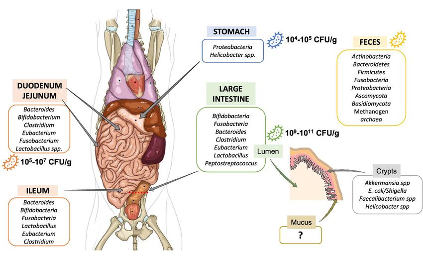
ALTEX, accepted manuscript
                                                                                            published January 13, 2022
                                                                                             doi:10.14573/altex.2109011

Fig. 1: Canine digestive compartments and associated mechanical, physicochemical and microbial processes
Key parameters of the oral, gastric, intestinal and colonic compartments from the medium healthy dogs are summarized and
compared in vivo data in healthy human. Lack of in vivo data are represented by “?”. BA = bile acid.

3    Canine digestion

3.1. Digestive anatomy
Because dogs are facultative carnivores, their GIT is adapted to high-protein and high-fat diets, i.e., relatively short and simple
compared to herbivores and even omnivores like human or swine, reflecting the lower retention time required for meat
comparatively to grass digestion (Moon et al., 2018) (Fig. 1). Part of dog’s digestive tract weight in the total body mass is
inversely correlated to canine size, representing 7 % and 2.8 % of the total body weight in 5 and 60 kg dogs, respectively
(Weber, 2006). The GIT length is a reflect of height at the shoulder with a 6:1 ratio (Morris and Rogers, 1989).
           Digestion in dogs starts in the mouth with the mechanic action of mastication, using 42 teeth and only 2000 taste
buds. Canine teeth are massive and form the crocs, able to cut meet into pieces. Molars are formed by a larger crown than in
humans and dotted with tubers allowing the grinding of bones. However, the mastication step is less important than in human
as most of dogs do not really chew but swallow large pieces. After swallowing, food bolus crosses the esophagus whose total
length is similar to that of human, i.e., around 30 cm for a medium dog (Kararli, 1995; Freiche and Hernandez, 2010). Like in
human, canine stomach is in form of a J-elongated bag, located in the abdomen, starting by cardia and extending to pylorus,
and composed of two parts, antrum and fundus (Kararli, 1995). Stomach compartment has a great dilatation capacity (0.5-8 L
volume), higher than in human. In dogs, gastric walls measured between 3 to 5 mm (Kararli, 1995; Freiche and Hernandez,
2010). Then, digestion continues along small intestine, which is anatomically divided in three segments as in human: the

                                                                                                                                 3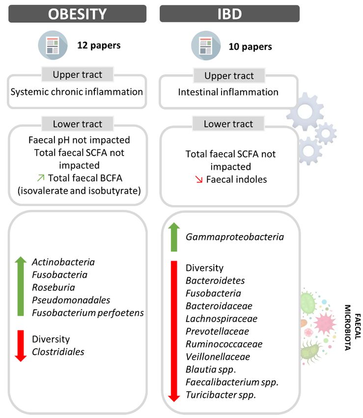
ALTEX, accepted manuscript
                                                                                            published January 13, 2022
                                                                                             doi:10.14573/altex.2109011

duodenum, jejunum and ileum, measuring from 1-1.5 to 3 m, with 10% length for duodenum, 85% for jejunum and 5% for
ileum, whereas ileum represents 60% of the total small intestine length in human (Kararli, 1995; Oswald et al., 2015). This
suggests that canine ileal function may be different between dog and human. Canine small intestine diameter is also shorter
than human (5 cm versus 1 cm). Duodenum thickness reaches 6 mm whereas intestinal loops measure around 2-3 mm. In
carnivores, the large intestine is shorter than in omnivores (20-80 cm), with 2-3 cm diameter measured in medium dogs
contrarily to human, which shows a higher length (90-150 cm) and diameter (5 cm) (Kararli, 1995). The large intestine is
constituted by 3 parts: the ascending, transverse, and descending colon. Finally, rectum forms the terminal section of large
intestine until anus. Unlike human, the three parts of the canine colon are not so well defined, with the particularity to be non-
sacculated and devoid of sigmoid colon (Kararli, 1995).
           As for humans, peripheral organs are implicated in dog digestion. Pancreas secretes the pancreatic juice into the
duodenum, and is involved in protein (trypsin, chymotrypsin, elastase, and carboxypeptidase), carbohydrate (α-amylase) and
lipid (lipase and phospholipase) digestion. Pancreatic juice also contains antimicrobial agents that contribute to intestinal
microbial balance. Liver is coupled with gallbladder and located near the stomach. Liver has a central role in digestion with
vitamin and glucose storage (glycogen), detoxification and deletion of toxic substance (e.g., urea), and lipid digestion in
allowing intestinal lipids saponification by bile acids (BA). Bile is produced by the liver, store in the gallbladder and discharged
into the duodenum.
           Of note, depending on body size and breed, dogs show an important diversity in anatomical features such as intestinal
length and volume, intestinal villus morphology and intestinal or colonic permeability (Zentek and Meyer, 1995; Oswald et al.,
2015). Because of the tight relationships between anatomy and digestive physicochemical parameters, these differences may
affect dog digestion and key parameters such as pH, digestive secretions, transit time and consequently, gut microbiota. To
avoid making this ground too complex, next sections (3.2 and 3.3) only focus on digestion processes in medium size dogs
(from 10 to 30 kg).

3.2. Physicochemical parameters
pH. Gastrointestinal pH changes along the dog digestive tract (Fig. 1). Mean salivary pH of medium dogs is around 7.3-7.8
(Smeets-Peeters et al., 1998). In the stomach, the arrival of food bolus induces the secretion of gastrin, which in turn stimulates
hydrochloric acid (HCl) production. Gastric pH of Beagles under fasted conditions is around 1.5 (range 0.9-2.5), quite similar
to that of human (range 1.4-2.1) (Dressman, 1986; Kararli, 1995; Mahar et al., 2012). Moreover, several studies observed a
higher gastric pH in fed dogs, ranging from 2 to 5.5 (Smith, 1965; Dressman, 1986; Kararli, 1995; Shinchi et al., 1996;
Martinez, 2002; Duysburgh et al., 2020). Small intestinal pH increases to value close to the neutrality because of the buffering
capacity of pancreatic juice and bile (Kararli, 1995). Small intestinal pH also increases from the proximal to the distal parts,
from 6.5 to 8 in medium size dogs (Koziolek et al., 2019). The few studies investigating the canine jejunal pH measured a
mean pH of 6.8 for medium dogs (Mentula et al., 2005; Kalantzi et al., 2006), which is quite similar to that of human (value of
6-7) (Kararli, 1995; Martinez, 2002). Colonic pH is, like in human, more acidic than the small intestine one, with values of 5-
6.5 (Smith, 1965; Koziolek et al., 2019). There are also plenty of data concerning the fecal pH of medium dogs, with a pH
range of 6-6.9 (Eisenhauer et al., 2019; Nogueira et al., 2019).
           Digestive secretions. Like in human, dog digestion is accelerated by digestive secretions containing various enzymes
(Fig. 1). Amylase, lactate dehydrogenase and adenosine deaminase are found in dog saliva (Lavy et al., 2012; Iacopetti et al.,
2017; Ricci et al., 2018). Gastric mucosa secretes gastric juice containing proteolytic (pepsin, chymosin) and lipolytic (lipase)
enzymes (Aspinall, 2004; Durand, 2010). As mentioned before, the canine pancreatic juice, discharged in the duodenum,
contains a large cocktail of enzymes including amylase, lipase, phospholipases, cholesterases, proteases and nucleases (Kienzle,
1988; Robin, 2007). Of note, gastric and pancreatic juices are poorly characterized in dogs, in contrast to bile for which more
data are available. Bile, produced by liver and partially stored in gallbladder, is discharged into the duodenum during
postprandial phases. In gallbladder, bile is up to 10-fold more concentrated than in liver with a total concentration higher than
in human, around 50 (40-90) mmol/L in dogs versus 3-45 mmol/L in human (Nakayama, 1969; Kararli, 1995; Kakimoto et al.,
2017; Nagahara et al., 2018; Larcheveque, 2019). Canine bile stored in gallbladder contains up to 15 BA but the three majors
found in healthy dogs count for 99% of total BA pool, with 72.8% taurocholic acid, 20.3% taurodeoxycholic acid and 6.2%
taurochenodeoxycholic acid (Washizu et al., 1994). Moreover, 0.15% of BA are conjugated to glycine and 0.81% are
unconjugated (Nagahara et al., 2018). BA concentrations measured in fecal samples show concentrations range of 5.8-7.5 µg
total BA per mg of dry feces (Schmidt et al., 2018; Blake et al., 2019; Manchester et al., 2019). Lastly, as observed in various
mammals including human, mucins are produced by goblet cells all along the canine GIT (Kararli, 1995). Mucus thickness has
been evaluated in dogs only in the stomach, measuring 425 and 576 μm respectively in the fundus and antrum in accordance
to human values (Bickel and Kauffman, 1981; Kararli, 1995; Etienne-Mesmin et al., 2019). This mucin layer allows protection
of the epithelium against acidic pH of stomach and withstands bone fragments (Moon et al., 2018). In the large intestine, the
mucus layer is colonized by a microbial biomass, as described in section 3.3.
           Gut motility and transit time. Using wireless motility capsule, Farmer and collaborators found that gut motility
indexes in dogs are higher in the large intestine (199 mmHg*second/min) compared to the small intestine (134
mmHg*second/min) and stomach (55 mmHg*second/min) (Farmer et al., 2018). Canine motility is quite different to that of
human. Maximum pressure under fed conditions is higher in human than in dog (241 and 119 mmHg in the human stomach
and small intestine, respectively, versus 52 and 75 mmHg for dog), but with a similar maximum frequency of 3.7
contractions/min in the gastric compartment (Boscan et al., 2013; Farmer et al., 2018). Data on transit time in the different
digestive compartments of dogs (Fig. 1) wary widely depending on the method used (e.g., radiopaque markers, plastic beads,
13C-octanic acid breath test, sulfasalazine-sulfapyridine method or wireless motility capsule), breed, age, feed (composition,

energy density or viscosity) and environment (laboratory or owners’ home, stress context or sedation). Gastric emptying time
varies from 2-19.8 h in fed medium dogs (Hinder and Kelly, 1977; Theodorakis, 1980; Ehrlein and Pröve, 1982; Meyer et al.,
1985; Lui et al., 1986; Gupta and Robinson, 1988; Hornof et al., 1989; Arnbjerg, 1992; Carrière et al., 1993; Cullen and Kelly,

                                                                                                                                  4
ALTEX, accepted manuscript
published January 13, 2022
doi:10.14573/altex.2109011
Fig. 2: Variations in gut microbiota composition along the canine digestive tract
Main bacteria populations found in the different compartments of the gastrointestinal tract from the medium dog are represented.
CFU: colony-forming unit.
1996; Sagawa et al., 2009; Boillat et al., 2010; Mahar et al., 2012; Boscan et al., 2013; Koziolek et al., 2019). There is also no
consensus on small intestinal transit time with a range of 1.1-3.6 h (Boillat et al., 2010; Oswald et al., 2015). Few studies
investigated large intestinal transit time on dogs from various weight and breeds: T1/2 values were 7.1-42.9 h for the first study
(dog’s weight from 19.6 to 81.2 kg) and 1.1-49.1 h for the second one (dog’s weight from 11.8 to 63.8 kg) (Boillat et al., 2010;
Warrit et al., 2017). Large weight variations as well as breed differences in these two studies may contribute to the important
ranges observed in large intestinal transit times.
3.3. Canine gut microbiota
Composition. Gut microbiota has been weakly described in dogs and most of available studies have been performed since 2003.
In addition, these studies have been mostly done on feces and not on samples collected from the different digestive
compartments due to invasive procedures. Nevertheless, it is now acknowledged that microorganisms colonize the entire GIT
of dogs from mouth to rectum. Like in human, there are in dogs longitudinal variations (i.e., along the GIT) in gut microbiota
composition due to changes in pH, substrate concentrations (including oxygen and nutrient availability) and transit time (Hooda
et al., 2012; Friedman et al., 2018; Etienne-Mesmin et al., 2019). Figure 2 gives an overview of available data regarding gut
bacteria composition depending on gastrointestinal regions in dogs. Stomach is the less colonized compartment with 104 to 105
colony forming units (CFU) per gram of content in medium dogs, mainly composed by Proteobacteria, including Helicobacter
spp. as in humans, that are potential pathogenic strains (Benno et al., 1992; Mentula et al., 2005; Hooda et al., 2012). Small
intestine contains 105 to 107 CFU/g of content (Benno et al., 1992; Mentula et al., 2005). Duodenum is colonized by Firmicutes
(calculated median on the three available publications: 47%), Proteobacteria (27%), Bacteroidetes (9%), Fusobacteria (3%)
and Actinobacteria (1%), whereas jejunum is characterized by a higher abundance in Proteobacteria (37%), Actinobacteria
(11%) and Fusobacteria (10%), together with lower percentages of Firmicutes (33%) and Bacteroidetes (7%) (Xenoulis et al.,
2008; Suchodolski et al., 2009; Garcia-Mazcorro et al., 2012). A single study on 6 dogs investigate ileal microbiota and
observed 31% Fusobacteria, 24% Firmicutes, 23% Bacteroidetes and 22% Proteobacteria (Suchodolski et al., 2008). Large
intestine is the most colonized part of the GIT, with up to 10 9-1011 CFU/g of content (Hooda et al., 2012). According to a
unique publication using 16S Illumina sequencing to investigate microbiota composition, colonic digesta is dominated by 37%
Firmicutes, 33% Bacteroidetes, 29% Fusobacteria and 1% Proteobacteria (Suchodolski et al., 2008a). It’s interesting to
highlight that majority of taxa colonizing the colon are also found in canine feces (Pilla and Suchodolski, 2020), which seems
to be rather different from the human situation where a significant number of mucus-adherent bacteria from the colon are not
found in feces (Pilla and Suchodolski, 2020). In addition to Bacteria (98%), canine fecal microbiota contains 1.1% Archaea,
0.4% Fungi and 0.4% viruses, mainly bacteriophages (Suchodolski, 2011; Swanson et al., 2011). Fecal microbiota of healthy
dogs is dominated by three main bacterial phyla: Firmicutes, Bacteroidetes and Fusobacteria (Pilla and Suchodolski, 2020).
Bacteria from Actinobacteria and Proteobacteria phyla are also found in canine feces but in a lesser proportion. Fusobacteria
and Proteobacteria seem to be more abundant in dogs than in humans, probably related to a carnivorous versus omnivorous
diet (Fig. 1) (Simon, 2019). Unlike in human where Fusobacterium is frequently associated to diseases, this genus is related in
dogs to non-stressful conditions and therefore probably a healthy state, especially because its abundance increases when dogs
have access to the outside (Oswald et al., 2015). In addition to longitudinal variations, scare data are suggesting variations in
5ALTEX, accepted manuscript
published January 13, 2022
doi:10.14573/altex.2109011
microbiota composition from the digestive lumen to the surface of the intestinal epithelium covered by a mucin layer. Only
two studies investigated the mucosa-associated bacteria on the outer mucus layer in the colon of healthy dogs, using targeted
FISH approach (Simpson et al., 2006; Cassmann et al., 2016). Cassmann et al. (2016) demonstrated that free colonic mucus is
mainly colonized by Bacteroidetes spp. and Eubacteria. Of interest, Akkermansia muciniphila, a well-known mucin degrading
bacterium in humans, inversely correlated to obesity, was not yet identified in canine feces (Garcia-Mazcorro et al., 2020).
Metabolic activities. Gut microbiota is known to play a key role in host homeostasis and health maintenance, as it is
implicated in many nutritional (e.g., fibre degradation and vitamin synthesis), immunological (immune system maturation) and
physiological processes (e.g., vascularization, epithelium integrity, “barrier” effect against pathogens and lipid digestion via
the metabolism of primary BA into secondary BA) (Durand, 2010; Andoh, 2016). Gut microbiota metabolic activity leads to
short-chain fatty acid (SCFA) and gas production as mean fermentation products resulting from degradation of soluble fibres.
In dog like in human, the three main SCFAs are acetate, propionate and butyrate, with relative percentages in fecal samples of
60:25:15 (Mondo et al., 2019). Non-digested proteins from diet and endogen proteins (e.g., from mucins) are also metabolized
by gut microbiota, leading to the production of branched chain fatty-acids (BCFA), ammonia, indoles and phenols (Weber et
al., 2017). Total fecal SCFA and BCFA were investigated, with values widely varying between studies, i.e., 91-423 and 4.7-
36.1 µmol/g of lyophilized stools, respectively (Beloshapka et al., 2012; Minamoto et al., 2015; Alexander et al., 2019;
Detweiler et al., 2019; Eisenhauer et al., 2019; Nogueira et al., 2019). Lastly, to our knowledge there is no in vivo data on gas
production in dogs and the two available studies on gas composition focused on malodorous compounds such as hydrogen
sulfide (Collins et al., 2001; Giffard et al., 2001).
4 From healthy to diseased dogs: impact on digestive physiology and gut microbiota?
Just as in humans (such master, such dog!), diseases such as obesity and IBD are more and more prevalent in dogs. Because
the etiology of such diseases in human is getting clearer, the emergence of similar pathologies in dogs raises awareness of
veterinary and petfood companies to develop new pharma and nutrition products. We can easily imagine that these diseases
would affect the entire GIT. However, most of available data on obese or IBD dogs are focusing on the lower gut and its
associated microbiota. Indeed, in many intestinal or extra-digestive diseases such as obesity and IBD, in dog like in human,
gut microbiota appears to be in a disrupted state called “dysbiosis” where both functional and compositional states are altered
(Pilla and Suchodolski, 2020).
4.1. Obesity
In Western countries, obesity is considered as the most common nutritional disorder in pets, due to imbalance between energy
intake and expenditure (Osto and Lutz, 2015). Dogs are considered as clinically obese when their body weight is at least 20-
30% above ideal weight and a universal body condition score (ranging from 1 to 9) defines overweighed dogs at 7 and obese
dogs at 8/9 (Apper et al., 2020). Both sexes have a similar incidence and all dogs are concerned whatever their size, even if
certain breeds seem to be more predisposed, like Labrador Retriever, Bernese mountain dog, cavalier King Charles or Beagles
(Osto and Lutz, 2015). Canine obesity is generally associated to insulin resistance, altered lipid profiles, hypertension,
orthopedic and cardiorespiratory diseases and development of low-grade systemic inflammation (Tvarijonaviciute et al., 2012).
While gene mutations are associated with increased body weight in Beagles and Labrador, diet is clearly a determinant of
obesity through excess energy intake (Zeng et al., 2014; Raffan et al., 2016).
Several mechanisms may implicate gut microbiome in the onset and evolution of dog obesity. Based on human and
mice data, this includes higher energy utilization from non-digestible carbohydrates, manipulation of host gene functions and
exacerbation of inflammation (Hamper, 2016). Five studies have compared fecal microbiota composition of obese and lean
dog (Fig. 3). Using 16S rRNA gene pyrosequencing in companion and laboratory dogs, dominance of Firmicutes (> 90%) was
observed in both obese and lean groups, but mean abundance of Actinobacteria and Roseburia was greater in obese dogs (Handl
et al., 2013). Using the same method, Salas-Mani et al. (2018) showed that Proteobacteria predominated in obese dogs (76%)
whereas fecal microbiota from lean dogs was mainly composed by Firmicutes (85%). In addition, Clostridiales appeared to be
less abundant in obese compared to lean dogs, while opposite result was found for Pseudomonadales. However, in a recent
study involving 17 healthy and 22 obese companion dogs, no significant difference in any taxa was highlighted when comparing
the two groups using Illumina 16S rRNA gene sequencing (Forster et al., 2018). With the same method, Bermudez Sanchez
and colleagues (2020) described a relative abundance of 92% of Firmicutes, 2% Fusobacteria, 1% Bacteroidetes and a median
Firmicutes/Bacteroidetes ratio of 0.123 in 20 obese dogs (Bermudez Sanchez et al., 2020). Lastly, in a very recent study, a
positive association between Fusobacteria level, especially Fusobacterium perfoetens, and body score condition in 24
overweight laboratory Beagles was established using metagenomic analysis (Chun et al., 2020). In dog like in human, microbial
diversity seems to decrease in obese compared to lean dogs, with Shannon index of 1.3 and 2.3, respectively (Park et al., 2015).
However, more investigations are required to characterize canine obese microbiota, in feces but also in other digestive
compartments, to determinate if some species should be used as obesity markers. As an example, Proteobacteria were recently
correlated to increased weight in overweighed dogs, as it has been suggested in human (Apper et al., 2020).
In addition to microbiota changes, other physiological modifications have been demonstrated in obese humans
compared to healthy individuals, including an increase in BA production by liver (coupled with high amounts of primary BA
in stool), and a higher SCFA production leading to a decrease in colonic pH (Rahat-Rozenbloom et al., 2014). Due to
similarities between dog and human GIT and lifestyle, it would be interesting to establish if similar phenomena yet poorly
investigated occur in obese dogs. Mean fecal pH of obese companion Beagles (6.6) was not significantly different from that of
lean dogs (6.8) whatever the diet, i.e., high-fat or low-fat diet (Xu et al., 2017). In the same study, total fecal SCFA concentration
was also equivalent between the two groups, whereas fecal BCFA such as isovalerate and isobutyrate were significantly more
concentrated in obese compared to lean dogs. According to our knowledge, there is until now no study that has investigated
6ALTEX, accepted manuscript
published January 13, 2022
doi:10.14573/altex.2109011
Fig. 3: Impact of obesity and inflammatory bowel disease on dog digestive physiology
Main variations in upper and lower digestive physiology and changes in fecal microbiota composition associated to canine obesity
and IBD are represented. Increased parameters are indicated by a rising green arrow, while decreased ones are symbolized by
a falling red arrow. BCFA: branched-chain fatty acids; IBD: inflammatory bowel disease; SCFA: short-chain fatty acids.
BA production in obese versus lean dogs. However, recent studies recommend to monitor fecal BA concentrations along with
microbiota in lean and obese dogs, as they appear to be interesting markers of glucose homeostasis failure in obese dogs (Forster
et al., 2018; Apper et al., 2020).
4.2. Inflammatory bowel disease
In dogs, IBD is classified in chronic enteropathies, but defining its prevalence remains difficult because diagnosis of the disease
is not easy. IBD is characterized by an alternation of clinically active (with pain and diarrhea) and insignificant phases that
occur irregularly. Outside active phases, there are recurring gastric symptoms with histopathological changes in mucosa of
small and large intestines (Malewska et al., 2011). The most predominant causes of canine IBD include bacterial and
environmental factors, genetic predisposition of selected breeds, food allergies and some drugs (Malewska et al., 2011).
Pathogenesis of IBD includes loss of tolerance for endogenous microbiota, chronic inflammation of the GIT associated with
an increase in intestinal permeability, and immune cells infiltration in the lamina propria (Junginger et al., 2014). Clinical
scenarios include decline in activity level and appetite, vomiting, increase in stool frequency, loss of stool consistency (increase
in fecal water content), and weight loss.
Recent reviews on IBD (Fig. 3) report in dogs modifications in gut microbiota structure (compared to healthy animals)
similar to that found in human, with a decrease in fecal abundance in Firmicutes and Bacteroides together with an increase in
Proteobacteria (Hooda et al., 2012). This was confirmed in a recent study by Pilla and Suchodolski (2020) who observed a
decrease in Firmicutes (i.e., Ruminococcaceae, Veillonellaceae and Lachnospiraceae), Bacteroidetes (i.e., Bacteroidaceae and
Prevotellaceae) and Fusobacteria. Nevertheless, there is no absolute consensus since Xu et al. (2016) didn’t observe any
significant difference in Firmicutes, Bacteroidetes and Enterobacteriaceae abundance between healthy and IBD groups. In
their study, Minamoto and collaborators used real-time PCR to quantify some underrepresented populations, such as Blautia
spp., Faecalibacterium spp., Turicibacter spp. and Escherichia coli (Minamoto et al., 2015). The authors observed that these
three first populations were less abundant in IBD group, whereas E. coli was non-significantly increased. They also employed
454-pyrosequencing analysis and showed a significant decrease in fecal diversity in IBD compared to healthy dogs, as also
described in humans (Minamoto et al., 2015). Furthermore, a dysbiosis index was designed using qPCR targeting eight bacterial
groups to assess fecal microbial changes associated to canine chronic enteropathies (AlShawaqfeh et al., 2017). This dysbiosis
index allowed a discrimination between healthy and diseased dogs with 95% confidence range. Other studies investigated gut
microbial changes directly in dog digestive compartments via mucosal biopsies. Molecular-phylogenetic studies have revealed
7ALTEX, accepted manuscript
published January 13, 2022
doi:10.14573/altex.2109011
a bacterial and/or fungal dysbiosis in the duodenum of dogs with idiopathic IBD (Suchodolski et al., 2008, 2010; Xenoulis et
al., 2008; Suchodolski et al., 2012a,b). Most of the time, Clostridiales and Fusobacteria proportions were decreased in IBD
compared to healthy dogs, whereas Proteobacteria increased. Fungal DNA was more frequently detected in dogs with chronic
enteropathies (76.1%) than in healthy animals (60.9%), but also more prevalent in mucosal (82.8%) than in luminal samples
(42.9%). In addition, Cassmann et al. (2016) showed from ileal and colonic biopsies a significantly increase in
Enterobacteriaceae and E. coli bacteria attached onto surface epithelia or invading intestinal mucosa in IBD compared to
healthy dogs, as observed in human. FISH analysis of colonic biopsies from Boxer with granulomatous colitis revealed mucosa
colonization by an unknown adherent and invasive E. coli strain (Simpson et al., 2006). Taken all together, these data indicate
that global microbial structure and diversity more than a single taxa should be followed to discriminate healthy and IBD dog
microbiota (Scarsella et al., 2020).
Concerning functional activity, there is no significant difference in fecal SCFA concentrations between IBD and
healthy dogs but a lower indole concentration was measured in diseased dogs (Xu et al., 2016; Pilla and Suchodolski, 2020).
This is an important point because indoles have well-known anti-inflammatory effects, strengthen epithelial barrier and
decrease E.coli attachment to epithelium wall (Chèvreton, 2018). Alterations in microbial functions associated with IBD were
estimated using a prediction tool (PICRUSt) from 16S rRNA gene data, highlighting a significant increase in secretion system
pathways and transcription factor (Minamoto et al., 2015). Other parameters modified in humans during IBD (Duboc et al.,
2013; Rana et al., 2013; Fitzpatrick and Jenabzadeh, 2020), such as transit time and BA dysmetabolism (increase in fecal
primary BA) have not been investigated yet in dogs.
5 In vitro canine models as an alternative to in vivo assays in dogs
5.1. Generalities on in vitro gut models: static versus dynamic and in vitro versus in vivo
A wide range of in vitro gut models has been already developed, from the simplest static mono-compartmental models to the
most complex dynamic and multi-compartmental models (Guerra et al., 2012; Payne et al., 2012). These in vitro models have
been primarily developed to mimic human digestion, but are more and more frequently adapted to simulate animal digestion,
mainly that of pig or piglet (Meunier et al., 2008; Tanner et al., 2014; Fleury et al., 2017; Dufourny et al., 2019), cat (Sunvold
et al., 1995; Van den Abbeele et al., 2020a) or dog (Sunvold et al., 1995; Smeets-Peeters et al., 1999; Tzortzis et al., 2004;
Hervera et al., 2007; Bosch et al., 2008; Cutrignelli et al., 2009; Panasevich et al., 2013; Lee et al., 2017; Vierbaum et al., 2019;
Oba et al., 2020; Van den Abbeele et al., 2020a; Verstrepen et al., 2021), as described in the next 5.2 section.
Simple static models of the upper gut (Minekus et al., 2014) reproduce in a single vessel maintained at body
temperature the successive oral, gastric and/or small intestinal phases of human digestion, by changing pH conditions and
adding appropriate digestive secretions (e.g., α-amylase in the oral phase, pepsin and/or lipase in the stomach and bile and/or
pancreatic juice in the intestinal phase). Simplest models of the colon compartment are thermostatic batch culture systems.
These models are inoculated with feces to simulate colonic fermentation and maintained under anaerobic conditions by flushing
with nitrogen or carbon dioxide, but without any renewal of nutritive growth medium until the end of experience. Such
approaches are therefore limited in time by substrate availability (24 to 72 h) and parameters like pH or redox potential are not
regulated. Compared to static systems, dynamic models reproduce changes in at least one parameter, such as pH kinetics,
variation in digestive secretions or chyme transit. They can be mono-compartmental or composed of sequential vessels
simulating the successive digestive compartments. Dynamic mono-compartmental models of the upper gut are only gastric
digester (Kong and Singh, 2010; Thuenemann et al., 2015), while multi-compartmental models include gastric and small
intestinal compartments, most frequently simulating the duodenal section (Tompkins et al., 2011; Ménard et al., 2015). All
these models only reproduce physicochemical parameters of the upper digestive tract, such as temperature, gastric and intestinal
pH, gastric and ileal deliveries, transit time, digestive secretions and passive absorption of nutrients and water. Dynamic large
intestine models are based on the principle of continuous or semi-continuous fermentation and just like batch systems are
inoculated with fecal samples. Such models are maintained under anaerobiosis and reproduce colonic temperature, pH and
transit time, and redox potential can be monitored. Moreover, a nutritive medium aiming to mimic ileal effluents and composed
of various complex sources of carbon and nitrogen, electrolytes, BA, and vitamins is continually added to the bioreactor, while
fermentation medium is regularly removed. This allows maintaining functional microbiota up to several weeks (or even several
months with specific adaptations) without microbial washout (Fehlbaum et al., 2015). Several configurations of these colon
models include the use of three-stage bioreactors in series to mimic the different sections of the human colon (Gibson et al.,
1988; Cinquin et al., 2006; Van de Wiele et al., 2015) or the addition of mucin beads to distinguish luminal from mucosal
colonic environments and their associated microbiota (Van den Abbeele et al., 2009; Deschamps et al., 2020). Up to now, if
the TNO gastroIntestinal Model (TIM-1) is probably the most complete in vitro system with its four compartments reproducing
the stomach and small intestine of monogastrics (Minekus et al., 1995; Meunier et al., 2008; Denis et al., 2016), only two
models simulate the whole digestive tract from the stomach to colon: the Simulator of Human Intestinal Microbial Ecosystem
(SHIME) (Molly et al., 1993; Roussel et al., 2020), and the SIMulator of the Gastro-Intestinal tract (SIMGI) (Barroso et al.,
2015).
Despite the obvious limitations of in vitro approaches, i.e., no input from nervous, endocrine or immune systems,
artificial gut models have many advantages in terms of low cost, technical flexibility and reproducibility. Especially, the spatial
compartmentalization of bi- and multi-compartmental models allows sample collection over time and in the desired segment
of the GIT, while in vivo studies mainly provide end-point measurements (e.g., in fecal samples), since access to the different
segments of the digestive tract (from the stomach to proximal colon) remains very restrictive. Besides, in canine in vivo assays
(like in human), there is frequently a huge discrepancy between studies due to different diets (e.g., homemade, canned or dry
foods), lifestyle (companion or laboratory animals), and sizes, breeds or genetic background. Therefore, biological
interpretation of in vivo data is complexified by this myriad of factors, among which inter-individual variability is one of the
8ALTEX, accepted manuscript
published January 13, 2022
doi:10.14573/altex.2109011
main challenges. On the contrary, in vitro approaches enable a high level of experimental control and reproducibility, excluding
confounding environmental or dietary factors, and therefore allow to carry out in-depth mechanistic studies on pharma and
food compounds.
5.2. Currently available canine in vitro gut models
Since 1995, twelve in vitro models of the canine gut have been developed (Tab. 1). There is no available model of the oral
phase and only three are mimicking the upper GIT (Smeets-Peeters et al., 1999; Hervera et al., 2007; Lee et al., 2017).
5.2.1 In vitro models of the upper gut
While Hervera and colleagues (2007) simulate stomach and small intestine digestion in batch vessels by adding crushed dog
food to pepsin and pancreatin secretions only, a very complete model of the canine upper gut has been developed in 2000 based
on the TIM-1 technology, first set-up to reproduce human digestive conditions. FIDO model (for Functional gastroIntestinal
Dog Model) integrates all the upper digestive compartments (stomach, duodenum, jejunum and ileum) and simulates body
temperature, kinetics of gastric and small intestine pH, half-time delivery of gastric and ileal compartment, transit time and
chyme mixing, sequential delivery of digestive secretions (gastric juice containing Rhizopus lipase and porcine pepsin, porcine
pancreatic juice, bovine trypsin, electrolytes and porcine bile), and intestinal passive absorption through dialysis hollow fibres
(Smeets-Peeters, 2000). FIDO was set-up to mimic canine digestive parameters of medium dogs according to a large review
of the literature (Smeets-Peeters et al., 1998). All the parameters were therefore adapted to in vivo data, except for temperature
that was kept at 37°C like in human and parameters of passive absorption, probably due to a lack of data. The model was
validated only for nutritional applications, following protein digestibility and calcium bioaccessibility, both in the FIDO model
and in vivo in ileal cannulated dogs (5 dogs). Even if TIM-1 model is still the most complete simulator of the upper gut, it only
reproduces physicochemical and not microbial digestive parameters. Another main limitation of this model is that tested food
should be finely mixed before digestion, which can widely influence nutrient digestibility. More recently, the Artificial
Stomach-Duodenum (ASD) dissolution model was adapted to dog digestion to allow mechanistic understanding through
formulation solubility studies (Lee et al., 2017). This bi-compartmental model, set at 37°C, simulate both the stomach and
duodenum with associated pH (6.8 and 6.8-7, respectively), transit time (adapted from in vivo data) and pancreatic secretions.
It’s interesting to note that in this model the gastric pH is particularly high compared to the two others (6.8 for the ASD model
versus pH 2 for Hervera’s model and 2-6 for FIDO). On the contrary, pH values set for the small intestine are quite similar
(i.e., 6.8-7, 6.8 and 6.2-7, respectively).
5.2.2 In vitro models of the lower gut: batch and continuous models
Eight other available devices are in vitro static models of the canine colon, based on batch fermentation. The simplest model
is that of Cutrignelli et al. (2009) which aimed to reproduce the colon of adult dogs. A single vessel was inoculated with diluted
feces from adult large dogs, maintained at 39°C under anaerobiosis without any addition of nutritional medium, except for
tested carbohydrates. Another batch model set-up by Sunvold et al. (1995) to reproduce the colon from adult medium dogs,
used a simulated growth medium supposed to be adapted to dog digestion. This simple model was validated based on in vivo
data from 30 medium dogs, regarding fibre digestibility and production of SCFAs. Another static model was very recently
developed by Van den Abbeele and collaborators (Van den Abbeele et al., 2020a). This batch system was inoculated with dog
fecal samples but there is no information in the paper on the age and size of animals. They introduced a nutritive medium
aiming to simulate dog ileal effluents but in fact, almost similar to that used for human experiments (Van den Abbeele et al.,
2018). The same limitation on the lack of adaptation of nutritive medium composition to dog situation could be raised on batch
models developed by other teams (Tzortzis et al., 2004; Panasevich et al., 2013; Vierbaum et al., 2019). Bosch et al. (2008)
developed a more complete batch fermentation model mimicking the ileum, proximal colon, transverse colon, or rectum
compartment in different vessels inoculated with corresponding digestive fluids from 3 adult small or large dogs. In this study,
the authors used a nutritive medium previously adapted for piglet fermentation, from another medium initially developed for
rumen bacteria maintenance, without any modification in relation to dog digestion (Williams et al., 2005). The main
disadvantage of this model is the requirement of in vivo digestive fluids to inoculate in vitro vessels, and inter-individual
variability associated with such approach. Finally, only one static batch model integrates mucin covered microcosms in order
to represent the intestinal mucus but they don’t discuss associated results (Oba et al., 2020). To conclude on these batch models,
apart for temperature setting, fecal inoculation and anaerobiosis, in vitro parameters were generally not adapted to canine
digestion. Even if there are nice in vitro/in vivo correlations that have been established for some of these models, in vitro
digestion conditions remained far from in vivo complexity, and do not consider nutrient composition of dog digestive fluids,
digestive secretions, nor pH variations between small and large intestinal compartments and residence time in each gut section.
The only dynamic in vitro model of the lower gut in dog was adapted from the SHIME system, first set-up to
reproduce human conditions. The SCIME model (for simulation of canine intestinal microbial ecosystem), is currently the only
one reproducing the entire canine GIT from the stomach to the large intestine (Duysburgh et al., 2020). SCIME is composed
of four bioreactors simulating the stomach, small intestine, proximal and distal colon. Only colonic compartments were
inoculated with fecal samples from medium dogs. In vivo parameters that are reproduced include body temperature,
regionalized gastric and intestinal pH, gastrointestinal transit time, digestive secretions (pancreatic juice and bile) and
anaerobiosis. The authors mentioned that most of these parameters have been adapted to medium dog digestion even if
associated in vivo data are not clearly mentioned in the publication. SCIME was validated through comparison with in vivo
data in ten Beagles when digesting fructooligosaccharides, regarding microbial composition and SCFA/BCFA production. Of
note, in vitro results in proximal and distal colon (even if clearly distinct profiles were obtained) were only compared to in vivo
data in fecal samples. Recently, SCIME was optimized with the addition of mucin-covering plastic beads, based on Mucosal-
SHIME (M-SHIME) technology (Van den Abbeele et al., 2009), to reproduce luminal and mucosal microenvironment of the
canine colon, resulting in the M-SCIME (Van den Abbeele et al., 2020b; Verstrepen et al., 2021). Due to the lack of in vivo
9ALTEX, accepted manuscript
published January 13, 2022
doi:10.14573/altex.2109011
Tab. 1: Main in vitro models developed to simulate the gastrointestinal tract of dogs and their characteristics
CO2: carbon dioxide; FIDO: Functional gastroIntestinal DOg model; GET: gastric emptying time; GI: gastrointestinal; LITT: large intestine transit time; N2: nitrogen; N/A: not applicable; ND: not
defined; M-SCIME: Mucosal Simulator of the Canine Intestinal Microbial Ecosystem; SITT: small intestine transit time
Dog’s generalities Adaptations Physicochemical parameters Microbiota
GI from
bibliographic Validation pH Presence
References Age Dog’s compartment Temper- Transit Food Digestive Absorp- Process Anaero-
research processes Agitation of Mucus
(years) size modelized ature pH time reproduction secretions tion type biosis
Value microbiota
kinetic
UPPER TRACT
MONO-COMPARTMENTAL
Gastric
Stomach: 2 digestion:
STATIC
In vivo Crushed dry
Hervera et Stomach and Magnetic Small pepsin
ND ND But no digestibility 39°C No No extruded No No N/A No No
al., 2007 small intestine bar intestine: Intestinal
reference to in trial (n=54) canine food
6.8 digestion:
vivo data pancreatin
Lee et al.,
2017 Adapted from Stomach:
MULTI-COMPARTMENTAL
Smeets- In vivo
Stomach and 6.8 Pancreatin
Artificial ND Medium Peeters et al., pharma trial 37°C Paddle No Yes No No No N/A No No
duodenum Duodenum: solution
Stomach- 1998; Carino (n=6)
6.8-7
Duodenu et al., 2006;
DYNAMIC
m Bhattachar et
al., 2011
Stomach: Gastric
In vivo 2-6 juice,
Smeets- GET: T1/2:
Stomach, digestibility Water Duodenum: pancreatic
Peeters et 1.5 h Crushed dry or
duodenum, Adapted from trial pressure 6.2 ± 0.2 juice,
al., 1999 “adult” Medium 37°C Yes canned dog Passive No N/A No No
jejunum and Smeets- (5 ileal on flexible Jejunum: intestinal
SITT: T1/2: food
ileum Peeters et al., cannulated wall 6.5 ± 0.2 electrolyte
FIDO 5h
1998 dogs) Ileum: solution, bile
7.0 ± 0.2 solution
LOWER TRACT
No dog food.
Continuou
Pre-reduced
MONO-COMPARTMENTAL
Vierbaum 4.3 ± No s shaking Yes, from Batch
Medium Large intestine 37°C No N/A No anaerobe No No Yes No
et al., 2019 0.9 validation at 370 feces 24 h
sterilized
rpm
medium
Based on a
STATIC
Cutrignelli Yes, from Batch
3 Large Large intestine literature 39°C No No N/A No No No No Yes No
et al., 2009 feces 48 h
review
Panase- In vivo No dog food
Periodic Yes, from Batch Yes
vich et al., ND ND Large intestine But no digestibility 39°C No N/A No Anaerobe No No No
mixing feces 12 h CO2-gas
2013 reference to in trial (n=10) medium
vivo data
10ALTEX, accepted manuscript
published January 13, 2022
doi:10.14573/altex.2109011
No dog food.
Nutritive
medium based
on several
In vivo
Sunvold et solutions of Yes, from Batch Yes
5-6 Medium Large intestine fermentation 39°C No No N/A No No No No
al., 1995 vitamins, oligo- feces 24 h CO2-gas
study (n=30)
elements,
supplemented
with fibrous
substrates
No dog food.
Van den Based on a Yes
Magnetic Colonic Yes, from Batch
Abbeele et ND ND Large intestine literature 39°C No N/A No No No N2-gas No
bar background feces 48 h
al., 2020a review flow
medium
No dog food.
Yes
Tzortzis et No Magnetic Colonic Yes, from Batch
ND ND Large intestine 37°C No 6.8 No No No N2-gas No
al., 2004 validation bar background feces 24 h
flow
medium
Mucin-
Continu-
Colonic Yes covere
Oba et al., No ous Yes, from Batch
4.6 Large Large intestine 39°C No N/A No background No No N2-gas d
2020 validation shaking feces 48 h
medium flow microc
at 90 rpm
osms
No dog food.
Filtered
digestive fluids
Ileum,
added to a
Adult proximal colon,
Bosch et Small and No nutritive Yes, from Batch Yes
and transverse 39°C No No N/A No No No No
al., 2008 large validation medium digesta 72 h CO2-gas
mature colon or
previously
rectum
adapted for
rumen bacteria
growth
SCIME
In vivo
fermentation Dog food
study (n=10) added at 9 g/L
MULTI-COMPARTMENTAL
Resi-
and in stomach
Stomach: 2 dence
literature juice. Small
Duysburgh Small times
and large Yes, from
et al., 2020 Stomach, intestine: Mucin-
DYNAMIC
M-SCIME intestine feces, in the
SCIME small intestine, 6.8 GET: 1 h Pancreatic Yes cov-
Comparison Magnetic effluents Passive large Continuou
ND Medium proximal colon But no 39°C No Proximal SITT: 4 h juice and N2-gas ered
with SCIME bar simulated by (feed) intestinal s
Verstrepen and distal reference to in colon: LITT: 6 h bile flow micro-
results addition of compart-
et al., 2021 colon vivo data 5.6-5.9 (trans- cosms
- digested ments only
M-SCIME Distal colon: verse)
Comparison suspension
6.65-6.9 10 h
of in vitro from the
(distal)
results with previous
faecal compartment
inoculum
(microbiota)
11ALTEX, accepted manuscript
published January 13, 2022
doi:10.14573/altex.2109011
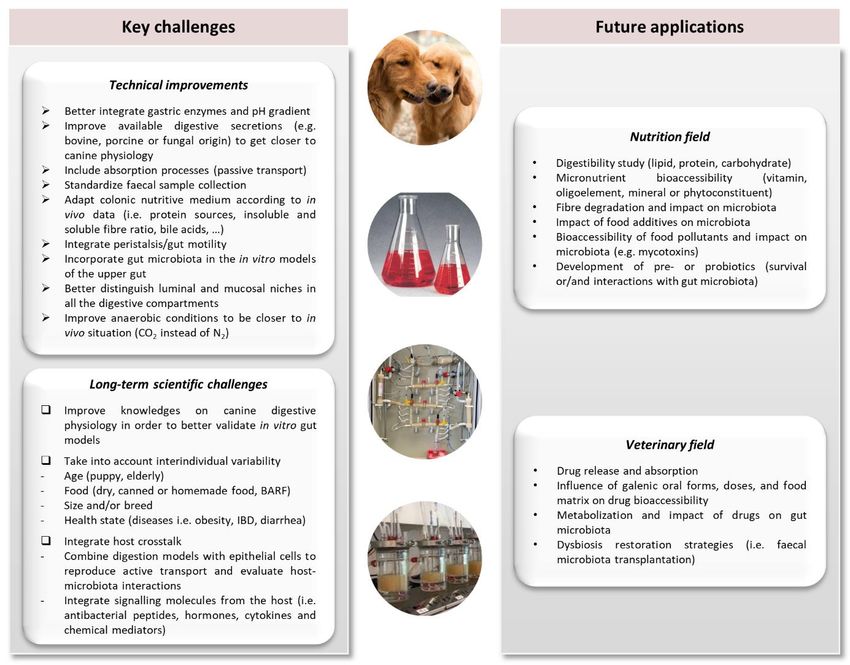
Fig. 4: Main challenges in the development of in vitro gut models of the canine digestive tract and their applications in
nutritional and veterinary fields
The figure gives an overview of the main technical and scientific challenges and applications of canine in vitro gut models, as
reliable tools to test or develop new products in the food and pharma fields.
data, this recent optimization was not validated compared to bacterial mucosal profiles in dog, but only compared to previous
results obtained in the M-SHIME. Once again, in vitro colonic pH varies widely between current available models, which could
be explained by the fact that authors generally base their model settings on a unique in vivo study, whereas large inter-individual
variations are observed in dogs, depending on age, size, and breed.
6 In vitro gut models as powerful tools to study canine digestion
6.1. Scientific and technical challenges to be addressed
As described before, parameters of in vitro models have been not fully adapted to in vivo data, probably due to the rarity of
information in dogs. Therefore, many scientific and technical challenges still need to be addressed to get closer to canine
digestion and consider the complexity of this environment (Fig. 4).
First, technical improvements should be considered to simulate more realistically canine digestive conditions at each
level of the GIT. Currently, as mentioned earlier, there is no canine chewing simulator, but such development is certainly not
a priority since most of dogs do not chew but swallow large pieces. Regarding the upper gut, the FIDO model already shows a
high level of complexity. The M-SCIME also possesses a gastric compartment that would merit improvements such as a
progressive acidification of the chyme (already made in the M-SHIME). This change in gastric pH during canine digestion is
certainly a key parameter in food disruption and digestion as it influences gastric pepsin and lipase activities (Carrière et al.,
1993; Sams et al., 2016). FIDO and M-SCIME also both homogenize food before in vitro digestion. In dogs, higher food
particle sizes seem to reach the stomach. Even if there is very few available data on this subject in dogs (unlike in human),
some canine studies showed no correlation between food size and density of particles on gastric emptying time, but also on the
entire upper gut digestion process (Gruber et al., 1987; Meyer et al., 1988; Chen et al., 2008; De Cuyper et al., 2018), while
others observed an increase gastric half emptying time with a higher meal viscosity and high-fat content (Ehrlein and Pröve,
1982; Palerme et al., 2020). Recently, a new gastric and small intestinal model, the Engineered Stomach and Small Intestine
(ESIN) has been developed to fill this gap and better handle both ingested liquids and real-size food particles in human
simulated digestion studies (Guerra et al., 2016). Even if this model has been primarily set-up for human applications, we can
easily imagine future developments to reproduce dog digestion. Another main issue in upper gut models is the use of porcine,
bovine, or fungal secretions/enzymes instead of canine ones. Of course, due to obvious ethical constrains, digestive secretions
collected from dogs cannot be used (and are not commercially available). Therefore, further investigations are needed to ensure
12You can also read