IL36 is a critical upstream amplifier of neutrophilic lung inflammation in mice
←
→
Page content transcription
If your browser does not render page correctly, please read the page content below

ARTICLE
                  https://doi.org/10.1038/s42003-021-01703-3                OPEN

                  IL36 is a critical upstream amplifier of neutrophilic
                  lung inflammation in mice
                  Carolin K. Koss1,2, Christian T. Wohnhaas1,2, Jonathan R. Baker3, Cornelia Tilp1, Michèl Przibilla1, Carmen Lerner1,
1234567890():,;

                  Silvia Frey1, Martina Keck1, Cara M. M. Williams1,5, Daniel Peter1, Meera Ramanujam4, Jay Fine4,
                  Florian Gantner1,2, Matthew Thomas1, Peter J. Barnes3, Louise E. Donnelly3 & Karim C. El Kasmi 1 ✉

                  IL-36, which belongs to the IL-1 superfamily, is increasingly linked to neutrophilic inflam-
                  mation. Here, we combined in vivo and in vitro approaches using primary mouse and human
                  cells, as well as, acute and chronic mouse models of lung inflammation to provide
                  mechanistic insight into the intercellular signaling pathways and mechanisms through which
                  IL-36 promotes lung inflammation. IL-36 receptor deficient mice exposed to cigarette smoke
                  or cigarette smoke and H1N1 influenza virus had attenuated lung inflammation compared with
                  wild-type controls. We identified neutrophils as a source of IL-36 and show that IL-36 is a key
                  upstream amplifier of lung inflammation by promoting activation of neutrophils, macrophages
                  and fibroblasts through cooperation with GM-CSF and the viral mimic poly(I:C). Our data
                  implicate IL-36, independent of other IL-1 family members, as a key upstream amplifier of
                  neutrophilic lung inflammation, providing a rationale for targeting IL-36 to improve treatment
                  of a variety of neutrophilic lung diseases.

                                                                   Konstanzer Online-Publikations-System (KOPS)
                                                           URL: http://nbn-resolving.de/urn:nbn:de:bsz:352-2-etkfqsxy95kt5
                  1 Boehringer Ingelheim Pharma GmbH & Co KG, Biberach, Germany. 2 Department of Biology, University of Konstanz, Konstanz, Germany. 3 Airway Disease,

                  National Heart and Lung Institute, Imperial College London, London, UK. 4 Boehringer Ingelheim Pharmaceuticals Inc., Ridgefield, CT, USA. 5Present address:
                  WRDM, Inflammation and Immunology Research Unit, Pfizer, Cambridge, MA, USA. ✉email: karim_christian.el_kasmi@boehringer-ingelheim.com

                  COMMUNICATIONS BIOLOGY | (2021)4:172 | https://doi.org/10.1038/s42003-021-01703-3 | www.nature.com/commsbio                                             1
ARTICLE                                                               COMMUNICATIONS BIOLOGY | https://doi.org/10.1038/s42003-021-01703-3

T
        he recently described interleukin (IL)-1 family cytokines           (Fig. 1a). These experiments indicated that IL-36 receptor
        IL-36α, IL-36β, and IL-36γ1–3 are emerging as contributors          (encoded by Il1rl2, herein designated as Il36r) signaling was an
        to acute and chronic tissue inflammation in human dis-               upstream driver of neutrophil recruitment and activation in mice
ease, particularly in the neutrophilic skin disease psoriasis4–6. In        exposed to CS smoke.
addition, experimental animal models of skin inflammation,                      To further determine whether IL-36 cytokines acted as
arthritis, and intestinal inflammation further implicate IL-36               upstream drivers of neutrophil recruitment in the lung, we
cytokines as important inflammatory mediators7–11. In the lung,              instilled IL-36γ (as one representative IL36 family cytokine
IL-36 has been suggested to be important in the pathogenesis of             previously described in the literature as a potent pro-
experimental bacterial and viral pneumonia in mice7,12. IL-36               inflammatory stimulus)32 intratracheally (i.t.) into naive mice
cytokines signal via a heterodimeric receptor comprised of an IL-           for 4 h (in order to examine early upstream events before the
36 receptor (IL-36R) chain and the IL-1 receptor accessory pro-             emergence of secondary effects). In contrast to AM numbers, IL-
tein (IL-1RAP) that is also shared with the IL-1 and IL-33                  36γ significantly increased BAL neutrophil numbers (Fig. 1b) to
receptors13,14. IL-36 cytokines can be expressed by skin epithelial         amounts comparable to those observed in CS-exposed mice
cells9,11, which correlates with increased mRNA and protein                 (Fig. 1a). Additionally, C-X-C chemokine ligand 1 (CXCL1; a key
concentrations of IL-36 cytokines in human psoriatic skin lesions           neutrophil chemoattractant protein) concentrations were
and experimental models of psoriasis-like diseases in mice,                 increased in the BAL relative to phosphate-buffered saline
including acanthosis and hyperkeratosis4,5,8–11,15. A common                (PBS)-exposed mice (Fig. 1c). IL-36γ also significantly increased
finding in diseases where IL-36 cytokines play a role is the pre-            concentrations of IL-1α, IL-1β, and granulocyte macrophages
sence of neutrophils8. Like other IL-1 family cytokines, IL-36              colony-stimulating factor (GM-CSF; cytokines typically generated
cytokines are produced as precursor proteins whose bioactivity is           by neutrophils, macrophages, or the epithelium33) in the BAL
1000-fold increased after proteolytic processing16,17. Typically,           relative to PBS-exposed mice (Fig. 1d). In addition, relative Cxcl1
activation of IL-36 cytokines is associated with increased                  and Il6 mRNA amounts were increased within the cell pellet
expression of multiple proteases released by neutrophils, such as           recovered from the BAL relative to PBS exposure (Suppl. Fig. 1a).
neutrophil-derived cathepsin G, elastase, and proteinase-318.                  We next determined whether IL-36 cytokines directly pro-
Consistent with the link between IL-36 cytokines and neutrophils,           moted pro-inflammatory activation of AMs and neutrophils.
IL-36 cytokines have been strongly linked to the pathogenesis of            Murine AMs obtained from BAL of naive mice were cultured in
generalized pustular psoriasis (GPP) and hidradenitis suppur-               the presence of GM-CSF (to maintain normal AM function34,35)
ativa, diseases in which neutrophils are a hallmark feature19.              and exposed to IL-36 cytokines for 24 h, after which Il1a and Il1b
Importantly, blocking IL-36 receptor with a monoclonal antibody             mRNA expression was determined. Both Il1a (19-fold) and Il1b
markedly reversed the skin lesions in human subjects with GPP20.            (2-fold) expression was significantly increased relative to that in
IL-36 signaling is also activated in intestinal inflammation such as         unstimulated AMs (Fig. 1e). Moreover, relative to unstimulated
inflammatory bowel disease and experimental colitis21,22. Fur-               AMs, IL-36 cytokine-stimulated AMs also had significantly
thermore, IL-36 also plays an important role in the joint syno-             increased mRNA expression for Il36g (4-fold) and Cxcl1 (30-
vium of patients with rheumatoid arthritis23,24. These studies              fold), indicating AMs as a source of CXCL1 and IL-36γ in the
have extended IL-36 and IL-36R expression to include fibroblasts             alveolar compartment. Importantly, a combination of IL-1α and
and macrophages25–28, and some reports have also suggested IL-              IL-1β did not induce transcription of Il1a, Il1b, or Cxcl1 and only
36 expression by lung epithelial cells29,30.                                mildly increased Il-36g in AMs (Fig. 1e). We also examined the
   Thus, there is a rationale for considering IL-36 cytokines as            effect of other lung-associated inflammatory mediators by
important contributors to the pathogenesis of lung diseases,                exposing AMs to lipopolysaccharide (LPS) or its canonical
specifically those that are characterized by the accumulation of             downstream cytokine tumor necrosis factor (TNF)-α. Exposing
neutrophils, such as severe non-T2 asthma, chronic obstructive              AMs to TNF-α did not stimulate mRNA expression for Il36g or
pulmonary disease (COPD), acute respiratory distress syndrome,              Cxcl1 in AMs (Suppl. Fig. 1b), while the fold induction of mRNA
and cystic fibrosis31. Associative data have linked IL-36 cytokine           encoding Cxcl1, Il36g, Il1b, Il1a, and Il6 after stimulating AMs
(s) to neutrophilic inflammation in multiple diseased tissues, yet           with LPS was similar to that observed in response to IL-36
the mechanisms by which IL-36 drives pathology remain elusive.              cytokines, indicating IL-36 cytokines as a potent pro-
Here, we used both wild-type (WT) and knockout (KO) in vitro                inflammatory stimulus on AMs (Suppl. Fig. 1c).
and in vivo murine experimental approaches together with pri-                  Considering that AMs were cultured in GM-CSF, we
mary human cells to deconvolute the network of stimuli and                  stimulated bone marrow-derived neutrophils from naive mice
responses that orchestrate neutrophilic lung disease. Here, we              in vitro with IL-36γ alone or in combination with GM-CSF.
provide the mechanistic rationale by which targeting IL-36 may              Combining GM-CSF with IL-36γ resulted in significantly
alleviate our most burdensome respiratory diseases.                         increased mRNA expression for Il36g (4-fold), Cxcl1 (10-fold),
                                                                            Il1a (600-fold), and Il1b (24-fold) relative to IL-36γ alone or GM-
                                                                            CSF alone when compared to unstimulated neutrophils (Fig. 1f).
Results                                                                     GM-CSF was capable of stimulating Il36g expression in mouse
IL-36 in neutrophilic lung inflammation. We exposed WT and                   neutrophils, suggesting a further mechanism by which IL-36γ can
IL-36 receptor KO mice (Il36r−/−) to cigarette smoke (CS) for               be induced. As was observed for AMs, IL-1α and IL-1β
3 weeks. Exposure of WT mice to CS resulted in significantly                 stimulation of neutrophils failed to induce gene expression for
increased bronchoalveolar lavage (BAL) neutrophil numbers                   these cytokines, even in combination with GM-CSF (Suppl.
(4.5 × 10⁵ neutrophils/mL). In contrast, alveolar macrophage                Fig. 1d). These findings prompted us to determine whether GM-
(AM) numbers did not change after exposure to CS. In Il36r−/−               CSF stimulation increased mRNA expression of the Il36r in
CS-exposed mice (1.7 × 10⁵ neutrophils/mL), we observed a 62%               neutrophils. Indeed, GM-CSF exposure resulted in an ~10-fold
reduction in BAL neutrophil numbers relative to WT mice. In                 increase in Il36r mRNA expression in neutrophils, while no
addition, myeloperoxidase (as an indicator of neutrophil acti-              change in the expression of Il1r1 was observed (Fig. 1g). GM-CSF
vation) concentrations in the BAL of Il36r−/− CS-exposed mice               also significantly induced the mRNA expression of the IL-1
were significantly reduced (to concentrations observed in mice               receptor antagonist (Il1rn), whereas the mRNA expression of the
exposed to room air (RA)) relative to CS-exposed WT mice                    IL-36 receptor antagonist (Il36rn) remained below the detection

2                         COMMUNICATIONS BIOLOGY | (2021)4:172 | https://doi.org/10.1038/s42003-021-01703-3 | www.nature.com/commsbio
COMMUNICATIONS BIOLOGY | https://doi.org/10.1038/s42003-021-01703-3                                                                           ARTICLE

Fig. 1 IL-36γ is an upstream inflammatory driver in mouse neutrophils and alveolar macrophages. a Neutrophil and macrophage counts and
myeloperoxidase concentration in the bronchoalveolar lavage fluid (BALF) from room air (RA) exposed (WT n = 9; Il36r−/− n = 6) and 3-week cigarette
smoke (CS) exposed mice (WT n = 10; Il36r−/− n = 10). b–d Neutrophil and macrophage counts, CXCL1, IL-1α, IL-1β, and GM-CSF protein concentrations
in BALF from untreated (n = 6) and IL-36γ-exposed mice (intratracheal instillation) (n = 7). e, f Cxcl1, Il1a, Il1b, and Il36g mRNA expression in either e naive
mouse alveolar macrophages (pooled n = 15 mice) and stimulated in vitro with no cytokines (−), IL-36γ, or IL-1α/IL-1β or f in mouse bone marrow-derived
neutrophils (from n = 4 mice) in vitro stimulated with no cytokines (−), IL-36γ, GM-CSF, or IL-36γ+GM-CSF. g Il36r and Il1r mRNA expression in mouse
bone marrow-derived neutrophils (from n = 4 mice) in vitro stimulated with no cytokines (−), IL-36γ, GM-CSF, and IL-36γ+GM-CSF. a, e, f *P ≤ 0.05,
**P ≤ 0.01, ***P ≤ 0.001, ****P ≤ 0.0001 vs all other groups by one-way ANOVA and Tukey’s correction. b, c, d, g **P ≤ 0.01, ***P ≤ 0.001, ****P ≤ 0.0001
vs untreated by t test. e AMs were pooled from 15 mice, data are presented as (mean ± SEM) of technical triplicates.

limit (Suppl. Fig. 1d). Thus, GM-CSF might skew the respon-                      inflammatory IL-1 family cytokines in neutrophils and macro-
siveness of neutrophils toward IL-36 relative to IL-1 through                    phages either alone or in combination with GM-CSF.
upregulation of the IL-36R.
   We also determined whether IL-36γ alone or in combination                     IL-36 acts as an upstream inflammatory driver of macrophages
with GM-CSF would increase the transcription of CXCL1, IL1A,                     and fibroblasts. We conditioned bone marrow-derived macro-
IL1B, and IL36G in human neutrophils. In contrast to mouse                       phages (BMDMs) from naive mice toward a putative lung inter-
neutrophils, GM-CSF and not IL-36γ induced mRNA expression                       stitial phenotype by exposing them to transforming growth factor
for CXCL1 (2-fold), IL1A (69-fold), IL1B (9-fold), and IL-36G (4-                (TGF)-β36,37 and GM-CSF34,35,38 in combination with IL-36
fold) relative to unstimulated neutrophils (Fig. 2a). Combining                  cytokines or IL-1α or IL-1β for 24 h. GM-CSF induced a 55-fold
IL-36γ stimulation with GM-CSF did not result in any further                     increase in mRNA for Il36r in BMDMs (Fig. 3a) and CXCL1
increase in the mRNA expression for these cytokines (Fig. 2a).                   protein release after IL-36 cytokine stimulation (77-fold) was
Furthermore, IL-36 did not increase IL36R gene expression in                     about 10-fold higher in GMCSF/TGF-β conditioned macrophages
human neutrophils relative to untreated cells (Fig. 2b). However,                relative to that observed in unconditioned BMDMs (Fig. 3b). We
exposing human neutrophils to GM-CSF resulted in a significant                    therefore continued using GM-CSF/TGF-β conditioned BMDMs.
increase of IL36R (encoded by IL1RL2 herein designated as Il36R)                 We detected a 140-fold increase in Il36g mRNA expression, a 10-
mRNA (Fig. 2b). IL1R mRNA was not induced after any of these                     fold increase in Cxcl1 mRNA expression, 4495-fold increase for
stimulations (Fig. 2b). Finally, we exposed human lung epithelial                Il1a mRNA expression, and a 235-fold increase for Il1b mRNA
cells to IL-36γ and found significantly increased protein secretion               expression in TGF-β/GM-CSF conditioned BMDMs stimulated
and mRNA expression of CXCL1, IL36G, GM-CSF, IL1A, and                           with IL-36 relative to unstimulated BMDMs (Fig. 3c). In contrast,
IL1B (Fig. 2c, d).                                                               BMDMs not conditioned in GM-CSF/TGF-β had a reduced
   Together, these findings indicated that in the alveolar                        responsiveness to IL-36 cytokines with Il36g (6-fold), Cxcl1 (1.4-
compartment IL-36γ can act as an upstream inflammatory driver                     fold), Il1a (1.7-fold), and Il1b (3.4-fold) only slightly increased
to promote neutrophil recruitment and production of pro-                         after IL-36 cytokine stimulation (Suppl. Fig. 2a). As we observed

COMMUNICATIONS BIOLOGY | (2021)4:172 | https://doi.org/10.1038/s42003-021-01703-3 | www.nature.com/commsbio                                                   3
ARTICLE                                                                  COMMUNICATIONS BIOLOGY | https://doi.org/10.1038/s42003-021-01703-3

Fig. 2 IL-36γ is an upstream inflammatory driver in human neutrophils and small airway epithelial cells. a, b CXCL1, IL1A, IL1B, and IL36G mRNA
expression in human peripheral blood-derived neutrophils (from n = 4 donors) in vitro stimulated with no cytokines (−), IL-36γ, GM-CSF, or IL-36γ+GM-
CSF. b IL36R and IL1R mRNA expression in human peripheral blood-derived neutrophils (from n = 4 donors) neutrophils in vitro stimulated with no
cytokines (−), IL-36γ, GM-CSF, and IL-36γ+GM-CSF. c, d CXCL1, GM-CSF, and IL1α protein concentrations and CXCL1, IL36G, GM-CSF, IL1A, and IL1B
mRNA expression in small airway epithelial cells (SAEC) stimulated with either (−) or IL-36αβγ. a, b *P ≤ 0.05, **P ≤ 0.01, ***P ≤ 0.001, vs all other groups
by one-way ANOVA and Tukey’s correction. c, d *P ≤ 0.05, **P ≤ 0.01, ***P ≤ 0.001, ****P ≤ 0.0001 vs untreated by t test.

in AMs and neutrophils in Fig. 1, stimulation with a combination                   When primary mouse fibroblasts obtained from the lungs of
of IL-1α and IL-1β did not affect mRNA expression for Cxcl1, Il1b,              naive mice were exposed in vitro to IL-36γ or IL-1α and IL-1β
and Il1a relative to untreated BMDMs (Fig. 3c and Suppl. Fig. 2a).              stimulation for 24 h, we observed significantly increased mRNA
Additionally, mouse BMDMs upregulated mRNA expression of                        expression for Il1a (284-fold), Il1b (337-fold), Cxcl1 (151-fold),
Il1r1 and Il1rn in response to IL-36γ stimulation, while Il36rn                 Il36g (50-fold), Il1r1 (1.3-fold), Il1Rn (5-fold), and Il36a (22-fold)
mRNA expression was undetected (Fig. 3c and Suppl. Fig. 2a).                    in response to IL-36γ relative to unexposed cells (Suppl. Fig. 2b).
   In human monocyte-derived macrophages (MDMs) from                            Stimulation with IL-1α and IL-1β did not result in any detectable
healthy volunteer donors, IL36R mRNA was not induced by IL-                     increases in mRNA expression for these genes and Il36b and Il36rn
36 cytokines or by GM-CSF stimulation (Fig. 4a). Stimulation                    were also not expressed in response to any of the stimulation
with IL-36 in combination with GM-CSF significantly increased                    conditions (Suppl. Fig. 2b). Conditioning fibroblasts with GM-CSF
relative mRNA amounts of IL1A (53-fold) and IL1B (30-fold)                      and TGF-β resulted in an additional increase in mRNA for Il1a
(Fig. 4a). Stimulation of human MDMs with IL-36γ also                           (1015-fold), Il1b (761-fold), Il36g (81-fold), and Il1rn (6.2-fold)
promoted mRNA expression for CXCL1 (Suppl. Fig. 3a).                            when exposed to IL-36 cytokines (Fig. 3d). Analogous to the
However, MDMs did not exhibit a significant increase in IL36G                    findings in mouse neutrophils and mouse BMDMs, GM-CSF also
mRNA expression after IL-36 stimulation relative to untreated                   increased mRNA expression for the Il36r in primary lung
MDMs, which was only slightly increased (1.3-fold) after                        fibroblasts, albeit with a lower fold induction. IL-36 also slightly
stimulation with GM-CSF. The combination of IL-36 cytokines                     increased the Il36r in primary lung fibroblasts (Fig. 3e).
and GM-CSF significantly increased the IL36G expression relative                    When primary human lung fibroblasts (from otherwise
to GM-CSF alone (Fig. 4a).                                                      healthy donors) were stimulated with IL-36 cytokines, GM-

4                            COMMUNICATIONS BIOLOGY | (2021)4:172 | https://doi.org/10.1038/s42003-021-01703-3 | www.nature.com/commsbio
COMMUNICATIONS BIOLOGY | https://doi.org/10.1038/s42003-021-01703-3                                                                      ARTICLE

Fig. 3 IL-36γ is an upstream amplifier in mouse macrophages and fibroblasts. a Relative mRNA amounts of Il36r in naive mouse bone marrow-derived
macrophages (BMDMs, n = 6) in response to no stimulation (−) or stimulation with GM-CSF and IL-36γ. b CXCL1 protein concentrations in supernatant of
BMDMs (n = 4) after no stimulation (−), or stimulation with IL-36αβγ. IL-1α, IL-1β alone, or in combination with GM-CSF and TGFβ c, d Relative mRNA
amounts of Il36g, Cxcl1, Il1a, Il1b, Il1r1, Il1rn, and Il36rn in BMDMs (n = 4) (c) and primary mouse fibroblasts (n = 4) (d) after no stimulation (−) or
stimulation with IL-36αβγ, IL-1α, IL-1β alone, or in combination with GM-CSF and TGF-β. e Relative mRNA amounts in primary mouse fibroblasts (n = 4) of
Il36r after no stimulation (−) or stimulation with IL-36αβγ or GM-CSF and TGF-β. Shown are the mean values ± SEM of biological replicates. a–d *P ≤ 0.05,
**P ≤ 0.01, ***P ≤ 0.001, ****P ≤ 0.0001 vs all other groups by one-way ANOVA and Tukey’s correction. e *P ≤ 0.05, vs untreated by t test.

Fig. 4 IL-36γ is an upstream amplifier in human macrophages and fibroblasts. a Relative mRNA amounts in human monocyte-derived macrophages
(MDM) (depicted are mean values ± SME of technical triplicates from one representative of four experiments) of IL-36G, IL-36R, IL1A, and IL1B and b IL-36γ,
IL-36R, IL1A, IL1B, GMCSF, and CXCL1 in primary human lung fibroblasts (n = 4) after no stimulation (−) or stimulation with IL-36αβγ, GM-CSF alone, or
with the combination of IL-36αβγ and GM-CSF. Shown are the mean values ± SEM of biological replicates. a, b *P ≤ 0.05, **P ≤ 0.01, ***P ≤ 0.001, ****P ≤
0.0001 vs all other groups by one-way ANOVA and Tukey’s correction.

CSF, or the combination thereof, we observed increased mRNA                      Inflammatory conditions in the lung also promote tissue
expression for IL36G, IL36R, IL1A, IL1B, GM-CSF, and CXCL1                     remodeling39, in which matrix metalloproteinase 9 (MMP9) has
after IL-36 stimulation relative to untreated fibroblasts                       been shown to be an important mediator40. MMP9 protein and
(Fig. 4b).                                                                     mRNA expression were increased in both primary lung

COMMUNICATIONS BIOLOGY | (2021)4:172 | https://doi.org/10.1038/s42003-021-01703-3 | www.nature.com/commsbio                                              5
ARTICLE                                                                 COMMUNICATIONS BIOLOGY | https://doi.org/10.1038/s42003-021-01703-3

Fig. 5 IL-36γ is critical in neutrophilic lung inflammation. a Neutrophil counts in BALF from room air (RA) (n = 18) exposed or from 1-week (n = 9), 2-
week (n = 9), and 3-week (n = 8) cigarette smoke (CS) exposed mice and from H1N1(4 days post treatment) exposed mice (n = 8) and from mice
exposed to 2 weeks CS followed by 48 h of H1N1 exposure (n = 8). b Relative mRNA amounts of IL-36g in the cellular BAL pellet from room air (RA) and
2 week cigarette smoke (CS)-exposed mice (n—4). c, d Neutrophil numbers in BALF and IL1β, CXCL1, and IL-6 protein concentrations in lung homogenate
of room air-exposed mice (RA; n = 4), 2-week CS-exposed mice (n = 4–8), and 2-week CS-exposed mice challenged with H1N1 for 48 h (CS + H1N1) (n =
4–8). e Neutrophil numbers in BALF and relative mRNA amounts of IL-36g and Cxcl1 in lung homogenate from room air-exposed (RA, n = 5) and 2-week
CS-exposed mice challenged with H1N1 for 48 h (n = 7–8) exposed WT and Il36r−/− mice. f, g IL1β, CXCL1, IL6, and TNF protein concentrations in
BALF and in lung homogenate from room air-exposed (RA, n = 5) and 2-week CS-exposed mice challenged with H1N1 for 48 h (n = 7–8) exposed WT and
Il36r−/− mice. Depicted are mean values ± SEM of biological replicates. a, c–g *P ≤ 0.05, **P ≤ 0.01, ***P ≤ 0.001, ****P ≤ 0.0001 vs all other groups by
one-way ANOVA and Tukey’s correction. b *P ≤ 0.05, **P ≤ 0.01, ***P ≤ 0.001, ****P ≤ 0.0001 vs untreated by t test.

fibroblasts and BMDMs after stimulation with IL-36 cytokines                   mice, CS + H1N1-exposed mice exhibited significantly higher
(Suppl. Fig. 2c).                                                             increases in Il36g mRNA in the BAL pellet (1759-fold increased
  In summary, these findings highlight the difference between                  relative to RA) compared to CS-exposed mice (40-fold relative to
IL-1α/β and IL-36 activation and pinpoint IL-36 as an upstream                RA) (Fig. 5b). In addition, as indicators of lung inflammation, we
amplifier of human and mouse macrophages and fibroblasts.                       found a significant increase of IL-1β, CXCL1, and IL-6 protein in
                                                                              lung tissue of CS + H1N1-exposed mice relative to CS-exposed
                                                                              mice (Fig. 5d). These findings showed that this model was sui-
IL-36 is important in neutrophilic lung inflammation in vivo.                  table to generate lung inflammatory conditions with high neu-
Thus far, the data suggested that IL-36 signaling was important               trophil recruitment to the alveolar space, which also correlated
for neutrophil recruitment and activation both in vivo (Fig. 1a–c)            with marked Il36g expression.
and in vitro (Figs. 1 and 3). We therefore predicted that IL-36                  We therefore utilized this model to determine the contribution
would play an important role in a lung inflammatory condition                  of IL-36 cytokine signaling to lung inflammation in vivo. Il36r−/−
associated with high neutrophil numbers. To test this hypothesis,             mice exposed to CS + H1N1 exhibited a significant decrease
we advanced our CS smoke model depicted in Fig. 1 to either CS                (~72%) of BAL neutrophil numbers relative to that observed in
for 1, 2, or 3 weeks or to CS (for 2 weeks) followed by exposure to           CS + H1N1-exposed WT mice (Fig. 5e). Il36r−/− mice exposed to
H1N1 influenza virus for 48 h (CS + H1N1). Neutrophil numbers                  CS + H1N1 also exhibited significantly decreased mRNA expres-
in recovered BAL were 1.8 × 10⁵/mL after 1 week CS exposure,                  sion for Cxcl1 in the lung homogenate relative to CS + H1N1-
8 × 10⁴/mL after 2 weeks of CS exposure, 3.6 × 10⁵/mL after                   exposed WT mice (Fig. 5e). Furthermore, Il36r−/− mice exposed
3 weeks of CS exposure, 8.2 × 10⁵/mL after H1N1, and 1.05 × 10⁶/              to CS + H1N1 also exhibited significantly decreased mRNA
mL after CS + H1N1 (Fig. 5a, c). Importantly, consistent with the             expression for Il36g in the lung homogenate (Fig. 5e). Finally, IL-
higher numbers of neutrophils in CS + H1N1 mice relative to CS                1β, CXCL1, IL6, and TNF-α protein concentrations were also

6                           COMMUNICATIONS BIOLOGY | (2021)4:172 | https://doi.org/10.1038/s42003-021-01703-3 | www.nature.com/commsbio
COMMUNICATIONS BIOLOGY | https://doi.org/10.1038/s42003-021-01703-3 ARTICLE
decreased in CS + H1N1-exposed Il36r−/− mice in both BAL 36 in activating fibroblasts (Fig. 6b and Suppl. Fig. 5e, f). The
fluid (Fig. 5f) and lung homogenate (Fig. 5g and Suppl. Fig. 4a) combination of poly(I:C) and IL-36 (7.5-fold) resulted in a 50%
relative to CS + H1N1-exposed WT. reduced Ifnb1 mRNA expression in BMDMs relative to poly(I:C)
We next used mice with genetic deficiency in IL-1RAP (the (15-fold) stimulation alone. IL-36 alone did not induce Infb1
common receptor chain for the receptors for IL-1α, IL1-β, IL-33, expression in BMDMs (Suppl. Fig. 5g). Neither poly(I:C) nor IL-
and IL-362) to define the effect of IL-36R signaling (reflecting the 36 stimulation resulted in increased Infb1 mRNA expression in
contribution of IL-36) within the IL-1 family in this model. CS + fibroblasts (Suppl. Fig. 5g).
H1N1-exposed Il1rap−/− exhibited a significant reduction in the Similar to mouse fibroblasts, human lung fibroblasts stimulated
expression of markers for lung inflammation (Suppl. Fig. 4b). with the combination of poly(I:C) and IL-36 cytokines exhibited a
Specifically, we observed a 61% reduction in BAL neutrophil significant increase in CXCL1, IL1A, IL1B, and GM-CSF mRNA
numbers relative to WT mice exposed to CS + H1N1. In relative to untreated controls (Fig. 7a). Neither poly(I:C) nor IL-
addition, IL-1β, CXCL1, and IL6 protein concentrations in lung 36 cytokine exposure promoted IL36G mRNA expression in
homogenate were significantly reduced in CS + H1N1-exposed human fibroblasts (Fig. 7a).
Il1rap−/− mice relative to CS + H1N1-exposed WT mice In human MDMs, IL-36 cytokine stimulation resulted in
(Suppl. Fig. 4b). Importantly, the percentage of reduction in significant increase of CXCLl1, IL1A, and IL1B mRNA expression
BAL neutrophil numbers was comparable between Il1rap−/− relative to untreated or poly(I:C) alone and was not changed by
and Il36r−/− CS + H1N1-exposed mice (Suppl. Fig. 4c). While combining IL-36 cytokines with poly(I:C) (Fig. 7b). Similar to the
Il1rap−/− CS + H1N1-exposed mice exhibited an 81% reduction fibroblast data, IL36G mRNA expression in MDMs was not
for CXCL1 and an 80% reduction for IL-1β protein concentra- increased after IL-36 cytokine, poly(I:C), or IL-36 cytokine + poly
tions in lung homogenate, Il36r−/− CS + H1N1-exposed mice (I:C) stimulation (Fig. 7b).
exhibited a 47% reduction for CXCL1 and 44% reduction for IL- Human basal lung epithelial cells exposed to poly(I:C)
1β. Notably, the percentage of reduction in IL-6 protein amounts exhibited significant and marked increases in protein concentra-
in the lung homogenate was equal between Il1rap−/− (45%) and tions of GM-CSF (63-fold), CXCL1 (2.1-fold), and IL-36γ (71-
Il36r−/− (48%) CS + H1N1-exposed mice (Suppl. Fig. 4c). These fold) and mRNA expression of GM-CSF (7-fold), CXCL1 (4-fold),
findings indicated that, within the IL-1 family of cytokines that IL36G (364-fold), IL1A (186-fold), and IL1B (49-fold) (Fig. 8a, b).
signal through IL-1RAP, IL-36 cytokines contribute to a large LPS, used as an additional TLR agonist did not induce mRNA
degree to the lung inflammatory response in CS + H1N1 mice. increases of these cytokines. (Fig. 8a, b). IL-1α protein
concentration was not increased after poly(I:C) or LPS stimula-
tion relative to untreated control and IL-1β was only detected
IL-36 cooperates with polyinosinic-polycytidylic acid (poly(I: after poly(I:C) stimulation (Fig. 8a).
C)) on macrophages and fibroblasts. Based on the observed These data indicated that poyl(I:C) increased the production of
attenuated inflammation in Il36r−/− mice in response to CS + pro-inflammatory cytokines in lung epithelial cells and coop-
H1N1, we next employed in vivo approaches in which we used erated with IL-36 to further amplify pro-inflammatory pathways.
the Toll-like receptor 3 (TLR3) agonist poly(I:C) as a viral
mimetic and focused on measuring neutrophils, GM-CSF,
CXCL1, IL-36, and MMP9 cytokine expression in the BAL of Neutrophils are a source of IL-36γ in acute lung injury. To
mice. Mice exposed to i.t. poly(I:C) had increased neutrophils determine neutrophils as a source of IL-36 not only in a lung
(but no increases in macrophages) in the BAL, 12 and 24 h post inflammation model generated by CS or CS and virus, we also
poly(I:C) exposure relative to PBS-exposed mice (Suppl. Fig. 5a). examined a lung inflammation model simulating exposure to
In addition, we found significantly increased protein concentra- bacterial stimuli. We therefore exposed mice to aerosolized LPS
tions for CXCL1 (at 4 and 24 h), GM-CSF (at 24 h), and MMP9 for 4 h before proceeding with single-cell RNA sequencing
(at 24 h) in BAL, as well as increased Il36g mRNA in the BAL (scRNA-seq) analysis of the BAL cells. We chose 4 h exposure as
pellet (at 12 h) relative to PBS-exposed mice (Suppl. Fig. 5a, b). it was determined to be the earliest time point with maximal
We next exposed AMs obtained by BAL and bone marrow- neutrophil numbers in the BAL fluid (2.12 × 10⁶ neutrophils/mL)
derived neutrophils from naive mice to poly(I:C) in vitro. Poly(I: in a pilot experiment (Suppl. Fig. 6a). After LPS challenge, the
C) induced small but significant increases in mRNA expression majority of the sequenced BAL cells represented neutrophils,
for Il36g (2-fold), Cxcl1 (2.3-fold), Il1a (1.16–fold), Il1b (2.17- which accounted for 77% of all BAL cells, while AMs represented
fold), and Il36r (1.11-fold) in AMs relative to untreated controls only 18% of the BAL cell population (Fig. 9a). Additionally, we
(Suppl. Fig. 5c). In mouse bone marrow-derived neutrophils, poly identified natural killer cells (4%) and few erythrocytes (1%)
(I:C) alone failed to induce increased mRNA expression for Il36g, based on their reported characteristic gene expression profiles41
Cxcl1 Il1a, and Il1b (Fig. 6a). Combining poly(I:C) stimulation (Fig. 9a and Suppl. Fig. 6b, c). We then determined the relative
with IL-36γ stimulation resulted in numerical increased mRNA transcriptional expression of Il36a, Il36b, and Il36g in comparison
expression for Il1b but had similar effects on Cxcl1 and trended to Il1a and Il1b in BAL neutrophils and macrophages. High-
toward increased expression of Il1a (Fig. 6a). resolution scRNA-seq analysis allowed us to identify neutrophils
Combining IL-36 cytokines with poly(I:C) resulted in sig- as the main source of Il36g. Il36g was detected in 84% of the
nificantly increased relative mRNA amounts in BMDMs and neutrophils and its transcript levels were 10.5-fold increased
primary mouse fibroblast for Il36g, Cxcl1, Il1a, Il1b, and Gmcsf relative to those detected in AMs (Fig. 9b, d). In contrast, Il1b was
(Fig. 6b) and increased protein concentrations for CXCL1, IL-36γ strongly expressed by both neutrophils and AMs. Nevertheless,
(pro-form), GM-CSF, and MMP9 relative to the individual Il1b was still increased 3.6-fold in neutrophils relative to AM
stimulations compared to untreated cells (Suppl. Fig. 5d, f). while Il1a was detected in both populations, whereas the overall
Interestingly, as observed for AMs (Suppl. Fig. 5c), poly(I:C) expression was significantly (1.5-fold) increased in neutrophils
promoted mRNA expression for the Il36r on BMDMs (Suppl. relative to AMs (Fig. 9b). In contrast to Il36g, Il36a was only
Fig. 5e). Primary mouse lung fibroblasts were comparable to detected in very few single cells (while Il36b was not detected in
BMDMs (Fig. 6b). Of note, poly(I:C) alone failed to induce Il36g, any cell type (Suppl. Fig. 6d)). Next, we investigated the expres-
Cxcl1, Il1a, Il1b, Gmcsf, and Il36r mRNA in primary lung sion of Cxcl1 across the different cell types. Cxcl1 expression was
fibroblasts, highlighting the requirement for cooperating with IL- predominantly restricted to AMs and increased 1.9-fold
COMMUNICATIONS BIOLOGY | (2021)4:172 | https://doi.org/10.1038/s42003-021-01703-3 | www.nature.com/commsbio 7ARTICLE COMMUNICATIONS BIOLOGY | https://doi.org/10.1038/s42003-021-01703-3 Fig. 6 IL-36γ cooperates with poly(I:C) on mouse macrophages and fibroblasts. a Relative mRNA amounts of Il36g, Cxcl1, Il1a, and Il1b in mouse neutrophils (n = 2–6) unstimulated with (−) or stimulated with IL-36αβγ, poly(I:C), or the combination of IL-36αβγ and poly(I:C) (depicted are mean values ± SEM of biological replicates). b Relative mRNA amounts of Il36g, Cxcl1, Il1a, Il1b, and Gmcsf in BMDMs (n = 4) and primary mouse fibroblasts (n = 4) (depicted are mean values ± SEM of biological replicates) unstimulated (−) or stimulated with IL-36αβγ, poly(I:C), or the combination of IL-36αβγ and poly(I:C). *P ≤ 0.05, **P ≤ 0.01, ***P ≤ 0.001, ****P ≤ 0.0001 vs all other groups by one-way ANOVA and Tukey’s correction. Fig. 7 IL-36γ cooperates with poly(I:C) on human macrophages and fibroblasts. a, b Relative mRNA amounts of IL-36g, CXCLl1, IL1A, IL1B, and GM-CSF in human fibroblasts and human MDMs (depicted are mean values ± SME of technical triplicates from one representative of three to four experiments) unstimulated (−) or stimulated with IL-36αβγ, poly(I:C), or the combination of IL-36αβγ, and poly(I:C). *P ≤ 0.05, **P ≤ 0.01, ***P ≤ 0.001, ****P ≤ 0.0001 vs all other groups by one-way ANOVA and Tukey’s correction. compared to neutrophils (Fig. 9b). We next determined mRNA A more detailed analysis of the neutrophil clusters in the expression for Il36a,b,g and Il1a,b as well as Cxcl1 by quantitative scRNA-seq analysis revealed two subpopulations that we polymerase chain reaction (qPCR) in mouse bone marrow- designated N1 and N2 (Suppl. Fig. 6f). To differentiate the derived neutrophils and mouse AMs from naive mice in response subclusters, we did a pathway analysis. Subcluster N2 showed to in vitro stimulation with LPS for 4 h to closely reflect the increased IL-1 family and IFN pathways, whereas subcluster N1 in vivo stimulation protocol. Consistent with the scRNA-seq data, exhibited increased remodeling pathways (Suppl. Fig. 6g). Finally, we found that neutrophils were more responsive to LPS stimu- increased differential expression of MMP9 was observed to be lation in terms of Il36g expression (48-fold increased) compared restricted to subcluster N1, suggesting that these neutrophils to the expression in AMs (8-fold increased) (Fig. 9c). In addition, could promote tissue remodeling (Suppl. Fig. 6h). induction of Cxcl1 mRNA expression upon LPS stimulation was A hypothetical model of how cells and mediators interplay with restricted to AM and not induced in stimulated neutrophils IL-36 to promote neutrophilic lung inflammation is depicted in (Fig. 9c). Il1a and Il1b were induced in both cell types after LPS Fig. 10. stimulation but more predominantly in AMs. AMs had increased Il36a (690-fold) mRNA expression after LPS stimulation and Il36b was undetectable (Suppl. Fig. 6e). Furthermore, IL-36α and Discussion IL-36β mRNA expression remained below the detection limit for Here, we combined chronic and acute lung inflammation mouse neutrophils. models induced by CS, viral (poly(I:C)), and bacterial (LPS) Human neutrophils and human MDMs exhibited a 100- and components with additional in vivo and in vitro approaches using 200-fold increase in IL36G mRNA expression after LPS primary mouse and human cells to provide mechanistic insight stimulation relative to unstimulated cells. Human fibroblasts that identifies IL-36 within the IL-1 family of cytokines as an early exhibited a 2.5-fold IL36G mRNA induction after LPS stimulation upstream innate immune driver and amplifier of acute and relative to untreated controls (Fig. 9e). chronic lung inflammation, particularly when associated with 8 COMMUNICATIONS BIOLOGY | (2021)4:172 | https://doi.org/10.1038/s42003-021-01703-3 | www.nature.com/commsbio
COMMUNICATIONS BIOLOGY | https://doi.org/10.1038/s42003-021-01703-3                                                                        ARTICLE

Fig. 8 Small airway epithelial cells as the main cellular source of GMCSF. a GM-CSF, CXCL1, IL-36γ, IL-1β, and IL-1α protein concentrations and GM-CSF,
CXCL1, IL36G, IL1A, and IL1B mRNA expression of lung epithelial basal cells from healthy donor stimulated with poly(I:C) and LPS (depicted are mean values
± SEM of technical triplicates from one representative of three experiments). *P ≤ 0.05, **P ≤ 0.01, ***P ≤ 0.001, ****P ≤ 0.0001 vs all other groups by one-
way ANOVA and Tukey’s correction.

neutrophils. Mechanistically, lung neutrophils were identified as                the phenotype of the Il1rap−/− mice was largely replicated in
both a cellular source and target of IL-36 and IL-36 promoted the               Il36r−/− mice. These data solidify IL-36 as a key upstream pro-
pro-inflammatory activation of lung macrophages, fibroblasts,                     inflammatory driver among the IL-1 family of cytokines that
and epithelial cells. IL-36 stimulated feed-forward signaling that              signal through IL-1RAP. Importantly, genetic ablation of IL-36
resulted in the generation of IL-36 and IL-1 in fibroblasts and                  signaling was sufficient to attenuate recruitment of neutrophils
macrophages. Moreover, IL-36 cooperated with viral analog poly                  to the same degree as seen in Il1rap−/− mice, while the protein
(I:C) and with GM-CSF to enhance the pro-inflammatory                            expression of IL-1β and CXCL1 was more attenuated in the
responses of macrophages and fibroblasts.                                        Il1rap−/− mice. These findings can be interpreted that blocking
   There is substantial evidence associating the pro-inflammatory                IL-36R allows for maintaining at least part of the IL-1 innate-
role of IL-36 with the presence of neutrophils in mouse models of               mediated immune response necessary to maintain some level of
skin inflammation and also in human skin diseases8,42, such as                   immune defense, while it abrogates the acute inflammatory and
GPP or hidradenitis suppurativa, where blockade of IL-36 signaling              tissue destructive effects of IL-36, which superimpose on the
can greatly reduce disease symptoms20. In addition, IL-36 has been              underlying inflammation and exacerbate immune pathology and
described to be increased in patients with COPD43, and in COPD,                 substantially compromise organ function in the face of acute
neutrophils are correlated with the severity of the symptoms and                pathogen challenge. The findings that Il36g expression within the
with microbial exacerbation, while high neutrophil numbers are                  alveolar immune cell compartment was restricted to neutrophils
correlated with poor prognosis and disease progression44,45. Pre-               in LPS-challenged mice and that stimulation of mouse AMs,
vious reports also showed GM-CSF release after pulmonary                        mouse neutrophils, and human neutrophils with LPS in vitro
infection and the importance of GM-CSF in inflammatory pro-                      increased IL-36γ production demonstrate that neutrophils can
cesses in the lung has been reported, including in COPD46,47.                   also be an important source of IL-36γ in humans and mice in
Finally, previous reports had suggested an important role for IL-36             neutrophilic inflammation associated with bacterial infection.
in mouse models of bacterial and viral pneumonia7,12. Yet, no                   Thus, we propose that IL-36R blockade in patients with chronic
studies had investigated the interplay between IL-36, GM-CSF, and               lung disease prone to microbial exacerbations will alleviate
neutrophils in chronic lung inflammation or in lung inflammation                  symptoms while preserving sufficient innate immune signaling
in which acute virus challenge is superimposed on chronic injury, a             mediated by IL-1α and IL-1β to conserve host defense.
reductionist mechanistic model for human lung disease secondary                    We also demonstrate that IL-36γ promoted generation of IL-
to smoking and virus infection.                                                 36γ itself and was upstream of both IL-1α and IL-1β in neu-
   Using a genetic approach employing mice with Il36r deficiency,                trophils, fibroblasts, and macrophages. IL-36γ was also capable of
we showed that IL-36 signaling was a critical component of the                  inducing CXCL1 in vivo and in a variety of cell types in vitro,
pro-inflammatory response in both CS- and CS + HN1N-                             including lung macrophages, lung fibroblasts, neutrophils, and
exposed mice, demonstrating a role for IL-36 in both low-grade                  lung epithelial cells. Notably, CXCL1, as well as IL-1α and IL-1β,
chronic lung inflammation (CS model) as well as high-grade                       expression in macrophages was restricted to IL-36γ stimulation
acute microbial lung inflammation super-imposed on pre-existing                  and not observed with the other IL-1 family cytokines IL-1α and
chronic injury, such as during acute viral exacerbations in COPD                IL-1β, placing an important role on IL-36 in an upstream
(CS + H1N1 model). Specifically, Il36r−/− mice had significantly                  amplification loop for the production of CXCL1 and IL-1 family
attenuated lung neutrophil influx and reduced alveolar IL-1β,                    cytokines in a variety of innate immune cells. Neutrophils
CXCL1, IL-6, and TNF-α concentrations. Making use of                            themselves also responded to IL-36 stimulation with CXLC1
IL1rap−/− mice, we isolated the contribution of IL-36 cytokines                 production, highlighting a feed-forward loop between IL-36,
within the IL-1 family of cytokines in this model and showed that               CXCL1, and neutrophils. Thus, the IL-36γ-triggered pro-

COMMUNICATIONS BIOLOGY | (2021)4:172 | https://doi.org/10.1038/s42003-021-01703-3 | www.nature.com/commsbio                                                9
ARTICLE COMMUNICATIONS BIOLOGY | https://doi.org/10.1038/s42003-021-01703-3
a)
b)
c) Il36g Il1a Il1b Cxcl1
1.5×10 2 ****
Relative mRNA
4×10 1 6×10 1 8×10 2
3×10 1 6×10 2
1×10 2 4×10 1
2×10 1 4×10 2
5×10 1 2×10 1
1×10 1 2×10 2
0 0 0 0
LPS (4 h) - + - + - + - + - + - + - + - +
AMs Neut AMs Neut AMs Neut AMs Neut
d) e)
Neutrophils MDM Fibroblast untreated
IL36G human cells
Relative mRNA
1.5×10 2 1.5×10 2 1.5×10 2 LPS
***
1×10 2 1×10 2 1×10 2
****
5×10 1 5×10 1 5×10 1
0 0 0
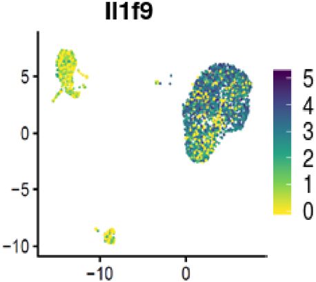
Fig. 9 Neutrophils are a source of IL-36γ in acute lung injury. a UMAP representation of the cell populations identified in BAL from LPS-exposed mice.
Cell identity is indicated by the color code. Relative frequency of the cell types is shown in the bar plot using the same color code. b Violin plots display
normalized expression levels of Il36g (Il1f9), Il1a, Il1b, and Cxcl1 across the different cell populations. c Relative mRNA amounts of the same genes used in b
after 4 h in vitro LPS stimulation of naive mouse alveolar macrophages (AM) and naive mouse bone marrow-derived neutrophils; depicted are mean values
± SEM of biological duplicates (each biological duplicate represents pooled AMs from 4 mice) and n = 8 from the neutrophils analyzed by one-way
ANOVA. d Visualization of normalized IL-36g (Il1f9) expression levels per single cell. e mRNA expression of IL-36g after LPS stimulation in vitro of human
neutrophils, MDMs (depicted are mean values ± SME of technical triplicates from one representative of three to four experiments) and human fibroblasts
n = 4. Data represents the depicted mean ± SEM of biological replicates; ***P ≤ 0.001, ****P ≤ 0.0001 vs untreated by t test.
inflammatory mediator output broadly facilitates feed-forward (IC) did not induce GM-CSF or CXCL1 in macrophages or
amplification of the inflammatory response by engaging a variety neutrophils, making the lung epithelium the most likely source of
of cells to increase neutrophil recruitment (CXCL1) and sensiti- GM-CSF and CXCL1 in vivo in response to poly(I:C). Therefore,
zation to IL-36γ or IL-1. poly(I:C) can promote IL-36 generation indirectly through
Our findings furthermore demonstrate that, downstream of epithelial-derived cytokines such as GM-CSF, which acts on
neutrophils, activated IL-36 cytokines subsequently cooperate neutrophils to produce IL-36. Another important finding of our
with additional pro-inflammatory signals, such as GM-CSF or study was that IL-36 also cooperated with poly(I:C) to directly
poly(I:C)/viruses as an upstream driver of IL-1, GM-CSF, and increase the expression of Il36g, Cxcl1, Il1a, Il1b, and Gmcsf in
CXCL1, further amplifying the pro-inflammatory interplay macrophages and fibroblasts. Thus poly(I:C) can also promote IL-
between neutrophils, IL-36, IL-1, and GM-CSF. Moreover, mice 36 signaling. Previous reports had suggested the cooperation
exposed to poly(I:C) had increased neutrophils and protein between TLR signaling and cytokine signaling with IL-36 in
concentrations of GM-CSF and CXCL1 in the BAL. Notably, poly promoting inflammation32 (Fig. 6).
10 COMMUNICATIONS BIOLOGY | (2021)4:172 | https://doi.org/10.1038/s42003-021-01703-3 | www.nature.com/commsbioCOMMUNICATIONS BIOLOGY | https://doi.org/10.1038/s42003-021-01703-3 ARTICLE
relationship between GM-CSF and neutrophils by the fact that
humans treated with GM-CSF had increased numbers of neu-
trophils52. Another study demonstrated that LPS-exposed GM-
CSF−/− mice had reduced infiltration of neutrophils compared to
the WT control53.
We also observed that IL-36γ- or IL-1-stimulated cells
increased the expression of the IL-1RN but not the IL-36RN,
suggesting unrestrained IL-36γ signaling while limiting IL-1 sig-
naling in these conditions. Intriguingly, severe neutrophilic skin
diseases in humans are associated with polymorphisms in IL-
36RN genes54,55.
Finally, inflammatory conditions in the lung are associated
with tissue remodeling. Therefore, we used fibroblasts as struc-
tural target cells reported to produce MMP9 and examined
MMP9 production as a surrogate for an important remodeling
factor after IL-36 cytokine stimulation56,57. Consistent with pre-
Fig. 10 Proposed signaling pathways involving IL-36 as an upstream vious reports, IL-36 was indeed capable of inducing MMP9
amplifier in neutrophilic lung inflammation. 1 Chronic (e.g., cigarette expression in primary lung fibroblasts57, involving IL-36 signal-
smoke) or acute (H1N1 or poly(I:C) injury to the lung epithelium promotes ing in pro-fibrotic fibroblast activity21,56.
release of CXCL1 and GMCSF. 2 CXCL1 promotes neutrophil recruitment In conclusion, we have used several mouse models to reflect
while GM-CSF upregulates the IL-36R and IL1RN by neutrophils and important triggers of lung inflammation that bear physiological
promotes IL-36γ expression. 3 IL-36γ activates neutrophils to generate IL1 relevance in humans with COPD, such as CS, CS + H1N1, poly(I:
and IL36. 4 IL-36γ subsequently activates alveolar and interstitial C), and LPS, allowing us to mechanistically investigate the
macrophages and fibroblasts to generate IL-1, CXCL1, GM-CSF, MMPs, and interplay between IL-36 and neutrophilic lung inflammation. We
more IL-36. GM-CSF also upregulates the lL36R on macrophages and on have shown that injured epithelium released GM-CSF, which
fibroblasts and sensitizes cells to IL-36. 5 Poly(I:C) as a TLR agonist increased IL-36 expression in the alveolar compartment. IL-36
cooperates with IL-36γ in amplifying activation of macrophages and subsequently activated alveolar and interstitial macrophages, as
fibroblasts. Note that IL-36RN is not induced while IL1RN is induced, well as fibroblasts, to generate IL-1, CXCL1, GM-CSF, MMPs,
shifting the balance toward heightened responsiveness to IL-36. and IL-36 (Fig. 10). Our study thus pinpoints IL-36 as a key
upstream pro-inflammatory driver and amplifier of neutrophilic
lung inflammation and provides a mechanistic explanation for
We also identified human lung epithelial cells as a source of IL- the link between IL-36 and neutrophilic inflammation that had
36γ in response to poly(I:C) but not LPS exposure (Fig. 8). There been observed previously in human diseases and in animal
is literature evidence for IL-36γ expression in the lung epithelium models19,29,57–59 (Fig. 10). Our data therefore provide an
in patients with lung disease31,48. Furthermore, IL-36γ is pro- experimental rationale for exploring the therapeutic potential of
minently expressed in keratinocytes of the skin9,11, particularly in IL-36 signaling blockade to attenuate pro-inflammatory events in
response to bacterial challenge or the presence of neutrophils8,49. human lung disease with a significant contribution of neutrophils,
Therefore, the lung epithelium might be an additional source of such as asthma and COPD.
IL-36γ in the face of viral exacerbation, while the LPS-mediated
IL-36 generation in neutrophils described here suggests neu- Methods
trophils as a major source of IL-36 in the face of bacterial Animals. Female and male WT C57BL/6J mice and Il1rap−/− mice on the C57BL/
exacerbation. 6 background or Il36r−/− mice or WT Balb/cAnCrl mice, all 8–13 weeks of age,
In human neutrophils, IL36G mRNA was increased in response were purchased from Charles River (Sulzfeld, Germany or the US). Animals were
housed in groups of 5 mice per cage under specific pathogen-free conditions in
to IL-1 rather than IL-36γ. Therefore, it is tempting to speculate isolated ventilated cages at 20–25 °C and a humidity of 46–65% with a dark/night
that in humans with underlying chronic lung inflammation the cycle of 12 h. Mice had free access to water and chow. All experiments were
increased levels of IL-150 induce IL-36 signaling to promote tissue approved by the animal welfare officers within Boehringer Ingelheim Pharma
inflammation. The finding that IL-36R expression in murine GmbH and Co KG, as well as by the local authorities for the care and use of
experimental animals (Regierungspräsidium Tübingen; TVV 12-009-G and 14-
neutrophils and human neutrophils was increased by GM-CSF and 016-G; 35/9185.81-8). Experiments with influenza virus were performed under
the observation that combining GM-CSF with IL-36 (mouse) or IL- biosafety level 2 conditions and were in accordance with German national
1 (human) amplified the production of IL-36 suggests that GM- guidelines and legal regulations.
CSF tailors responsiveness of mouse and human cells toward IL-36
and IL-1, respectively. This suggests a tightly regulated and context- Reagents. Human and mouse primary cells in this study were stimulated with the
dependent restriction of IL-1 family cytokine generation and sig- cytokines and the concentrations depicted in Table 1. Unless otherwise described,
naling. Context-dependent activity of IL-36 has been described the cells were stimulated with macrophage media containing Dulbecco’s modified
Eagle’s medium (DMEM) (1×)+GlutaMAXTM-I (GIBCO #31966-021); 10%
previously by showing that the activity of IL-36 cytokines is ~1000- Spezial-HI fetal calf serum (FCS; GIBCO #16140-071); 1%NEAA (100x GIBCO
fold increased after extracellular processing by neutrophil-derived #11140-035); 1% P/S (10,000 U/mL Penicillin, 10,000 µg/mL Streptomycin GIBCO
proteases, enabling IL-36 activity in the presence of neutrophils16. #15140-122) and recombinant human MCSF.
Furthermore, it was recently reported that neutrophil extracellular
traps facilitate generation of IL-1 family members, particularly IL- Poly(I:C) administration. For i.t. administration of poly(I:C), female C57BL/6
3651. Thus neutrophils are important for both generating and mice, 8–10 weeks old (Javier Laboratories), were anesthetized with isoflurane for 3
activating IL-36, placing neutrophils as an early upstream pro- min before instilling 2 mg/kg poly(I:C) (LMW) (#tlrl-picw/tlrl-picw-250, Invivo-
Gen, Lot: PIW-40-03) diluted in NaCl (0.9%)) in a total volume of 50 µL/animal
inflammatory event in the IL-36 signaling cascade. with a 1-mL syringe during inspiration. BAL was performed 4, 12, 24, and 48 h
Here, we made an additional observation that GM-CSF was an after the poly(I:C) administration.
inducer of IL-36R in mouse and human neutrophils. Intriguingly,
GM-CSF promoted the expression of IL-36R but not the IL-1 IL-36 i.t. administration. For i.t. administration of IL-36γ, female Balb/c mice
receptor on macrophages and fibroblasts. There is evidence for a were anesthetized with isoflurane for 3 min and 1 µg IL-36γ (6996-IL/CF, Lot
COMMUNICATIONS BIOLOGY | (2021)4:172 | https://doi.org/10.1038/s42003-021-01703-3 | www.nature.com/commsbio 11ARTICLE COMMUNICATIONS BIOLOGY | https://doi.org/10.1038/s42003-021-01703-3
Table 1 Cytokines used in this study.
Species Cytokine Concentration Catalog number Manufacturer
Human/mouse rhMCSF 100 ng/mL (macrophage differentiation) 216-MCC/CF R&D Systems
10 ng/mL (macrophage stimulation)
Human/mouse Salmonella LPS 100 ng/mL L6143-1MG Sigma
Mouse rmIL-36α 33 ng/mL 7059-ML/CF R&D Systems
Mouse rmIL-36β 33 ng/mL 7060-ML/CF R&D Systems
Mouse rmIL-36γ 33 ng/mL 6996-IL/CF R&D Systems
Mouse rmIL-1α 10 ng/mL 400-ML/CF R&D Systems
Mouse rmIL-1α 50 ng/mL (Suppl. Fig. 1c) 400-ML/CF R&D Systems
Mouse rmIL-1β 10 ng/mL 401-ML/CF R&D Systems
Mouse rmIL-1β 50 ng/mL (Suppl. Fig. 1c) 401-ML/CF R&D Systems
Mouse rmTNFα 10 ng/mL 410-MT/CF R&D Systems
Mouse rmGM-CSF 10 ng/mL 415-ML/CF R&D Systems
Human/mouse Poly(I:C) (LMW) 1 µg/mL tlrl-picw InvivoGen
Human rhTGFβ 10 ng/mL 7666-M/CF R&D Systems
Human rhIL-36α 33 ng/mL 6995-IL-010/CF R&D Systems
Human rhIL-36β 33 ng/mL 6834-ILB-025/CF R&D Systems
Human rhIL-36γ 33 ng/mL 2320-IL-025/CF R&D Systems
Human rhIL-1α 10 ng/mL 200-LA-010/CF R&D Systems
Human rhIL-1β 10 ng/mL 201-LB-010/CF R&D Systems
Human rhTNFα 10 ng/mL 410-MT/CF R&D Systems
Human rhGMCSF 10 ng/mL 7954-GM-010/CF R&D Systems
DAQQ041703A, R&D) that was diluted in PBS in a total volume of 50 µL/animal lung homogenate were measured by using MSD multiplex technology (Meso Scale
was administered with a 1-mL syringe during inspiration. BAL was performed 4 h Discovery, Gaithersburg, MD, USA) according to the manufacturer’s instructions.
after the IL-36γ administration. For time course analysis, mice were sacrificed 10,
20, or 30 min after the IL-36gγ administration (BAL was pooled from 2 to 3
animals from each group). Murine bone marrow-derived neutrophil isolation and stimulation. Neutrophils
were isolated from the suspension of bone marrow of male C57BL/6J mice
(8–13 weeks) according to the manufacturer’s instructions (Miltenyi Biotec, Neu-
LPS aerosol exposure. Male Balb/c mice were exposed once to a single dose of trophil Isolation Kit mouse; #130-097-658). The neutrophils were resuspended and
aerosolized Escherichia coli LPS (Serotyp 055:B5, Sigma Aldrich) in PBS (1 mg/mL) plated at 5 × 10⁵ cells/mL/well in a 24-well plate in macrophage media without
for 30 min. LPS was administered by a nebulizer (Parimaster®) connected to a self- MCSF and stimulated with Salmonella LPS, IL-36α, IL-36β, IL-36γ, IL-1α, IL-1β,
made Plexiglas box. After pre-flooding the box and all tubes for 30 min with the GM-CSF, and poly(I:C) (LMW) in vitro for 4 h. Cells were cultured in an incubator
LPS aerosol, mice were transferred into the box and were exposed to a continuous at 37 °C at 5% CO2.
flow of LPS aerosol for 25 min and left to remain for another 5 min after the aerosol
was discontinued60–62. Mice were sacrificed 2, 4, 6, 8, 12, 18, 24, 36, 48, and 60 h
after LPS exposure and BAL was performed as described in the section “Bronch- Bronchoalveolar lavage. Mice were euthanized by i.p. administration of an
oalveolar lavage.” overdose of pentobarbital (400–800 mg/kg). The trachea was cannulated and BAL
performed by flushing lungs twice with 0.8 mL of lavage buffer (Hanks’ balanced
salt solution (HBSS) containing 0.6 mM EDTA). Cell counts per mL BAL fluid
CS exposure model. Female and male C57BL/6J mice were either exposed to RA (neutrophils, AMs) were measured and differentiated using the Sysmex XT-1800i
or CS in a heated (38 °C) perspex box (homemade, whole-body exposure chamber) automated hematology analyzer as described previously63,64. The BAL fluid was
for 4 days (4–5 cigarettes/day), with the experimental readout on day 5 (1-week CS centrifuged for 5 min at 200 × g at 4 °C. Supernatant was decanted and cytokines
exposure model) or for 5 days/week (4–5 cigarettes/day) with the experimental were measured using MSD (Meso Scale Discovery, Gaithersburg, MD, USA) or
readout on day 11 (2-week CS exposure model)63. For the 3-week CS exposure enzyme-linked immunosorbent assays (ELISAs) following the manufacturers’
model, Il36r−/− mice and littermate WT control C57BL/6J mice were CS exposed instructions. BAL pellet was lysed using RLT buffer (350 µL, #1053393, Qiagen) to
5 days per week with the experimental readout on day 19. Mice were exposed to isolate RNA for downstream cDNA generation and gene expression analyses
five cigarettes (Roth-Händle without filters, tar 10 mg, nicotine 1 mg, carbon by qPCR.
monoxide 6 mg, Imperial Tobacco) per day. A semi-automatic cigarette lighter and
smoke generator with an electronic timer was used to control CS exposure
(Boehringer Ingelheim Pharma GmbH & Co. KG, Biberach, Germany) as pre- AM isolation and stimulation. BAL pellet was isolated as described in the section
viously described64. Eighteen hours after the last CS exposure, mice were eutha- “Bronchoalveolar lavage” and resuspended and 2.5 × 10⁵ cells/0.5 mL/well were
nized on either day 5 or day 19 with an overdose of pentobarbital (400 mg/kg plated in a 24-well plate in macrophage media with GM-CSF and stimulated with
intraperitoneal (i.p.)) and BAL was performed. Salmonella LPS (4 h), IL-36α, IL-36β, IL-36γ, IL-1α, IL-1β, TNF-α, and poly(I:C)
(LMW) in vitro. Cells were cultured in an incubator at 37 °C at 5% CO2.
CS exposure and H1N1 infection model. Il36r−/− and Il1rap−/− mice and lit-
termate WT control C57BL/6J mice received RA or were infected with Influenza Bone marrow-derived macrophages. C57BL/6J mice (male, 8–13 weeks) were
virus in combination with CS exposure for 2 weeks as described in the 2-week CS sacrificed by i.p. administration of an overdose of pentobarbital (400–800 mg/kg),
exposure model in the section “CS exposure model.” For viral infection, mice were and bone marrow cells were harvested from femurs and tibiae by flushing the
anesthetized with 3% isoflurane and infected 2 h after smoke exposure by respective bone with PBS. Isolated cells were centrifuged at 200 × g for 5 min,
administering 3 Infections Units (IU) of H1N1 for IL1rap−/− and WT controls and supernatant was decanted, and pellets were resuspended in 100 mL of macrophage
30 IU of H1N1 for Il36r−/− mice and WT controls in PBS in a total volume of 50 media. Cells were differentiated to macrophages in an incubator at 37 °C at 5% CO2
µL intranasally, 25 µL per nostril on day 7. Influenza virus A/PR/8/34 (H1N1) was for 6–7 days before scraping, counting, and plating for downstream experiments.
obtained from Boehringer Ingelheim in Laval, Canada.
Stimulation of BMDMs. In all experiments, 4 × 10⁵ macrophages/mL were seeded
Lung homogenate. Lungs were removed and washed with PBS and snap frozen in in a 12-well plate in macrophage media. Macrophages were allowed to adhere for
liquid nitrogen immediately after extraction and weighing, and they were stored at 16–24 h before stimulating for 24 h with Salmonella LPS (4 h), IL-36α, IL-36β, IL-
−80 °C. For homogenization, lungs were thawed and a FastPrep-24 Sample Pre- 36γ, IL-1α, IL-1β, TNF-α, and poly(I:C) (LMW) in vitro. Cells were cultured in an
paration System following the manufacturer’s instructions (MP Biomedicals, incubator at 37 °C at 5% CO2. To mimic the lung milieu, BMDMs were stimulated
Irvine, CA, USA) was used. Homogenates were then centrifuged for 10 min at with TGF-β and GM-CSF in combination with IL-36α, IL-36β, IL-36γ, IL-1α, and
300 × g and 4 °C and the supernatant was stored at −20 °C. Cytokine amounts in IL-1β for 24 h.
12 COMMUNICATIONS BIOLOGY | (2021)4:172 | https://doi.org/10.1038/s42003-021-01703-3 | www.nature.com/commsbioYou can also read