Hyperglycemia induced VEGF and ROS production in retinal cells is inhibited by the mTOR inhibitor, rapamycin
←
→
Page content transcription
If your browser does not render page correctly, please read the page content below
www.nature.com/scientificreports
OPEN Hyperglycemia‑induced VEGF
and ROS production in retinal cells
is inhibited by the mTOR inhibitor,
rapamycin
Teruyo Kida*, Hidehiro Oku, Sho Osuka, Taeko Horie & Tsunehiko Ikeda
Determine the impact of the mTOR inhibitor, rapamycin, on the hyperglycemia-induced expression
of vascular endothelial growth factor (VEGF) and the production of reactive oxygen species (ROS) in
retinal cells. Rats made hyperglycemic for 8 weeks by streptozotocin, as well as control rats, received
i.p. rapamycin (1 mg/kg) for 3 days prior to immunostaining of their retinas with anti-VEGF and
anti-glial fibrillary acidic protein (GFAP) and measuring retinal protein levels of VEGF and GFAP by
Western blotting. In other experiments, flow cytometry analysis of ethidium fluorescence determined
intracellular ROS levels in the absence or presence of rapamycin (1 μM) under normoglycemic (5.5 mM)
and hyperglycemic (25 mM) conditions in a rat retinal Müller cell line (TR-MUL5) and primary human
retinal microvascular endothelial cells (HRMECs). In the diabetic retina, VEGF was elevated and
colocalized with the glial marker, GFAP, whose level was also elevated. Treatment with rapamycin
inhibited the diabetes-induced VEGF and GFAP increases. We also found that raising extracellular
glucose from 5.5 mM to 25 mM resulted in significant rapamycin-sensitive increases in the ROS
levels of TR-MUL5 cells and HRMECs. In rat retina, rapamycin attenuates the diabetes-induced VEGF
overexpression, and in cultured Müller cells and HRMECs, inhibits the hyperglycemia-induced boost
ROS.
Hyperglycemia is the primary cause of vascular complications occurring in individuals with d iabetes1–3. An
important microvascular complication is sight-threatening diabetic r etinopathy4–6 in which chronic hypergly-
cemia is associated with increased expression of vascular endothelial growth factor (VEGF), which is known to
be synthesized in the retinal Müller glial cells, and elevated levels of reactive oxygen species (ROS), which are
produced by retinal cells including the glia and vascular endothelium7–12.
These responses of the diabetic retina are likely to be interrelated since exposure to VEGF is known to boost
the production of ROS by the Müller cells and also the cells of the vascular endothelium13,14. Even though the
physiological roles of VEGF in the retina remain uncertain, its upregulation is well-established to also cause a
breakdown of the blood-retinal barrier, whose integrity is essential for optimal retinal function. Furthermore,
although redox signaling is plays a key role in the physiological regulation of blood fl ow9,15,16, ROS overpro-
duction is thought to contribute to the endothelial dysfunction and neurodegeneration observed in diabetic
retinopathy3,17–19.
In this study, we considered the working hypothesis that the mTOR signaling pathway plays a role in the
pathological upregulation of retinal VEGF and ROS observed in the course of diabetes. Not only has elevated
mTOR activity been observed in diabetes20, but the mTOR pathway is involved in the upregulation of VEGF. To
begin assess our working hypothesis, we first performed immunohistochemistry to compare the expression of
VEGF and the glial marker, GFAP, in the diabetic retina and the effects of rapamycin, an immunosuppressive
and anti-cancer drug that is reported to suppress the action of m TOR21,22. We also used immunoblots to deter-
mine whether VEGF and GFAP expression were increased in the retina of a streptozotocin (STZ)-induced rat
model of diabetes. In addition, we conducted a flow cytometry study to determine the rapamycin effects on ROS
levels in cultured TR-MUL5 cells, a rat Müller cell line, and primary human retinal microvascular endothelial
cells (HRMECs) as well, following exposure to physiological (5.5 mM) and high glucose (25 mM) conditions.
In this study we show that rapamycin effectively attenuates the hyperglycemia-induced overexpression
of VEGF and GFAP in the rat retina. In addition, this mTOR inhibitor suppressed the high glucose-induced
Department of Ophthalmology, Osaka Medical College, 2‑7 Daigaku‑machi, Takatsuki‑City, Osaka 569‑8686, Japan.
*
email: opt038@osaka‑med.ac.jp
Scientific Reports | (2021) 11:1885 | https://doi.org/10.1038/s41598-021-81482-3 1
Vol.:(0123456789)
www.nature.com/scientificreports/
increases in ROS in cultured Müller cells as well as HRMECs whose dysfunction and degeneration is a well-
established feature of diabetic retinopathy.
Materials and methods
Animals. Twenty-four 9-week-old male Wistar rats weighing between 180 and 200 g were purchased from
Japan SLC Inc. (Shizuoka, Japan) and housed in an air-conditioned room at approximately 23 °C with 60%
humidity. They were maintained on a 12-h light/dark cycle and were provided with food and water ad libitum.
They were handled in accordance with the ARVO Statement for the Use of Animals in Ophthalmic and Vision
Research. Our experimental protocols conformed to the Animal Research: Reporting In Vivo Experiments
(ARRIVE) guidelines23 and was approved by the Osaka Medical College Committee on the Use and Care of
Animals (Approval number: 2019-100).
Chemicals. All chemicals were obtained from Sigma-Aldrich (St. Louis, MO, USA) unless otherwise speci-
fied. Rapamycin was purchased from Tokyo Chemical Industry (Tokyo, Japan). A rapamycin dose of 1 mg/kg has
been confirmed as neuroprotective and r enoprotective24,25, so we chose this concentration for our experiments.
In addition, a rapamycin concentration of 1 μM, which was converted from the dose of the rapamycin confirmed
in vivo study, was used for in vitro study.
Anesthesia and euthanasia. All procedures were performed anesthesia using intraperitoneal injection of
reviously26. Rats
medetomidine (0.75 mg/kg), midazolam (4 mg/kg), and butorphanol (5 mg/kg) as described p
were euthanized by exposure to CO2 with wood shaving bedding.
Induction of diabetes. Diabetic and non-diabetic rats were produced as described p reviously27. Diabetes
was induced by administering a single 60 mg/kg intraperitoneal injection of streptozotocin in 10 mM sodium
citrate buffer, pH 4.5, to each rat after an overnight fast. As controls, nondiabetic animals received an injection
of citrate buffer only. Animals with blood glucose levels higher than 250 mg/dL at 24 h after injections were con-
sidered diabetic. All experiments were conducted 8 weeks after the induction of diabetes. The blood glucose was
checked again at the time of the experiments to ensure that the rats showed hyperglycemia. The blood glucose
level at the time of sacrifice was 384.8 ± 23.9 mg/dL.
Intraperitoneal injection of rapamycin. Rapamycin (1 mg/kg) (Tokyo Chemical Industry, Tokyo,
Japan) was injected intraperitoneally once a day for 3 days in STZ-induced diabetic and normoglycemic rats.
Animals received general anesthesia as previously described, and perfused fixation was performed 24 h after the
intraperitoneal injections.
Immunohistochemistry of retinal slices. All procedures were performed as described previously27.
Twelve rats were deeply anesthetized, and saline was perfused through the heart, followed by 4% paraformalde-
hyde (PFA) in 0.1 M phosphate-buffered saline (PBS, pH 7.4). The retinas were carefully removed and post-fixed
in 4% PFA in PBS overnight. After washing with PBS, the retinal tissues were immersed in 30% sucrose for
2–3 days at 4 °C, embedded in OCT compound (Sakura Finetechnical, Tokyo, Japan), and cut into 10 μm-thick
frozen sections with a cryostat. After blocking with 1% normal goat serum plus 1% BSA and 0.1% triton X-100
in PBS, the retinal sections were incubated overnight at 4 °C with the following primary antibodies: mouse
monoclonal anti-VEGF (1:500, sc-7269, Santa Cruz Biotechnology, Inc., Dallas, TX, U.S.A.) and rabbit poly-
clonal anti-GFAP (1:500, AB5804, Merck Millipore, Billerica, MA, U.S.A.). These sections were incubated for
2 h at 37 °C with the appropriate Alexa 594- (A11012) or Alexa 488- (A11001) conjugated secondary antibodies
(1∶500; Invitrogen, Carlsbad, CA, U.S.A.). The processed sections were photographed with a fluorescent micro-
scope (BZ-X700, Keyence, Osaka, Japan) by using Z-scan of 35 images at 0.5 μm intervals in each retinal section
under the same exposure time.
Protein levels of VEGF and GFAP in retinas by western blotting. All procedures were performed
as described previously27,28. Retinas were excised from the eyes and homogenized in lysis buffer containing
1 mM phenyl methanesulfonyl fluoride, 10 μM pepstatin A, 10 μM leupeptin, 10 μM aprotinin, 0.1% sodium
dodecyl sulfate (SDS), 1% Nonidet P-40, 5% sodium deoxy cholate, 50 mM Tris–HCl (pH 7.6), and 150 mM
sodium chloride. The suspension was centrifuged, and the total protein concentration in the supernatant was
determined using the Lowry method (DC Protein Assay Reagent, Bio-Rad, Hercules, CA, USA). Samples were
separated on a 10–12% SDS-polyacrylamide gel and blotted onto PVDF membranes. The membranes were then
blocked with 5% skim milk in Tris-buffered saline, pH 7.4, with 0.1% Tween 20 (TBS-T) followed by an over-
night incubation at 4 °C with a rabbit polyclonal anti-VEGF (1:1000, sc-507, Santa Cruz Biotechnology) or
GFAP (1:1000, 04-1031, Merck Millipore). Tubulin (α-tubulin, 1:1000; Merck Millipore) was used as an inter-
nal control. The membranes were washed in TBS-T followed by incubation with a peroxidase-conjugated goat
anti-rabbit (W401B) or mouse IgG (W402B, 1:2500, Promega, Madison, WI, USA) secondary antibody for 2 h
at 37 °C. The protein bands were visualized following the addition of an ECL Plus Western blotting detection
system (GE Healthcare Life Sciences, Little Chalfont, UK). Protein band densities were measured with a lumi-
nescent image analyzer (LAS-3000, Fujifilm, Tokyo, Japan). Relative protein levels were quantified using the
embedded software (Multi Gauge version 3.0) and standardized according to α-tubulin protein levels.
Scientific Reports | (2021) 11:1885 | https://doi.org/10.1038/s41598-021-81482-3 2
Vol:.(1234567890)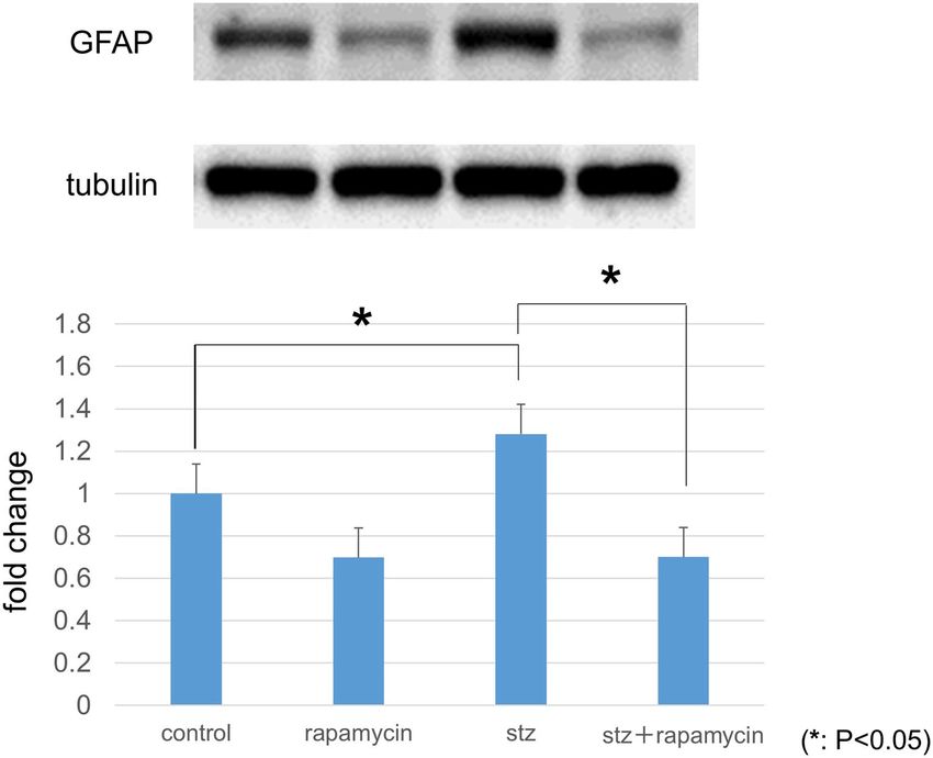
www.nature.com/scientificreports/
Cell cultures. A rat retinal Müller cell line (TR-MUL5) was obtained from Fact, Inc., Sendai, Japan29,30, and
primary human retinal microvascular endothelial cells (HRMECs) were purchased from Cell Systems Corpora-
tion, Kirkland, WA, USA. The TR-MUL5 cells and the HRMECs were cultured in Dulbecco’s Modified Eagle’s
Medium (DMEM) containing a physiological concentration of 5.5 mM glucose supplemented with 10% fetal
bovine serum (FBS) at 37 °C in a humidified atmosphere of 5% CO2/air. TR-MUL5 cells at passages 18 and 21,
and HRMECs at passages 4 and 6 were used in this study. These cells were maintained in DMEM that contained
5.5 mM glucose. Before reaching confluence, the media was changed to the control media (5.5 mM glucose in
DMEM) or the high glucose media (25 mM glucose in DMEM, D6429) to compare ROS levels, as described
later. Detailed procedures and treatment protocols for each experiment are described in subsequent sections.
Immunostaining of cultured Müller cells. To determine the effect of the mTOR inhibitor rapamycin
on the expression of VEGF and GFAP in the TR-MUL5 cells under high glucose condition, the cells that were
incubated in high glucose (25 mM) media with and without rapamycin were examined by immunocytochemis-
try. After fixation by 4% formaldehyde, the cells were incubated with primary antibodies of mouse monoclonal
anti-VEGF (sc-7269, 1:100, Santa Cruz Biotechnology, Inc., Dallas, TX, U.S.A.) and rabbit polyclonal anti-GFAP
(AB5804, 1:100, Merck Millipore, Billerica, MA, U.S.A.) overnight at 4 °C. After rinsing by PBS and blocking,
these cells were incubated for 2 h at room temperature in Alexa 594 (A11012) or Alexa 488- (A11001) conju-
gated to the appropriate secondary antibodies (Invitrogen, Carlsbad, CA, U.S.A.) diluted by 1:500. Nuclei were
stained with 4′,6-diamidino-2-phenylindole (DAPI, 1:1000, Dojindo, Inc. Kumamoto, Japan). The processed
samples were photographed with a fluorescence microscope (BZ-X700, Keyence, Osaka, Japan).
Flow cytometry analyses of ROS formation in müller cells and HRMECs. We examined the gen-
eration of ROS in cultured Müller cells (TR-MUL5) and HRMECs by treating them with hydroethidine and
measuring ethidium fluorescence by flow cytometry (EC800 cell analyzer, SONY, Tokyo, Japan). After a few pas-
sages of TR-MUL5 and HRMECs, the media was changed to the control media (5.5 mM glucose in DMEM) or
the high glucose media (25 mM glucose in DMEM, D6429) with 10% fetal bovine serum (FBS). The cells were
incubated overnight at 37 °C with or without rapamycin (1 μM) under physiological (5.5 mM) and high-glucose
(25 mM) conditions for 48 h. The cells were cultured in a medium lacking 10% FBS and were serum-deprived
overnight before the following FACS assay. Cells were harvested via trypsinization, centrifuged at 1000×g for
5 min, re-suspended in phenol red-free DMEM, and incubated with hydroethidine, a fluorogenic probe, for
20 min at 37 °C. Cell densities were adjusted to 2.0 × 10⁵ cells/mL. Hydroethidine is oxidized by ROS to form
ethidium, a fluorescent product, which is then retained intracellularly to allow a semi-quantitative estimation
of intracellular ROS levels31,32. The intracellular levels of ROS were measured using an EC800 cell analyzer at
488 nm excitation and 590–610 nm emission wavelengths. The acquisition and analysis software programs on
the EC800 was used to acquire and quantify the fluorescence intensities. This assay was performed to determine
the levels of ROS generated by the cultured cells in control and high glucose media in the presence or absence
of rapamycin (1 μM). In addition, the cultured Müller cells (TR-MUL5) were also incubated overnight with or
without VEGF (5 ng/mL) and rapamycin under physiological (5.5 mM) and high-glucose (25 mM) conditions to
determine ROS production by VEGF and the effects of rapamycin on this ROS production. The ethidium fluo-
rescence in TR-MUL5 cells exposed to VEGF and/or rapamycin under physiological (5.5 mM) and high-glucose
(25 mM) conditions was also measured by flow cytometry.
Statistical analyses. The means and standard deviation of the means were calculated. Unless otherwise
noted, two-tailed Student’s t-tests were used. The level of significance was set at P < 0.05. Multiple groups in
Figs. 4 and 5 were compared ANOVA analysis; Bonferroni correction.
Ethical approval. All applicable guidelines of ARVO Statement for the care and use of animals were fol-
lowed. Our experimental protocols conformed to the Animal Research: Reporting In Vivo Experiments
(ARRIVE) guidelines23.
All procedures performed in studies involving animals were in accordance with the ethical standards of the
Osaka Medical College Committee on the Use and Care of Animals (Approval number: 2019-100) at which the
studies were conducted.
Results
Immunohistochemistry of retinal slices. Compared with the nondiabetic control rats, immunoreactiv-
ity to VEGF was increased in the STZ-induced diabetic rats. As shown in Fig. 1, VEGF immunoreactivity was
seemed to be higher in the diabetic rat retinas than in the controls. Injection of rapamycin into the nondiabetic
rats seemed to decrease VEGF immunoreactivity. In the diabetic retina, VEGF expression seemed to be intensi-
fied mainly in these internal retinal layers. Rapamycin likely decreased the intensity in diabetic rats.
Immunoreactivity to GFAP seemed to be higher in the diabetic rat retina than in the control retina. The
observed longitudinal pattern of immunoreactivity to GFAP in the inner plexiform layer (IPL), the inner nuclear
layer (INL), the outer plexiform layer (OPL), and the outer nuclear layer (ONL) suggested that these GFAP
expression may involve Müller cells. Double staining with VEGF and GFAP demonstrated that the expression
of VEGF was co-localized with GFAP expression especially in the diabetic rat retinas. Rapamycin seemed to
inhibit the increases in GFAP expression in the diabetic retina.
Scientific Reports | (2021) 11:1885 | https://doi.org/10.1038/s41598-021-81482-3 3
Vol.:(0123456789)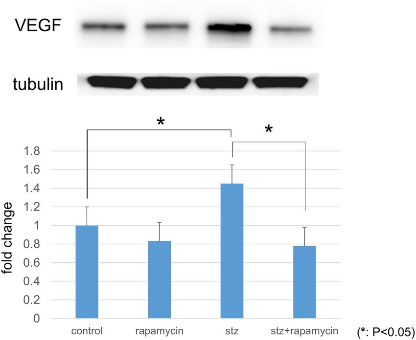
www.nature.com/scientificreports/

                                            Figure 1.  Representative photomicrographs of retinal tissues stained immunohistochemically with anti-VEGF
                                            (green) and anti-GFAP (red) antibodies. VEGF immunoreactivity was higher in the diabetic retina sections than
                                            in the control retinas, and expression of VEGF was colocalized with GFAP.

                                            Figure 2.  Immunostaining of VEGF and GFAP in TR-MUL5 cells. Expression of VEGF and GFAP in
                                            cells cultured in high glucose medium (HG) seemed to be reduced by the mTOR inhibitor rapamycin
                                            (HG + rapamycin).

                                            Immunostaining of cultured Müller cells. Representative photomicrographs of immunostaining with
                                            anti-VEGF (green) and anti-GFAP (red) in the TR-MUL5 cells are shown in Fig. 2. Immunoreactivities to VEGF
                                            and GFAP in high glucose condition seemed to be depressed by the mTOR inhibitor rapamycin.

          Scientific Reports |   (2021) 11:1885 |               https://doi.org/10.1038/s41598-021-81482-3                                              4

Vol:.(1234567890)
www.nature.com/scientificreports/
Figure 3. Protein levels of VEGF in retinas determined with Western blotting. VEGF expression was
significantly higher in retinas obtained from STZ-induced diabetic rats than from nondiabetic controls. The
increased VEGF expression was suppressed by treatment with the mTOR inhibitor rapamycin (P < 0.05, n = 3
each).
Protein levels of VEGF and GFAP in retinas by western blotting. The protein levels of VEGF
in the STZ-induced diabetic retina were significantly up-regulated to 145.2% compared to the control retina
(P < 0.0001). These increases were significantly suppressed by rapamycin (P < 0.0001) (Fig. 3, n = 3 each).
Protein levels of GFAP in the diabetic retina were significantly higher (28.1%) than in the control retina
(P = 0.0004). The increases were significantly suppressed by rapamycin (P < 0.0001) (Fig. 4, n = 3 each).
Flow cytometry analyses of intracellular production of ROS in TR‑MUL5 and HRMECs. Fig-
ure 5 shows the effects of rapamycin (1 μM) treatment on hydroethidine fluorescence in cultured rat Müller
cells (TR-MUL5) under physiological (5.5 mM) and high-glucose (25 mM) conditions. Flow cytometry analyses
showed significant increase in intracellular levels of ROS in TR-MUL5 under the high- glucose condition, and
these increases were significantly suppressed by rapamycin. This figure also shows the effects of rapamycin on
VEGF in ROS production of TR-MUL5 under physiological (5.5 mM) and high-glucose (25 mM) conditions.
We performed this assay to determine the effect of rapamycin on the ROS production of cultured Müller cells
(TR-MUL5) exposed to VEGF. Flow cytometry analyses showed significant increases in the intracellular levels
of ROS in TR-MUL5 cells exposed to VEGF, and these increases were significantly suppressed by rapamycin
(P < 0.0018, the Bonferroni correction).
Figure 6 shows the intracellular ROS levels in HRMECs exposed to rapamycin under physiological (5.5 mM)
and high-glucose (25 mM) conditions. Significant increases in the ROS levels in HRMECs under high-glucose
condition were observed, and these increases were significantly suppressed by rapamycin (P < 0.0083, the Bon-
ferroni correction).
Discussion
A key finding of this study is that the diabetes-induced increase in VEGF expression in the diabetic rat retina is
inhibited by the mTOR inhibitor, rapamycin. This enzymatic inhibitor also blocked the up-regulation of GFAP
by glial cells of the diabetic retina. An additional novel observation is that rapamycin inhibits the hyperglycemia-
induced increase in ROS in cultured Müller cells as well as HERMCs. We also found that under hyperglycemic
conditions, the VEGF-induced increase in Müller cell ROS was also suppressed by rapamycin.
The increase in VEGF levels in the diabetic retina have been previously reported19,27,33–35, and this response was
suppressed by rapamycin. Generally, mTOR coordinates protein synthesis, mitochondrial energy production36.
Scientific Reports | (2021) 11:1885 | https://doi.org/10.1038/s41598-021-81482-3 5
Vol.:(0123456789)www.nature.com/scientificreports/
Figure 4. Protein levels of GFAP in retinas determined with Western blotting. GFAP expression was
significantly higher in retinas obtained from STZ-induced diabetic rats than from nondiabetic controls. The
increased GFAP expression was suppressed by rapamycin (P < 0.05, n = 3 each).
Figure 5. ROS levels in cultured Müller cells (TR-MUL5) exposed to rapamycin and/or VEGF under
physiological (5.5 mM) and high-glucose (25 mM) conditions. Flow cytometry analyses showed significant
increases in intracellular ROS levels in TR-MUL5 under a high-glucose condition, and these increases were
significantly suppressed by rapamycin. In addition, flow cytometry analyses showed significant increases in
intracellular levels of ROS in TR-MUL5 exposed to VEGF, and these increases were significantly suppressed by
rapamycin (P < 0.0018, the Bonferroni correction).
Scientific Reports | (2021) 11:1885 | https://doi.org/10.1038/s41598-021-81482-3 6
Vol:.(1234567890)www.nature.com/scientificreports/
Figure 6. ROS levels in primary human retinal microvascular endothelial cells (HRMECs) exposed to
rapamycin under physiological (5.5 mM) and high- glucose (25 mM) conditions. Flow cytometry analyses
showed significant increases in intracellular ROS levels in HRMECs under a high-glucose condition, and these
increases were significantly suppressed by rapamycin (P < 0.0083, the Bonferroni correction).
In STZ-injected hyperglycemic rats which VEGF increases in the retina, this mTOR is a mplified20. In the present
study, rapamycin suppressed the increase of VEGF in the diabetic retina (Figs. 1, 2, 3) and prevented the hyper-
glycemia-induced ROS level in both cultured Müller cells (Fig. 5) and primary HRMECs (Fig. 6). This indicated
that rapamycin seemed to decrease ROS levels by inhibiting the mTOR pathway in retinal Müller cells; however,
we did not assess its effects on non-Müller rat retinal cells. Our results are consistent with co-localization being at
least in part within the Müller cells, and it is not excluded from the present study that some non-Müller cells may
also co-express VEGF and GFAP. In the retina, Müller cells and vascular endothelial cells might express mTOR.
ROS production, oxidative stress, and cellular death can be altered by several signaling pathways triggered
by the hyperglycemia that causes diabetic r etinopathy37. Our data indicated that VEGF-induced increase of the
ROS levels in Müller cells under high-glucose conditions and this increase was suppressed by rapamycin. We
also analyzed the intracellular ROS levels in primary HRMECs which have the potential to cause retinal vascu-
larization observed in human diabetic retinopathy. Our findings indicate that rapamycin might be an effective
alternative drug for the treatment of diabetic macular edema.
We also showed that GFAP, a glial marker, increased in the diabetic retina. Our findings showed that rapa-
mycin can reduce the increase in GFAP expression in the diabetic retina, suggesting that mTOR inhibition may
inhibit glial activation and/or proliferation. In non-diabetic rats, the effect of rapamycin on GFAP levels was
observed as well. Rapamycin might inhibit the activity of retinal glia even also under the physiological condition.
In diabetic retinopathy, neurodegeneration starts even at an early stage of the disorder where reactive gliosis plays
a crucial r ole17,38. It is known that there are two types of mTOR, mTORC1 (mainly retinal ganglion cells) and
mTORC2 (mainly glial cells) in the r etina39–41. The effects of rapamycin in this study seem to be caused mainly
by mTORC2 inhibition in the diabetic rat retina.
A limitation of this study is that although the chief focus was on oft-studied rat models of diabetes, it will be
of importance for future analyses to extend analyses to the retinas of animals and humans exhibiting anatomi-
cal and functional manifestations of diabetic retinopathy. Another possible limitation is that although mTOR is
reported to increase in the retinas of rats with experimental d iabetes20, this remains to be confirmed in retinas
of rats made hyperglycemic by STZ for 8 weeks. Thirdly, although FACS was used to quantify ROS in cultured
retinal Müller cells and HRMECs, it remains to be demonstrated in future studies that rapamycin suppresses
excessive ROS production in vivo within the diabetic retina.
In conclusion, our findings from in vivo and in vitro experiments showed that the mTOR inhibitor, rapamy-
cin, blocks the up-regulation of VEGF and GFAP in the diabetic rat retina and also inhibits the hyperglycemia-
induced increase in ROS in cultured Müller cells and HERMCs. These findings suggest that rapamycin may be
a potential candidate drug for ameliorating retinal complications in diabetes.
Received: 24 June 2020; Accepted: 7 January 2021
References
1. Targher, G., Bertolini, L., Zoppini, G., Zenari, L. & Falezza, G. Increased plasma markers of inflammation and endothelial dysfunc-
tion and their association with microvascular complications in Type 1 diabetic patients without clinically manifest macroangiopa-
thy. Diabet. Med. 22, 999–1004. https://doi.org/10.1111/j.1464-5491.2005.01562.x (2005).
2. Kohner, E. M. Microvascular disease: What does the UKPDS tell us about diabetic retinopathy?. Diabet. Med. 25(Suppl 2), 20–24.
https://doi.org/10.1111/j.1464-5491.2008.02505.x (2008).
3. Rask-Madsen, C. & King, G. L. Vascular complications of diabetes: Mechanisms of injury and protective factors. Cell Metab. 17,
20–33. https://doi.org/10.1016/j.cmet.2012.11.012 (2013).
4. Whitmire, W., Al-Gayyar, M. M., Abdelsaid, M., Yousufzai, B. K. & El-Remessy, A. B. Alteration of growth factors and neuronal
death in diabetic retinopathy: What we have learned so far. Mol. Vis. 17, 300–308 (2011).
5. Busch, S. et al. Systemic treatment with erythropoietin protects the neurovascular unit in a rat model of retinal neurodegeneration.
PLoS ONE 9, e102013. https://doi.org/10.1371/journal.pone.0102013 (2014).
Scientific Reports | (2021) 11:1885 | https://doi.org/10.1038/s41598-021-81482-3 7
Vol.:(0123456789)www.nature.com/scientificreports/
6. Bandello, F. et al. Retinal layer location of increased retinal thickness in eyes with subclinical and clinical macular edema in diabetes
type 2. Ophthalmic Res. 54, 112–117. https://doi.org/10.1159/000438792 (2015).
7. Zhou, X., Ai, S., Chen, Z. & Li, C. Probucol promotes high glucose-induced proliferation and inhibits apoptosis by reducing reactive
oxygen species generation in Müller cells. Int. Ophthalmol. 39, 2833–2842. https://doi.org/10.1007/s10792-019-01130-8 (2019).
8. Wu, M. Y., Yiang, G. T., Lai, T. T. & Li, C. J. The oxidative stress and mitochondrial dysfunction during the pathogenesis of diabetic
retinopathy. Oxid. Med. Cell. Longev. 2018, 3420187. https://doi.org/10.1155/2018/3420187 (2018).
9. Toda, N. & Nakanishi-Toda, M. Nitric oxide: ocular blood flow, glaucoma, and diabetic retinopathy. Prog. Retin Eye Res. 26,
205–238. https://doi.org/10.1016/j.preteyeres.2007.01.004 (2007).
10. Cheng, Z. et al. Betulinic acid derivatives can protect human Müller cells from glutamate-induced oxidative stress. Exp. Cell Res.
383, 111509. https://doi.org/10.1016/j.yexcr.2019.111509 (2019).
11. Wang, J., Xu, X., Elliott, M. H., Zhu, M. & Le, Y. Z. Müller cell-derived VEGF is essential for diabetes-induced retinal inflammation
and vascular leakage. Diabetes 59, 2297–2305. https://doi.org/10.2337/db09-1420 (2010).
12. Pierce, E. A., Avery, R. L., Foley, E. D., Aiello, L. P. & Smith, L. E. Vascular endothelial growth factor/vascular permeability fac-
tor expression in a mouse model of retinal neovascularization. Proc. Natl. Acad. Sci.U S A 92, 905–909. https://doi.org/10.1073/
pnas.92.3.905 (1995).
13. Lim, Y. C. et al. Prevention of VEGF-mediated microvascular permeability by C-peptide in diabetic mice. Cardiovasc. Res. 101,
155–164. https://doi.org/10.1093/cvr/cvt238 (2014).
14. Ma, M. et al. High glucose-induced TRPC6 channel activation decreases glutamate uptake in rat retinal müller cells. Front. Phar-
macol. 10, 1668. https://doi.org/10.3389/fphar.2019.01668 (2019).
15. Chidlow, G., Wood, J. P. M. & Casson, R. J. Investigations into hypoxia and oxidative stress at the optic nerve head in a rat model
of glaucoma. Front. Neurosci. 11, 478. https://doi.org/10.3389/fnins.2017.00478 (2017).
16. Yabana, T. et al. The relationship between glutathione levels in leukocytes and ocular clinical parameters in glaucoma. PLoS ONE
14, e0227078. https://doi.org/10.1371/journal.pone.0227078 (2019).
17. Simó, R., Stitt, A. W. & Gardner, T. W. Neurodegeneration in diabetic retinopathy: Does it really matter?. Diabetologia 61, 1902–
1912. https://doi.org/10.1007/s00125-018-4692-1 (2018).
18. Antonetti, D. A., Klein, R. & Gardner, T. W. Diabetic retinopathy. N. Engl. J. Med. 366, 1227–1239. https://doi.org/10.1056/NEJMr
a1005073 (2012).
19. Araki, E. & Nishikawa, T. Oxidative stress: A cause and therapeutic target of diabetic complications. J, Diabet. Investig. 1, 90–96.
https://doi.org/10.1111/j.2040-1124.2010.00013.x (2010).
20. Wei, J., Jiang, H., Gao, H. & Wang, G. Blocking mammalian target of rapamycin (mTOR) attenuates HIF-1alpha pathways
engaged-vascular endothelial growth factor (VEGF) in diabetic retinopathy. Cell Physiol. Biochem. 40, 1570–1577. https://doi.
org/10.1159/000453207 (2016).
21. Benjamin, D., Colombi, M., Moroni, C. & Hall, M. N. Rapamycin passes the torch: A new generation of mTOR inhibitors. Nat.
Rev. Drug Discov. 10, 868–880. https://doi.org/10.1038/nrd3531 (2011).
22. Ozdemir, G., Kilinc, M., Ergun, Y. & Sahin, E. Rapamycin inhibits oxidative and angiogenic mediators in diabetic retinopathy.
Can. J. Ophthalmol. 49, 443–449. https://doi.org/10.1016/j.jcjo.2014.07.003 (2014).
23. Kilkenny, C., Browne, W. J., Cuthill, I. C., Emerson, M. & Altman, D. G. Improving bioscience research reporting: The ARRIVE
guidelines for reporting animal research. PLoS Biol. 8, e1000412. https://doi.org/10.1371/journal.pbio.1000412 (2010).
24. Guo, N. et al. The protective effect and mechanism of rapamycin in the rat model of IgA nephropathy. Ren. Fail. 41, 334–339. https
://doi.org/10.1080/0886022x.2019.1577257 (2019).
25. Erlich, S., Alexandrovich, A., Shohami, E. & Pinkas-Kramarski, R. Rapamycin is a neuroprotective treatment for traumatic brain
injury. Neurobiol. Dis. 26, 86–93. https://doi.org/10.1016/j.nbd.2006.12.003 (2007).
26. Oku, H. et al. Tau is involved in death of retinal ganglion cells of rats from optic nerve crush. Invest. Ophthalmol. Vis. Sci. 60,
2380–2387. https://doi.org/10.1167/iovs.19-26683 (2019).
27. Kida, T. et al. Implication of VEGF and aquaporin 4 mediating Muller cell swelling to diabetic retinal edema. Graefe Arch. Clin.
Exp. Ophthalmol. 255, 1149–1157. https://doi.org/10.1007/s00417-017-3631-z (2017).
28. Suzuki, H. et al. Changes in expression of aquaporin-4 and aquaporin-9 in optic nerve after crushing in rats. PLoS ONE 9, e114694.
https://doi.org/10.1371/journal.pone.0114694 (2014).
29. Yanai, N. & Obinata, M. Apoptosis is induced at nonpermissive temperature by a transient increase in p53 in cell lines immortal-
ized with temperature-sensitive SV40 large T-antigen gene. Exp. Cell Res. 211, 296–300. https://doi.org/10.1006/excr.1994.1090
(1994).
30. Abukawa, H. et al. Modulation of retinal capillary endothelial cells by Müller glial cell-derived factors. Mol. Vis. 15, 451–457 (2009).
31. Bindokas, V. P., Jordan, J., Lee, C. C. & Miller, R. J. Superoxide production in rat hippocampal neurons: Selective imaging with
hydroethidine. J. Neurosci. 16, 1324–1336 (1996).
32. Zhao, H. et al. Detection and characterization of the product of hydroethidine and intracellular superoxide by HPLC and limita-
tions of fluorescence. Proc. Natl. Acad. Sci. U S A 102, 5727–5732. https://doi.org/10.1073/pnas.0501719102 (2005).
33. Arden, G. B. & Sivaprasad, S. Hypoxia and oxidative stress in the causation of diabetic retinopathy. Curr. Diabetes Rev. 7, 291–304
(2011).
34. Kida, T. et al. NADPH oxidase-mediated ROS production determines insulin’s action on the retinal microvasculature. Invest.
Ophthalmol. Vis. Sci. 56, 6754–6761. https://doi.org/10.1167/iovs.15-17534 (2015).
35. Islam, M. T. Oxidative stress and mitochondrial dysfunction-linked neurodegenerative disorders. Neurol. Res. 39, 73–82. https://
doi.org/10.1080/01616412.2016.1251711 (2017).
36. Morita, M. et al. mTOR coordinates protein synthesis, mitochondrial activity and proliferation. Cell Cycle 14, 473–480. https://
doi.org/10.4161/15384101.2014.991572 (2015).
37. Volpe, C. M. O., Villar-Delfino, P. H., Dos Anjos, P. M. F. & Nogueira-Machado, J. A. Cellular death, reactive oxygen species (ROS)
and diabetic complications. Cell Death Dis. 9, 119. https://doi.org/10.1038/s41419-017-0135-z (2018).
38. Tzekov, R. & Arden, G. B. The electroretinogram in diabetic retinopathy. Surv. Ophthalmol. 44, 53–60. https://doi.org/10.1016/
s0039-6257(99)00063-6 (1999).
39. Fort, P. E. et al. mTORC1-independent reduction of retinal protein synthesis in type 1 diabetes. Diabetes 63, 3077–3090. https://
doi.org/10.2337/db14-0235 (2014).
40. Nakahara, T., Morita, A., Yagasaki, R., Mori, A. & Sakamoto, K. Mammalian target of rapamycin (mTOR) as a potential therapeutic
target in pathological ocular angiogenesis. Biol. Pharm. Bull. 40, 2045–2049. https://doi.org/10.1248/bpb.b17-00475 (2017).
41. Losiewicz, M. K. et al. mTORC1 and mTORC2 expression in inner retinal neurons and glial cells. Exp. Eye Res. 197, 108131. https
://doi.org/10.1016/j.exer.2020.108131 (2020).
Acknowledgements
The authors thank Prof. Donald Puro for helpful comments on this paper.
Scientific Reports | (2021) 11:1885 | https://doi.org/10.1038/s41598-021-81482-3 8
Vol:.(1234567890)www.nature.com/scientificreports/
Author contributions
T.K., S.O., and T.H. performed the experiments. T.K. wrote the main manuscript text and T.K. and T.H. prepared
all figures. H.O. and T.I. oversaw the project and assisted with the writing of the manuscript.
Funding
Financial support included Grant-in-Aid for Scientific Research (C) (Researcher No. 90610105) from the Japan
Society for the Promotion of Science (Tokyo, Japan), and funding from the Osaka Eye Bank Association Fund
(Osaka, Japan). The sponsors had no role in the design or conduct of this research.
Competing interests
The authors declare no competing interests.
Additional information
Supplementary Information The online version contains supplementary material available at https://doi.
org/10.1038/s41598-021-81482-3.
Correspondence and requests for materials should be addressed to T.K.
Reprints and permissions information is available at www.nature.com/reprints.
Publisher’s note Springer Nature remains neutral with regard to jurisdictional claims in published maps and
institutional affiliations.
Open Access This article is licensed under a Creative Commons Attribution 4.0 International
License, which permits use, sharing, adaptation, distribution and reproduction in any medium or
format, as long as you give appropriate credit to the original author(s) and the source, provide a link to the
Creative Commons licence, and indicate if changes were made. The images or other third party material in this
article are included in the article’s Creative Commons licence, unless indicated otherwise in a credit line to the
material. If material is not included in the article’s Creative Commons licence and your intended use is not
permitted by statutory regulation or exceeds the permitted use, you will need to obtain permission directly from
the copyright holder. To view a copy of this licence, visit http://creativecommons.org/licenses/by/4.0/.
© The Author(s) 2021
Scientific Reports | (2021) 11:1885 | https://doi.org/10.1038/s41598-021-81482-3 9
Vol.:(0123456789)You can also read