Genetic engineering of marine cyanophages reveals integration but not lysogeny in T7-like cyanophages - Nature
←
→
Page content transcription
If your browser does not render page correctly, please read the page content below
www.nature.com/ismej

ARTICLE                  OPEN

Genetic engineering of marine cyanophages reveals integration
but not lysogeny in T7-like cyanophages
Dror Shitrit1, Thomas Hackl 2, Raphael Laurenceau2, Nicolas Raho2, Michael C. G. Carlson                                  1
                                                                                                                              , Gazalah Sabehi1, Daniel A. Schwartz             1
                                                                                                                                                                                 ,
                                            ✉
Sallie W. Chisholm 2 and Debbie Lindell 1

© The Author(s) 2021

    Marine cyanobacteria of the genera Synechococcus and Prochlorococcus are the most abundant photosynthetic organisms on earth,
    spanning vast regions of the oceans and contributing significantly to global primary production. Their viruses (cyanophages)
    greatly influence cyanobacterial ecology and evolution. Although many cyanophage genomes have been sequenced, insight into
    the functional role of cyanophage genes is limited by the lack of a cyanophage genetic engineering system. Here, we describe a
    simple, generalizable method for genetic engineering of cyanophages from multiple families, that we named REEP for
    REcombination, Enrichment and PCR screening. This method enables direct investigation of key cyanophage genes, and its
    simplicity makes it adaptable to other ecologically relevant host-virus systems. T7-like cyanophages often carry integrase genes and
    attachment sites, yet exhibit lytic infection dynamics. Here, using REEP, we investigated their ability to integrate and maintain a
    lysogenic life cycle. We found that these cyanophages integrate into the host genome and that the integrase and attachment site
    are required for integration. However, stable lysogens did not form. The frequency of integration was found to be low in both lab
    cultures and the oceans. These findings suggest that T7-like cyanophage integration is transient and is not part of a classical
    lysogenic cycle.

    The ISME Journal; https://doi.org/10.1038/s41396-021-01085-8

INTRODUCTION                                                                                for other viruses in the environment [30]. Some of these genes are
Marine cyanobacteria of the genera Synechococcus and Prochlor-                              conserved in multiple cyanophage types [16, 18, 21, 22] whereas
ococcus are abundant primary producers that span vast areas of                              others have no database match, and are termed “ORFans” [30, 31].
the oceans [1]. Their viruses (cyanophages) are also highly                                    Elucidating cyanophage gene functions and their role in
abundant [2–4] and influence cyanobacterial ecology and evolu-                               infection is key for understanding their ecological and evolu-
tion [5–7], as well as global biogeochemical cycles [8–10]. All                             tionary interactions with their cyanobacterial hosts. Heterologous
currently known cyanophages are tailed, double-stranded DNA                                 expression experiments [32–34], in-vitro biochemical assays
viruses belonging to the Myoviridae, Podoviridae and Siphoviridae                           [32, 35] and comparisons between phage strains with different
families [3, 11–15]. Over the last two decades, dozens of                                   gene contents [28, 36] have shed light on the function of some
cyanophage genomes from isolates and metagenomes have been                                  cyanophage genes. However, to elucidate the role of these genes
sequenced [13–21]. Analyses of these genomes have contributed                               in the infection process and to determine their contribution to
insights into the general properties and lifestyle of marine                                cyanophage fitness and evolution it is necessary to generate
cyanophages. For example, the T4-like cyanophages from                                      phage mutants and directly compare infection between phages
the Myoviridae family and the T7-like cyanophages from the                                  with and without the gene of interest. Thus, a genetic engineering
Podoviridae family share sets of core genes with the Escherichia coli                       system that is simple and easily transferable between host-virus
T4 and T7 phages, respectively [13, 15, 18, 22]. These genes are                            systems will provide a powerful tool for investigating the function
involved in virion formation, DNA replication and packaging,                                of a wide range of genes in a suite of ecologically relevant
transcription and other fundamental processes of the lytic                                  cyanophage-cyanobacterial systems.
infection cycle. In addition, cyanophage genomes contain many                                  Lysogeny is a well-known phenomenon in which a virus
intriguing and unusual features. In particular, they encode a                               remains in a non-lytic state within the host, typically after
variety of “auxiliary metabolic genes” (AMGs) [10, 14, 22–25] of                            integrating its DNA into the host chromosome. After integration,
(cyano)bacterial origin, including those linked to photosynthesis                           phages can remain latent within the host cell as prophages until
[26, 27], carbon metabolism [28] and nutrient utilization [18, 29], as                      an environmental trigger leads to their excision from the genome
determined from homology-derived functional assignments.                                    and entry into a lytic cycle inside the cell [37]. The presence of
Furthermore, a large reservoir of diverse genes with no putative                            the prophage suppresses infection by additional phages of the
function is present in cyanophage genomes [16–18, 22], as found                             same strain and can sometimes even prevent infection by other

1
 Faculty of Biology, Technion—Israel Institute of Technology, Haifa, Israel. 2Department of Civil and Environmental Engineering, Department of Biology, Massachusetts Institute of
Technology, Cambridge, MA, USA. ✉email: dlindell@technion.ac.il

Received: 14 March 2021 Revised: 2 August 2021 Accepted: 3 August 2021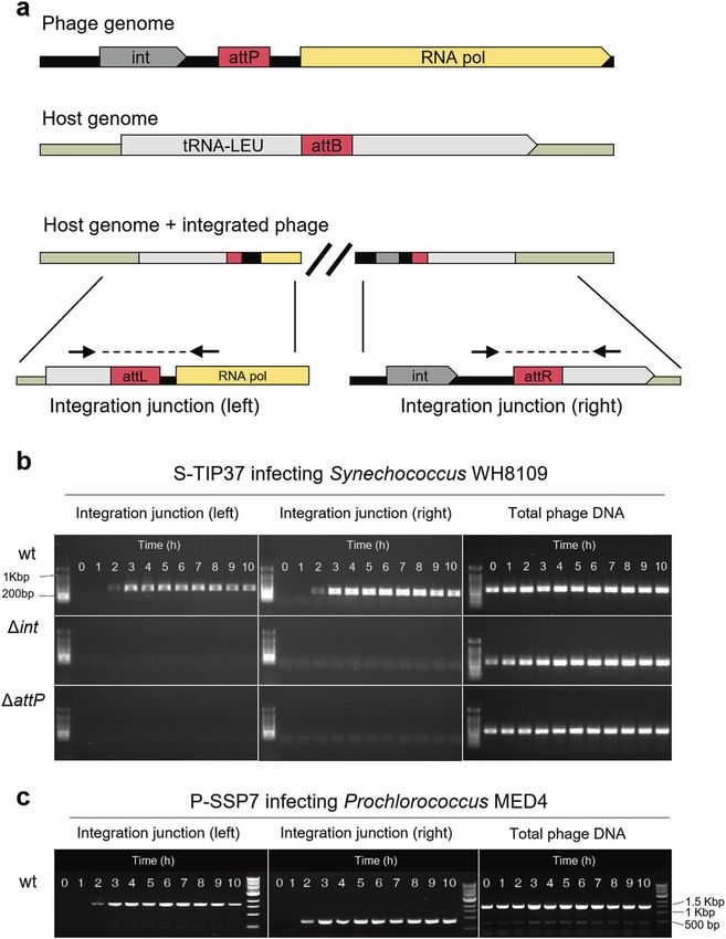
D. Shitrit et al.
                  2
                      phage strains [38]. This superinfection immunity is a result of the         on replicative plasmids inside cyanobacterial hosts (Fig. 1a).
                      transcriptional repression of phage lytic genes [39] or, in some            Recombination templates consist of two regions, 200–300 bp long
                      cases, inhibition of adsorption by additional phages [40].                  (Table 1), that are homologous to those flanking the target gene in
                         In recent years, the potential importance of lysogeny in the             the phage genome. This length of homologous regions was
                      marine environment has come to the fore and its extent and                  chosen to be long enough to increase the probability for
                      significance is under debate [37, 41, 42]. Integrated phages have            homologous recombination but without cloning an entire phage
                      been observed in many sequenced bacterial genomes [41–43] and               gene into the recombination template, which may be lethal when
                      single cells from the environment [41]. Integrase genes, used for           introduced into the host. A short foreign DNA TAG sequence
                      integration, are commonly found in viromes and cellular                     (20–60 bp long) is positioned between the homologous regions
                      metagenomes [42, 44, 45] and their presence is often considered             and replaces the target gene with minimal interruption to the
                      to infer the ability to integrate and partake in a lysogenic lifestyle      compact phage genome. This TAG sequence is used later for
                      [42, 45]. Integration and excision are key processes mediating              detection of recombinant phages by PCR. The recombination
                      horizontal gene transfer that shape the genomes of phage and                templates are cloned into a broad host-range, mobilizable plasmid
                      microbial populations in the oceans [37, 46], including cyano-              in E. coli and inserted into marine Synechococcus by conjugation
                      phages and cyanobacteria [27, 47, 48].                                      [56] to produce recombination host strains (see “Methods” for
                         T7-like cyanophages are generally described as lytic, due to             details). Infection of a recombination host with the wild-type
                      their resemblance to the lytic archetype Escherichia coli phage T7          phage results in lysates with a mixture of wild-type and
                      and their lytic behavior during infection [5, 49, 50]. Thus, it is          recombinant phages (recombinant-containing lysates).
                      surprising that many T7-like cyanophages carry an integrase gene,              We tested this recombination system for cyanophages belong-
                      in some cases followed by putative attachment sites (attP)                  ing to the two major cyanophage families and investigated loci
                      [13, 16, 17], two lysogeny-associated features that are known to            not expected to be essential for phage reproduction. The host
                      facilitate site-specific integration in lysogenic phages and are             strain used was Synechococcus sp. strain WH8109. The generation
                      absent from T7 itself. This raises the possibility of lysogeny in the       of recombinant-containing phage lysates was successful for all
                      T7-like cyanophages. However, intact prophages are absent from              three cyanophages tested: S-TIP37 and Syn5 from the T7-like
                      marine unicellular Synechococcus and Prochlorococcus isolates,              cyanophages, and Syn9 from the T4-like cyanophages (Table 1).
1234567890();,:

                      despite evidence for remnants of phage integration [47, 51, 52]. In         Recombinant phages were produced at an average frequency of
                      addition, efforts to isolate marine Synechococcus or Prochlorococ-          1.31 × 10⁻³ (0.1%) for five recombinant phages, ranging from
                      cus lysogens of any type have failed [3, 5, 52], making it unclear          3.61 × 10⁻⁴ to 3.71 × 10⁻³, as determined by quantitative PCR
                      whether this phenomenon occurs for cyanophages in these                     (qPCR) (Table 1). This range of frequencies was observed for
                      cyanobacteria.                                                              various deletion and insertion sizes and was not dependent on the
                         Here, we developed a method for genetic engineering of                   family or strain of phage investigated (Table 1). These recombina-
                      cyanophages that is highly effective for two distinct and dominant          tion frequencies were high enough to suggest that screening,
                      cyanophage families that infect marine Synechococcus and                    rather than selection systems, could be used to isolate recombi-
                      Prochlorococcus, the T7-like and T4-like cyanophages. Employing             nant phages. For comparison, recombination rates reported
                      this method, we provide experimental evidence for the integra-              previously for other phages range from nearly 10−3 [57] to as
                      tion of T7-like cyanophages into their host’s genome despite a              low as 10−9 [58].
                      lytic infection cycle, and show that both the integrase gene and
                      attP site are required for this process. We found that the frequency        A generalizable method for the screening and isolation of
                      of integration was low both in the lab and the oceans. Stable               recombinant phages
                      lysogens of T7-like cyanophages were not found, however, despite            The next stage in a genetic engineering method is the isolation of
                      repeated attempts in an experimental setup designed to obtain               recombinants. Although the recombination step described above
                      them even in the absence of superinfection immunity. These                  is quite efficient, detecting recombinant phages by PCR screening
                      results suggest that lysogeny does not occur in T7-like cyano-              thousands of individual plaques would be very time consuming
                      phages and that integration is less regulated and more transient            and labor intensive. To overcome this problem, we developed a
                      than that described for classical lysogenic phages.                         procedure that combines an enrichment step for recombinant
                                                                                                  phages and a high throughput PCR screening procedure for
                                                                                                  detection of recombinant-enriched lysates (Fig. 1b). In this
                      RESULTS AND DISCUSSION                                                      procedure, hundreds of host cultures, grown in 96-well plates,
                      REEP: a genetic engineering system for cyanophages                          are infected with subsamples of the recombinant-containing
                      Genetic engineering of phages requires two stages. The first is the          lysate at a concentration of 5–10 phages per well. This
                      generation of mutations in the phage genome and the second is               concentration is pre-determined using the most probable number
                      the isolation of the mutant phages. The genetic engineering                 method (MPN) and ensures lysis of all infected wells. Though most
                      system presented here is based on naturally high rates of                   wells contain only wild-type phages, wells with a recombinant
                      homologous recombination between plasmid and phage DNA                      phage generate lysates that are generally enriched more than
                      during infection. Following recombination, an enrichment proce-             100-fold to contain 10–20% recombinants rather than the ~0.1%
                      dure and a PCR screen are used for the isolation of mutant phages.          recombinants present in the initial recombinant-containing lysate
                      We name this method REEP for REcombination, Enrichment and                  (Table 1).
                      PCR screening. This is a simple and generalizable method                       Once lysis of the infected cultures is visible, a PCR screen is
                      particularly well suited for ecologically relevant host-virus systems       performed to detect wells enriched with recombinant phages that
                      for which genetic tools are limited.                                        are identified based on the inserted TAG sequence (Fig. 1b). It is
                                                                                                  generally sufficient to screen 3–10 96-well plates (180–600 wells,
                      High frequency of recombination between cyanophage and                      infected with 900–6000 phages) to detect several wells highly
                      plasmid DNA during infection                                                enriched with recombinant phages. PCR can be performed on
                      The first stage of any genetic engineering system is the generation          each plate individually or after pooling samples from each row of
                      of mutations. In the REEP method, as in various other phage                 10 plates into a single plate and screening the 10 individual wells
                      genetic engineering methods [53–55], phage mutations are                    that went into a positive pooled well. This reduces the number of
                      generated during infection by homologous recombination                      PCR reactions tenfold, although may reduce the sensitivity of
                      between cyanophage DNA and recombination templates carried                  detecting recombinant phages. The contents of the PCR-positive

                                                                                                                                                     The ISME Journal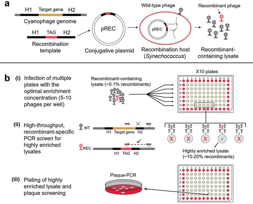
D. Shitrit et al.
                                                                                                                                                   3

Fig. 1 Diagram of the REEP genetic engineering system for cyanophages. a The process of producing recombinant-containing phage
lysates. A recombination template is composed of homologous regions (H1, H2, 200–300 bp each) that flank the target gene to be deleted in
the phage genome and a short TAG sequence that will be inserted in place of the target gene. This is cloned into the pREC plasmid and
inserted into Synechococcus to produce the recombination host. This host is infected with wild-type cyanophages. Homologous
recombination that occurs during infection produces a lysate containing wild-type (black) and recombinant (red) phages. b Enrichment
and screening for the isolation of recombinant phages. (i) Multiple 96-well plates are infected with the recombinant-containing lysate at the
optimal enrichment dilution, which is 5–10 phages per well. (ii) Once lysis is complete, the plates are screened by recombinant-specific PCR to
detect recombinant phages based on their TAG sequence. Wells in which a recombinant phage was present among the initial 5–10 phages
used for infection will now contain a lysate that is highly enriched with recombinants (>100-fold) and will be detected in the PCR screen. (iii)
The highly enriched lysates from PCR positive wells are plated for plaque screening and isolation of the recombinant phage.

wells are plated on cyanobacterial lawns for plaque formation and         phages, similar to those used in bacteria, such as antibiotic
~10–50 individual plaques are screened by PCR to obtain an                resistance genes. Specialized systems have been developed for
isolated recombinant phage that is further purified and taken for          well-studied model phages that often rely on strain-specific
validation (see “Methods”).                                               characteristics [62, 63]. For example, host genes that are essential
   The REEP method’s power lies in its simplicity. It requires only       for phage infection but not for host growth can be used as
the ability to insert a plasmid into the bacterial host and               selection markers. CRISPR-Cas based methods have been pro-
homologous recombination, which is a frequent process in many             posed as a generalizable approach for counter-selection against
DNA and RNA viruses [59, 60]. Even for viruses where homologous           wild-type phages [53, 64], as have the use of reporter genes to
recombination is less frequent, it can be enhanced by expression          isolate recombinant phages by visual screening [54, 55]. However,
of a recombination system commonly used for genetic engineer-             setting up such systems can be difficult and time consuming,
ing (e.g., the Lambda RED system) [61]. Furthermore, this method          especially as they require expression of foreign genes in the host,
does not require any specialized equipment or tools and the               often entailing the development and optimization of suitable
enrichment and screening is relatively high throughput when               regulatory elements and codon optimization. This is no trivial task
using 96-well plates and multi-channel pipettes. It generates             when working with non-model organisms with a limited set of
mutants with minimal disruption to the phage genome, using a              molecular tools and is likely to require specific adjustments for
very short sequence to replace the target gene, and can                   each host-virus system of interest. In addition, overexpression of a
potentially be used to generate scarless mutations (i.e., without         reporter gene, as well as the insertion of a large fragment into the
insertion of a TAG, with screening done based on differences in           compact phage genome, may have an effect on the phage
amplicon size). The REEP method was successfully employed on all          phenotype beyond that of the deletion of the gene of interest,
three cyanophages tested in this study and was efficient in                confounding results from infection experiments with such mutant
generating a range of deletions and insertions in cyanophages             phages.
belonging to two distinct phage families with different character-           Prior to the development of the REEP method, we examined
istics, including vast differences in genome size and content. This       several other approaches for isolation of recombinant phages
demonstrates the flexibility of this method, a valuable feature for        without success (see supplementary information for details). First,
studying a set of phage strains, as often done in ecologically            three different CRISPR-Cas based methods were constructed for
relevant systems.                                                         targeting and eliminating non-recombinant cyanophages [53, 64].
   Many methods for genetic engineering of bacteriophages exist           None of these systems were efficient against cyanophages,
[61–63]. Although the generation of mutations is quite straightfor-       despite being adjusted for use in cyanobacteria. In parallel, we
ward, a major obstacle is the isolation of the mutant phages. This        attempted to optimize several genes encoding fluorescent
is largely due to the lack of generalizable selection markers for         proteins for use as reporter genes for visual screening of

The ISME Journal
D. Shitrit et al.
4
recombinant phages, yet none were successful in generating a
detectable signal above the autofluorescence of the cyanobacter-
ial host. Another approach was screening for recombinant phages
by plaque-hybridization [65], which led to the detection of
Recombinant phage
8.48E-04 ± 8.61E-04
3.71E-03 ± 3.11E-04
3.61E-04 ± 2.09E-04
6.75E-04 ± 1.77E-04
9.57E-04 ± 3.77E-04
1.31E-03 ± 1.22E-03
recombinants, but did not yield viable recombinant cyanophages
frequency (n = 3)
despite many attempts. Last, cloning of whole phage genomes
into fosmid vectors and inserting them into Synechococcus by
conjugation were attempted, failing in the step of transfer into
Synechococcus. These unsuccessful attempts further emphasize
the advantages of our straightforward method. It has no gene
expression involved, is free of potentially confounding effects on
mutant phage fitness and phenotype, and is unlikely to require
Homology length (left/
changes to be transferred to other host-virus systems. These
advantages make this method an ideal candidate for genetic
engineering in viruses for which genetic methods are currently
unavailable, especially for ecologically relevant host-virus systems
266/290 bp
234/234 bp
268/268 bp
251/226 bp
251/274 bp
that are not well characterized and have limited genetic tools.
Average
right)
T7-like phage integration into marine cyanobacteria
Integrase genes are often detected in the genomes of cultured
phages and bacteria as well as in viromes and metagenomes
Inserted sequence (size)
collected from the oceans [13, 16, 22, 37, 43]. Based on this, many
marine phages are hypothesized to have the ability to integrate
into the genome of their hosts and to have a lysogenic life cycle
Frequency of recombinant phages in recombinant-containing lysates for various mutations in different cyanophages.
DR-TAG1 (23 bp)
DR-TAG1 (23 bp)
proCAT (961 bp)
pRL-TAG (57 bp)
pRL-TAG (57 bp)
[37, 46]. With a genetic system in hand, we set out to
experimentally test these hypotheses for T7-like cyanophages.
This entailed assessing whether: (1) T7-like cyanophages integrate
into the genome of their host; (2) the integrase gene is functional
and, together with the attP site, mediate integration; and (3) stable
integration ensues and superinfection immunity is conferred.
Site-specific integration of T7-like cyanophages occurs during
Non-coding region downstream of
Non-coding region downstream of
attP - putative integration (67 bp)
lytic infection
The presence of an integrase gene and a putative phage
attachment site (attP) in T7-like cyanophages was first reported
gp17 - integrase (508 bp)
Deleted sequence (size)
gp11- integrase (836 bp)
for the P-SSP7 phage that infects Prochlorococcus sp. strain MED4
[13]. The potential attP site is downstream of the integrase gene
and is identical to a region in a tRNA-Leu gene of the host.
Similarly, we found an integrase gene and a comparable putative
gp45 (22 bp)
attP site in the genome of S-TIP37 (Fig. 2a), a T7-like cyanophage
gp6 (14 bp)
that infects Synechococcus sp. strain WH8109 [66].
We first tested the ability of these two wild-type phages to
integrate into the genomes of their hosts over a 10-hour period
after phage addition (Fig. 2b, c). Intracellular genomic DNA was
subjected to PCR assays designed to amplify across expected
Phage type
integration junctions when the phages integrate into the
cyanobacterial tRNA-Leu gene. Both the S-TIP37 and the P-SSP7
T7-like
T7-like
T7-like
T7-like
T4-like
phages were found to integrate into their hosts’ genomes at the
expected site from 2 hours after infection onwards (Fig. 2b, c).
We then tested whether the integrase gene and the attP sites
Parent cyanophage strain
are functional and are involved in the integration process. This is
particularly important because marine cyanobacteria, including
both Synechococcus WH8109 and Prochlorococcus MED4, code for
integrase genes [47, 67], and host enzymes have been shown to
mediate phage integration in other systems [68]. We generated
two mutant strains of S-TIP37 in which either the integrase gene
(Δint) or the attP site (ΔattP) were knocked out. Neither left nor
S-TIP37
S-TIP37
S-TIP37
right integration junctions were formed during infection with
Syn5
Syn9
these mutant S-TIP37 phages (Fig. 2b). These results indicate that
both the integrase gene and the attP site are essential for
integration.
Interestingly, infection by both wild-type and integrase-mutant
S-TIP37 phages exhibited infection dynamics and phage progeny
S-TIP37-ΔattP
production typical of classic lytic phages (Fig. S1). The length of
strain name
S-TIP37-Δint
S-TIP37-Cm
the latent period and phage fitness were no different in the
Syn5-Δint
Syn9-Δ6.5
Mutant
Table 1.
mutant and wild-type S-TIP37 phages (Fig. S1, S2a). Nutrient
availability has previously been proposed to affect the frequency
of lysogeny [37, 69, 70]. We therefore hypothesized that fewer
The ISME JournalD. Shitrit et al.
5
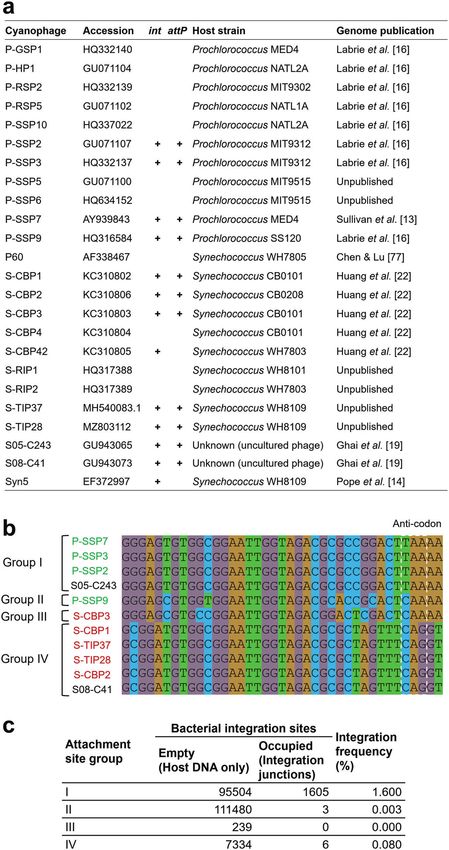
Fig. 2 Site-specific integration of T7-like cyanophages. a Organization of the integration elements found in cyanophages S-TIP37 and P-
SSP7, and in their host genomes. Integration junctions formed due to phage integration and the location of the primers (black arrows) used to
detect them by PCR are also shown. Int- integrase, attP/B – attachment sites on the phage/bacterial genomes, respectively, attL/attR – left and
right attachment sites after integration, respectively. Primers for total phage DNA assessment are located elsewhere on the phage genome
(see Table S5). b Evidence for phage integration from a PCR assay for detection of integrated phages during infection of Synechococcus
WH8109 with S-TIP37 phage strains: wild-type (wt), integrase deleted (Δint) and attP deleted (ΔattP) strains. c Integration of wild-type phage
P-SSP7 (wt) during infection of Prochlorococcus MED4. All results shown here are representative of 3 independent experiments.
phages would be produced (i.e., lower fitness would result) if more Thus, the frequency of integrated phages was low, ranging from
cells were lysogenized under nutrient deprivation. However, even 0.3–1% of infected cells for most of the infection cycle (Fig. 3c).
when the host was grown in nutrient-depleted medium, wild-type These findings indicate that integration is a relatively rare event,
and integration mutants demonstrated similar progeny produc- and may be transient, rather than a fundamental part of lytic
tion (Fig. S2). infection in these cyanophages.
The above results raised the question of whether phage
integration is a basic part of the lytic infection cycle, occurring S-TIP37 integration is not part of a classic lysogenic cycle
in every infected cell. To assess this we quantified the number of Hallmark features of lysogeny include stable integration into the
infected cells containing an integrated phage using the iPolony host genome, a regulatory system that controls the lytic/lysogenic
method, a method capable of detecting single molecules of viral life cycles and superinfection immunity [37, 38, 72]. To test
DNA in infected cells [71]. The percentage of integrated phages whether integrated T7-like cyanophages possess these features,
correlated with the number of infected cells, increasing as more we first searched for homologs of repressors of lytic infection
phages adsorbed and entered the cell and decreasing as phages present in known lysogenic phages. No such genes were
were released and cells lysed (Fig. 3). However, phage integration identified in T7-like cyanophages. Next, we performed a series
occurred only in a small fraction of cells, ranging from 0.04 to of experiments to test whether stable integration of S-TIP37
0.32%, even though up to 60% of the cells were infected (Fig. 3b). occurs when infecting its Synechococcus WH8109 host. Classic
The ISME JournalD. Shitrit et al.
6
prophage-containing colonies that express the chloramphenicol
resistance gene to grow. Roughly 3 × 10⁹ cells were plated in five
different experiments, which was expected to yield up to 9 × 10⁵
colonies considering the integration frequency of ~0.3% found in
the previous experiment (Fig. 3b). However, no chloramphenicol
resistant colonies were found. Note that the proCAT gene provides
chloramphenicol resistance in our pREC replicating plasmids
(Fig. 1a) and when expressed from the chromosome of
Synechococcus strains [75]. This suggests that despite phages
integrating into the genome of their host this integration is not
stable or that superinfection immunity is not provided by the
integrated phage.
We then asked whether stable integration occurs but super-
infection immunity is not provided, i.e. that prophage-containing
cells are killed as a result of subsequent infections. To address this
question, we repeated the above experiments in a manner that
removes and separates free phages from host cells, preventing a
second round of infection (see “Methods”). Here too, no colonies
grew on chloramphenicol plates, despite growth of colonies in the
absence of chloramphenicol, which confirmed that a second
round of infection and lysis was indeed prevented. Finally, to rule
out the possibility that prophages exist in these latter colonies but
did not provide resistance to chloramphenicol, we tested 2000
colonies for the presence of prophages by PCR and found none.
These findings indicate that stable integration did not occur,
irrespective of whether superinfection immunity ensues or not.
We note that these experiments do not enable us to ascertain
whether superinfection immunity is provided for the short period
during which phages are integrated into the host genome.
Our combined findings suggest that the site-specific integration
observed here in T7-like cyanophages is not a part of a classical
lysogenic lifestyle, but rather is a transient process. This intrinsic
lack of stable prophage integration may be due to the absence of
regulatory genes that, in well studied lysogen systems, suppress
Fig. 3 Dynamics of S-TIP37 infection and integration. a Phage
growth curve of the wild-type S-TIP37 phage during infection of the expression of lytic cycle genes and maintain the phage
Synechococcus WH8109. b Dynamics of infected cells (blue circles, genome integrated into the host genome [38, 72]. Their absence
left Y-axis) and of cells containing integrated wild-type phages can result in spontaneous excision of the integrated phage and
(yellow triangles, right Y-axis; only left junctions were tested), induction of the lytic cycle, or in lysis by a superinfecting phage. In
determined by single-cell, solid phase PCR. Percent infection and addition, lytic infection may be initiated by the integrated phage
integration were calculated from the number of polonies divided by from within the host genome. Such a phenomenon was recently
the number of cells added to the slide. c Average ratio of cells with reported in the classic lysogenic phage Lambda, with lytic
integrated phages to total number of infected cells, calculated by infection observed from a phage integrated into the host genome,
dividing average % integration by average % infection from each
possibly due to low expression of the CII gene coding for the
timepoint. Integration at timepoint zero was below the limit of
detection and was thus not used to calculate the integration/ repressor of the lytic cycle [76].
infection ratio. No integration was detected with the S-TIP37
integrase mutant, which served as a negative control in this T7-like cyanophage integration in the oceans
experiment. We sought to estimate the frequency of integrated T7-like
cyanophages in cyanobacteria in the oceans. First, we searched
for attP sites in 24 publicly available genomes in this group of
lysogens (e.g., Lambda lysogens of E. coli) can be isolated based cyanophages [14, 16, 19, 22, 77] by searching for homologous
on their superinfection immunity, by simply plating an infected sequences downstream of integrase-containing cyanophage
culture on agar plates and testing the colonies that grow for the genomes in cyanobacteria (Table S1). We found that of the 13
presence of a prophage [73]. However, when we attempted this cyanophage genomes with integrase genes, 11 also carried an
previously for various marine cyanobacteria and their integrase- attP site (Fig. 4). Consistent with the attP sites for P-SSP7 [13] and
containing cyanophages (including P-SSP7 and S-TIP37), none of S-TIP37 (this study), all attP sites were perfect matches for regions
the resistant colonies were found to contain a prophage [6, 74]. in cyanobacterial tRNA-Leu genes, which can thus be considered a
We therefore predicted that if stable integration occurs it is general cyanobacterial attachment (attB) site. Based on their
extremely rare. To overcome this we used our genetic system to sequences, we divided the attP sites into four groups, with groups
generate a strain of S-TIP37 carrying a chloramphenicol resistance I and II belonging to phages that infect Prochlorococcus and
gene (proCAT) in a non-coding region of the phage genome. This groups III and IV belonging to phages that infect Synechococcus
engineered phage strain, S-TIP37-Cm (Table 1), was allowed to (Fig. 4b). Second, we analyzed publicly available metagenomic
adsorb to Synechococcus WH8109 cultures which were then plated datasets from six different projects (Table S2), searching for reads
on chloramphenicol-containing agar plates. If stable lysogens that of cyanobacterial origin that contain these T7-like cyanophage
provide superinfection immunity exist then chloramphenicol attachment sites. The number of reads containing “empty”
resistant colonies are expected to grow. This approach allows integration sites (i.e., cyanobacterial DNA only) or phage-host
testing a vast number of phage-infected cells because it integration junctions were used to calculate the integration
eliminates colonies that originate from non-infected cells as well frequency. For the two dominant groups, I and IV, an integration
as from spontaneous phage resistant mutants, allowing only frequency of 1.6% and 0.08% was found, respectively, whereas
The ISME JournalD. Shitrit et al.
7
Fig. 4 Lysogeny- associated elements present in T7-like cyanophage genomes. a Analysis of available T7-like cyanophage genomes for the
presence of integrase genes (int) and attP sites. b Sequence comparison and group division of the attP sites found in the analyzed genome.
Cyanobacterial host genus is indicated by font color of the phage name (green- Prochlorococcus, red- Synechococcus, black- unknown).
c Frequency of integration found for cyanophages in the different attachment site groups in oceanic samples.
The ISME JournalD. Shitrit et al.
8
group II and III showed only 0.003% and 0% integration, Transient integration in up to 1% of infections may enhance the
respectively (Fig. 4c). Thus, similar to lab findings, T7-like frequency of the introduction of new genes into microbial hosts as
cyanophage integration is rare but detectable in the oceans. well as the capture of microbial genes by viruses in the
Cell density has been hypothesized to impact lysogeny. environment.
However, this is still under debate due to conflicting evidence
(summarized by Brum et al. [78]). Nonetheless, our combined
findings of rare integration at high cell densities in lab MATERIALS AND METHODS
experiments (5 × 10⁷ cells·ml−1) as well as at the considerably Culture growth
lower cyanobacterial densities found in the oceans (ranging from Cultures of Synechococcus sp. strain WH8109 were grown in artificial
~104 to 3 × 10⁵ cells·ml−1 [1]) argues against density being a factor seawater (ASW) medium [85], with modifications as described elsewhere
for the T7-like cyanophages. [86], at 21 °C and a light intensity of 30 µmol photons m−2 s−1.
Prochlorococcus sp. strain MED4 was grown in Pro99 medium [87], at
18 °C and 70 µmol photons m−2 s−1. All cultures were grown under a 14:10
light-dark cycle. Chlorophyll a fluorescence (excitation at 440 nm, emission
CONCLUSIONS at 660 nm) was used as a proxy for cell density and was measured in 96-
The discovery of phages as highly abundant and diverse members well plates using a Synergy Mx Microplate Reader (Biotek). Pour-plating
of the community in virtually every habitat on Earth [9, 10, 79, 80] was performed as previously described [56], using the ASW growth
has fueled recognition of their ecological importance. Over the medium with low-melting point agarose at a final concentration of 0.28%
past two decades there has been an explosion in the sequence [87, 88] and the addition of 1 mM sodium sulfite [88]. For isolation of
information available for environmental viruses. Yet a substantial cyanobacterial colonies, a heterotrophic helper strain, Alteromonas sp.
portion of viral genomes contains genes of unknown function EZ55, was added to the pour-plate mixture [89]. All bacterial and phage
strains used in this work are listed in Table S3. Experimentation was carried
[16, 17, 22, 30, 36], as well as other genes with putative functions
out at cell abundances of ~3–5 × 10⁷ cells/ml for Synechococcus WH8109
that were unexpected in viral genomes, leading to intriguing and Prochlorococcus MED4.
hypotheses as to their role in infection [13–15]. The lack of a
genetic system broadly applicable to ecologically relevant models
has precluded gaining fundamental understanding of the many Lysate preparation and phage growth curves
novel features of these significant members of the natural world. Lysates were prepared by infecting cyanobacterial cultures at early
logarithmic phase with phage from either a single plaque or a pre-
This method now provides the opportunity, in the post-genomics existing liquid lysate. Once full lysis of the culture was observed, lysates
era, to assess the function of interesting virus genes directly in the were filtered through 0.22 µm syringe filters (Millex-GV, Millipore) and
infection process and their impact on virus fitness and evolution. stored in glass tubes at 4 °C. Plaque assays were used to determine
Thus, the REEP genetic engineering method developed here infective phage concentrations. Lysate samples were diluted and plated in
places marine cyanophages among the few model organisms that agarose plates containing cyanobacteria at concentrations sufficient to
are culturable, genetically engineerable and ecologically produce lawns, allowing formation of plaques. Phage growth curve
significant. experiments were performed by infecting cyanobacterial cultures at a
The possibility of integration and a potential lysogenic lifestyle in multiplicity of infection (MOI) of 1-3, with subsamples collected over a
T7-like phages was first hypothesized 15 years ago for T7-like period of 10 h. To measure the concentration of infective phages in the
extracellular medium samples of 0.1 ml were collected, added to 0.9 ml of
cyanophages [13, 14, 16, 22] and more recently for T7-like medium, and filtered through a 0.22 µm syringe filter to remove
pelagiphages [81, 82], based on the presence of integrase genes cyanobacterial cells and the filtrate used in plaque assays.
and attP sites in their genomes. In our first use of the REEP method
we addressed this hypothesis for the T7-like cyanophages. Our
findings unequivocally indicate that these elements are used for Construction of plasmids and bacterial conjugation
The plasmid pDS-proCAT (Fig. S3) was used as a backbone for the
integration into the host genome even though these are essentially construction of all recombination plasmids used in this work. This
lytic phages. Indeed, no evidence for stable integration nor lysogeny replicative plasmid is a derivative of the broad host-range mobilizable
was found, although we cannot categorically rule it out for all vector pRL1342 (received as a gift from Peter Wolk [90]). It was modified by
members of this family or under all environmentally relevant replacing the original chloramphenicol resistance gene with a
conditions. Nonetheless, the absence of intact prophages in cyanobacterial-optimized copy, proCAT. This gene was codon-optimized
cyanobacterial genomes, including hundreds of single-cell genomes for expression in several Prochlorococcus strains and was found to
from diverse environments [83, 84], provides independent evidence work well in Synechococcus strains. For efficient expression, the proCAT
for the lack of classical lysogeny in these phages. Furthermore, gene was fused to the rnpB promoter and the atpB ribosome-binding site
lysogeny-derived immunity was never the mechanism of resistance of Prochlorococcus MED4. Recombination templates were constructed
either by the PCR overlap extension method [91] or by a two-step
among hundreds of experimentally selected resistant cyanobacterial cloning procedure. A short TAG sequence (20–60 bp) was inserted
strains, including against the P-SSP7 [6] and S-TIP37 [74] phages, between two regions of homology on the phage genome (~250 bp
even though integration occurs at a 1000-fold greater frequency long) (H1 and H2) that flank the region to be deleted (Fig. 1a).
than the appearance of resistance mutations [6]. Thus, although the Recombination plasmids (pREC) were inserted by electroporation into E.
presence of the integrase gene facilitated integration when an attP coli strain DH10b, an efficient recipient of large plasmids. Plasmids were
site was also present, we caution against automatically equating the extracted from PCR-positive colonies using a miniprep kit
presence of the gene with a lysogenic lifestyle. (NucleoSpin Plasmid EasyPure, MACHEREY-NAGEL) and transformed into
The physiological or evolutionary benefit that marine T7-like E. coli SM10 or S17.1, conjugation donor strains carrying the λpir gene [92].
Conjugation of plasmids into Synechococcus was done as described
cyanophages obtain from carrying these elements and transiently
previously [56] and resulting recombination hosts were grown in the
integrating into host genomes remains uncertain. One feasible presence of 1 μg/ml chloramphenicol. Plasmids used in this study are listed
scenario is that integration provides a safety net for phages that in Table S4.
cannot complete their lytic cycle. This may be important when
environmental conditions are not suitable for lytic infection.
Production of recombinant-containing cyanophage lysates
Alternatively, this process may allow defective phages to be
and estimating recombinant frequencies
rescued through complementation or recombination with other Recombination plasmids were conjugated into Synechococcus sp. strain
phages infecting the same host cell, and perhaps even facilitate WH8109 to form recombination hosts. Recombination hosts were infected
the formation of new genetic combinations, accelerating phage with wild-type phages at a low MOI (~0.01). After lysis of the cultures,
evolution. Clearly, phage integration is important for host and lysates were filtered and the 0.2 µm filtrate was stored as described above.
phage evolution through horizontal gene transfer [37, 46]. The presence of recombinant cyanophages was tested by PCR, using
The ISME JournalD. Shitrit et al.
9
primers that anneal to the TAG sequence and specifically detect cytometer (BD) based on their chlorophyll a fluorescence (excitation at
recombinants (Table S5) using 2x Taq PCR mix (Tiangen) and 2 μl of the 488 nm, emission at 580/30 nm) and forward scatter to remove free
lysate (in 20 μl reactions). Recombinant phage frequency was determined phages and excess glutaraldehyde [71]. A known number of cells
for several representative lysates by real-time qPCR (see below). (quantified using the same flow cytometer) were added to iPolony
reactions designed for intracellular DNA detection [71]. Briefly, a mixture of
cells, PCR reagents and polyacrylamide gel were poured into custom-made
Isolation of recombinant cyanophages by enrichment and PCR glass slides (11.6 µl final volume). A 15 min cell lysis step was followed by
screening in-gel PCR in a slide thermal cycler (DNA Engine with dual-block slide
Recombinant phages were enriched in 96-well plates containing cultures chamber, Bio-Rad). An acrydite-modified primer (Table S5) covalently binds
of Synechococcus sp. strain WH8109 at early logarithmic growth phase. The one PCR strand to the gel, creating local DNA amplification clusters,
optimal phage enrichment concentration was determined by the MPN termed polonies (PCR-colonies) [96]. Polonies were hybridized with a Cy5-
method. Synechococcus cultures grown in a 96-well plate were infected modified fluorescently labeled DNA probe (Table S5) after removal of the
with 10-fold serial dilutions of the recombinant-containing lysate, with unbound PCR strand. Hybridized polonies were identified using a GenePix
each dilution used to infect a single row of wells. The optimal enrichment 4000B microarray scanner (Axon Instruments). Plasmids harboring the
concentration is operationally determined to be the highest tenfold serial tested amplicons (with left integration junction or total phage) served as
dilution at which lysis is observed in an entire row of wells. This positive controls for PCR amplification and efficiency. Percent integration
concentration is typically 5–10 phages per well, and is used in the was adjusted for the efficiency of detection of single virus genome copies
subsequent enrichment step to infect the internal 60 wells of three to ten which was 33% for S-TIP37 [71].
96-well plates containing Synechococcus cultures. Once the cultures lyse,
the wells were screened by PCR for the presence of recombinant phages in
20 µl reactions using 10 µl Taq PCR MasterMix (Tiangen), 0.4 µM primers Testing for stable integration and superinfection immunity
and 2 µl of phage lysate (without prior filtration). Samples were run on The S-TIP37-Cm strain was constructed by inserting the proCAT chlor-
1.5% agarose gels with ~0.2 µg·ml−1 ethidium bromide in large electro- amphenicol resistance gene into the non-coding region between g45 and
phoresis trays containing 200 wells each. Lysates from PCR-positive wells g46 of the S-TIP37 phage. This engineered phage strain was used to infect
were filtered through a 0.22 µm syringe filter and plated. Plaques Synechococcus WH8109 cultures at a high MOI of 3. After a 2 h incubation
were purified twice and verified by PCR to obtain pure recombinant to allow phage adsorption, 108 cells per plate were plated in the presence
phage mutants. Full genome sequencing of all phage strains (Table S3), of 1 μg·ml−1 chloramphenicol. A total of 3 × 10⁹ cells was plated in 5
including the wild-type parental strains, were performed to verify the independent experiments.
mutation and to ensure an identical genetic background to the wild- To differentiate between lack of stable integration and lack of
type phage. superinfection immunity, in the event of no chloramphenicol resistant
colonies, we carried out the above experiment after physically preventing
subsequent infection of cells by free phages. Cells were diluted to 105
Real-time quantitative PCR to measure recombination cells·ml−1 and sorted to remove free phages [71] remaining after the 2 h
frequencies adsorption step. In addition, cells were plated at various dilutions with and
Recombination frequencies were determined by real-time qPCR, in which without chloramphenicol to physically distance potential prophage-
the copy-number of recombinant and non-recombinant phage DNA was containing cells from phages newly released by lytically infected cells. A
quantified. DNA templates were extracted from phage lysates using Wizard total of 15 plates were plated at each dilution in 3 independent
PCR Preps DNA Purification Resin and Minicolumns (Promega), as experiments. The presence of colonies in the absence of chloramphenicol
previously described [93]. Template DNA and primers (0.2 µM each) were indicated that this procedure prevented infection by remaining or newly
added to LightCycler 480 SYBR Green I Master mix (Roche) and amplified released phages. The efficiency of plating of these phage-exposed cultures
using a LightCycler 480 Real-Time PCR System (Roche). The LightCycler was approximately one-third of the non-infected control cultures, as
480 software was used to calculate the number of cycles required to reach expected from the percent infection measured two hours post infection
the optimal fluorescence threshold (Ct). All amplicons tested by qPCR were (see Results and Discussion). Over 2000 of these colonies, that grew in the
first amplified by PCR and cloned into plasmids using the TOPO TA Cloning absence of chloramphenicol and originated either from non-infected cells
Kit (Thermo Fischer). Plasmid DNA carrying the tested amplicons was or from lysogens, were tested for the presence of prophage by PCR.
extracted and used to generate calibration curves for calculation of the
absolute DNA copy number in each qPCR reaction.
Fitness assays
To assess the fitness of the different phage strains, Synechococcus cultures
Detection of integrated phages using PCR were infected over a 2-day period beginning with a low MOI of 0.001.
Cells were collected over the course of phage growth curve experiments Phage abundances were determined by plaque assay at the beginning and
for PCR detection of phage DNA integrated into the cyanobacterial end of experiment. A 2-day period was used to ensure host cultures did
genome. Samples were filtered onto polycarbonate 0.22 µm pore-size not become a limiting resource for phage production [97]. Phage fitness,
membrane filters (GE), washed twice with growth medium to remove free expressed as the number of population doublings per day, was calculated
phages, and washed once with 3 ml of preservation solution (10 mM Tris, as (log2(Nt/N0))/t, where t is the assay time in days and N0 and Nt are the
100 mM EDTA, 0.5 M NaCl, pH 8) [94, 95]. The filter was flash-frozen in phage concentrations at the beginning and end of the experiment,
liquid nitrogen and stored at −80 °C. A heat lysis method was used to respectively [97, 98]. Growth of the host culture was measured by
extract DNA from cells collected on filters [94, 95]. Filters were chlorophyll a fluorescence (see above). For growth under nutrient
resuspended in 10 mM Tris-HCL solution (pH = 8), shaken in a bead- deprivation (Fig. S2), a low-nutrient ASW medium was used, containing
beater (Mini-BeadBeater, Biospec) for 2 min (3450 oscillations/min) without all ingredients but at 10% nutrient concentrations for NaNO3, NaH2PO4,
beads. Samples were heated at 95 °C for 15 minutes and the supernatant NaHCO3 and trace metals. Exponentially growing Synechococcus cultures
collected. For detection of integrated phage DNA, 2 µl of intracellular were centrifuged for 5 min at 5200 RCF, and resuspended in this low
template DNA were used in 20 µl PCRs (Taq PCR MasterMix, Tiangen), as nutrient ASW medium. Nutrient deprivation was verified by reduced
described above. Primers specific for detection of total phage DNA and chlorophyll a fluorescence relative to growth in 100% ASW medium.
integration junctions were used (Table S5).
Analyses of attP sites and estimation of cyanophage
Quantification of infected cells and integrated phages using integration frequencies in the oceans
the iPolony method To identify potential attachment sites between cyanophages and their
For single-cell quantification of integrated phages, cultures of the putative hosts we compared 727 Prochlorococcus and Synechococcus
Synechococcus WH8109 host were infected with the wild-type S-TIP37 genomes (Table S1) against 12 integrase-carrying cyanophage genomes
phage (MOI = 3). The S-TIP37 integrase mutant was used as a negative (Fig. 4) using BLAST (BLAST v2.6.0 + : blastn -task blastn -reward 1 -penalty −4
control in infection experiments with the host strain. Samples from phage- -gapopen 5 -gapextend 2 -perc_identity 94 -evalue 10e-5). We filtered hits of
infected cultures were collected at different time points, fixed in 0.1% 39-44 bp length, collapsed those overlapping by at least 20 bp, and only kept
glutaraldehyde, incubated for 20 min in darkness, flash-frozen in liquid the ones present in more than 10 host genomes and directly downstream of
nitrogen and stored at −80 °C. Samples were sorted using an Influx flow the integrase gene (bedtools v2.27.1) [99]. From the curated alignment of the
The ISME JournalD. Shitrit et al.
10
corresponding sequences, we identified four distinct 39 bp attachment site 20. Ma Y, Allen LZ, Palenik B. Diversity and genome dynamics of marine cyano-
motifs, all of which match the first half of a tRNA-Leu gene. phages using metagenomic analyses. Environ Microbiol Rep. 2014;6:583–94.
To estimate how many cyanobacteria in the wild carry integrated 21. Sabehi G, Shaulov L, Silver DH, Yanai I, Harel A, Lindell D. A novel lineage of
phages, we screened 1093 publicly available metagenomic libraries from myoviruses infecting cyanobacteria is widespread in the oceans. Proc Natl Acad
six different marine sequencing projects [100–104] (Table S2) for reads Sci USA. 2012;109:2037–42.
containing a match to one of the four attachment site motifs with up to 22. Huang S, Zhang S, Jiao N, Chen F. Comparative genomic and phylogenomic
two differences (BBMap v38.16: bbduk.sh k = 39 edist = 2) [105]. We analyses reveal a conserved core genome shared by estuarine and oceanic
determined the locations of putative attachment sites in the cyanobacter- cyanopodoviruses. PLoS One. 2015;10:1–17.
ial host genomes by BLAST best hit against the four attachment site motifs 23. Ignacio-espinoza JC, Sullivan MB. Phylogenomics of T4 cyanophages: lateral
with a minimum bit score of 52. Based on those data, we generated a gene transfer in the ‘core’ and origins of host genes. Environ Microbiol.
database of fragments containing the attachment site motif and 500 bp of 2012;14:2113–26.
up- and downstream flanking regions for all combinations of cyanobac- 24. Crummett LT, Puxty RJ, Weihe C, Marston MF, Martiny JBHH. The genomic
teria and phages containing the same site. We then mapped the pre- content and context of auxiliary metabolic genes in marine cyanomyoviruses.
screened metagenomic reads to this database using bwa mem v0.7.16a- Virology. 2016;499:219–29.
r1181 and filtered the results using a custom Perl script (alignment length 25. Millard AD, Zwirglmaier K, Downey MJ, Mann NH, Scanlan DJ. Comparative
≥ 100 bp, aligned read fraction ≥95%, overlap with flanking regions genomics of marine cyanomyoviruses reveals the widespread occurrence
≥ 20 bp) [106, 107]. Finally, we obtained counts for “empty” and “occupied” of Synechococcus host genes localized to a hyperplastic region: Implica-
integration sites by counting reads matching to “host-att-host” and “host- tions for mechanisms of cyanophage evolution. Environ Microbiol.
att-phage”/“phage-att-host” fragments using a custom R script. 2009;11:2370–87.
26. Mann NH, Cook A, Millard A, Bailey S, Clokie M. Bacterial photosynthesis genes
in a virus. Nature 2003;424:741–741.
REFERENCES 27. Lindell D, Sullivan MB, Johnson ZI, Tolonen AC, Rohwer F, Chisholm SW. Transfer
1. Flombaum P, Gallegos JL, Gordillo RA, Rincón J, Zabala LL, Jiao N, et al. Present of photosynthesis genes to and from Prochlorococcus viruses. Proc Natl Acad Sci
and future global distributions of the marine cyanobacteria Prochlorococcus and USA. 2004;101:11013–8.
Synechococcus. Proc Natl Acad Sci USA. 2013;110:9824–9. 28. Puxty RJ, Millard AD, Evans DJ, Scanlan DJ. Viruses inhibit CO2 fixation in the
2. Goldin S, Hulata Y, Baran N, Lindell D. Quantification of T4-like and T7-like most abundant phototrophs on Earth. Curr Biol. 2016;26:1585–9.
cyanophages using the polony method show they are significant members of 29. Kelly L, Ding H, Huang KH, Osburne MS, Chisholm SW. Genetic diversity in
the virioplankton in the North Pacific Subtropical Gyre. Front Microbiol. cultured and wild marine cyanomyoviruses reveals phosphorus stress as a
2020;11:1210. strong selective agent. ISME J. 2013;7:1827–41.
3. Waterbury JB, Valois FW. Resistance to co-occurring phages enables marine 30. Yin Y, Fischer D. Identification and investigation of ORFans in the viral world.
Synechococcus communities to coexist with cyanophages abundant in seawater. BMC Genomics. 2008;9:24.
Appl Environ Microbiol. 1993;59:3393–9. 31. Clokie MR, Millard AD, Mann NH. T4 genes in the marine ecosystem: studies of
4. Marston MF, Sallee JL. Genetic diversity and temporal variation in the cya- the T4-like cyanophages and their role in marine ecology. Virol J. 2010;7:291.
nophage community infecting marine Synechococcus species in Rhode Island’s 32. Rihtman B, Bowman‐Grahl S, Millard A, Corrigan RM, Clokie MRJ, Scanlan DJ.
coastal waters. Appl Environ Microbiol. 2003;69:4639–47. Cyanophage MazG is a pyrophosphohydrolase but unable to hydrolyse magic
5. Clokie MRJ, Mann NH. Marine cyanophages and light. Environ Microbiol. spot nucleotides. Environ Microbiol Rep. 2019;11:448–55.
2006;8:2074–82. 33. Dammeyer T, Bagby SC, Sullivan MB, Chisholm SW, Frankenberg-Dinkel N.
6. Avrani S, Wurtzel O, Sharon I, Sorek R, Lindell D. Genomic island variability Efficient phage-mediated pigment biosynthesis in oceanic cyanobacteria. Curr
facilitates Prochlorococcus -virus coexistence. Nature. 2011;474:604–8. Biol. 2008;18:442–8.
7. Marston MF, Pierciey FJ, Shepard A, Gearin G, Qi J, Yandava C, et al. Rapid 34. Roitman S, Hornung E, Flores-Uribe J, Sharon I, Feussner I, Béjà O. Cyanophage-
diversification of coevolving marine Synechococcus and a virus. Proc Natl Acad encoded lipid desaturases: Oceanic distribution, diversity and function. ISME J.
Sci USA. 2012;109:4544–9. 2018;12:343–55.
8. Fuhrman JA. Marine virueses and their biogeochemical and ecological effects. 35. Thompson LR, Zeng Q, Kelly L, Huang KH, Singer AU, Stubbe J, et al. Phage
Nature. 1999;399:541–8. auxiliary metabolic genes and the redirection of cyanobacterial host carbon
9. Suttle CA. Marine viruses - major players in the global ecosystem. Nat Rev metabolism. Proc Natl Acad Sci USA. 2011;108:E757–64.
Microbiol. 2007;5:801–12. 36. Puxty RJ, Perez-Sepulveda B, Rihtman B, Evans DJ, Millard AD, Scanlan DJ.
10. Breitbart M, Bonnain C, Malki K, Sawaya NA. Phage puppet masters of the Spontaneous deletion of an “ORFanage” region facilitates host adaptation in a
marine microbial realm. Nat Microbiol. 2018;3:754–66. “photosynthetic” cyanophage. PLoS One. 2015;10:e0132642.
11. Suttle CA, Chan AM. Marine cyanophages infecting oceanic and coastal strains 37. Howard-Varona C, Hargreaves KR, Abedon ST, Sullivan MB. Lysogeny in nature:
of Synechococcus: abundance, morphology, cross-infectivity and growth char- mechanisms, impact and ecology of temperate phages. ISME J.
acteristics. Mar Ecol Prog Ser. 1993;92:99–109. 2017;11:1511–20.
12. Sullivan MB, Waterbury JB, Chisholm SW. Cyanophages infecting the oceanic 38. Ranade K, Poteete AR. Superinfection exclusion (sieB) genes of bacteriophages
cyanobacterium Prochlorococcus. Nature. 2003;424:1047–51. P22 and λ. J Bacteriol. 1993;175:4712–8.
13. Sullivan MB, Coleman ML, Weigele P, Rohwer F, Chisholm SW. Three Pro- 39. Fogg PCM, Allison HE, Saunders JR, McCarthy AJ. Bacteriophage Lambda: a
chlorococcus cyanophage genomes: Signature features and ecological inter- paradigm revisited. J Virol. 2010;84:6876–9.
pretations. PLoS Biol. 2005;3:0790–806. 40. van Houte S, Buckling A, Westra ER. Evolutionary ecology of prokaryotic immune
14. Pope WH, Weigele PR, Chang J, Pedulla ML, Ford ME, Houtz JM, et al. Genome mechanisms. Microbiol Mol Biol Rev. 2016;80:745–63.
sequence, structural proteins, and capsid organization of the cyanophage Syn5: 41. Tuttle MJ, Buchan A. Lysogeny in the oceans: lessons from cultivated model
a “horned” bacteriophage of marine Synechococcus. J Mol Biol. 2007;368:966–81. systems and a reanalysis of its prevalence. Environ Microbiol. 2020;22:4919–33.
15. Weigele PR, Pope WH, Pedulla ML, Houtz JM, Smith AL, Conway JF, et al. 42. Knowles B, Silveira CB, Bailey BA, Barott K, Cantu VA, Cobian-Guëmes AG, et al.
Genomic and structural analysis of Syn9, a cyanophage infecting marine Pro- Lytic to temperate switching of viral communities. Nature. 2016;531:466–70.
chlorococcus and Synechococcus. Environ Microbiol. 2007;9:1675–95. 43. Touchon M, Bernheim A, Rocha EPC. Genetic and life-history traits associated
16. Labrie SJ, Frois-Moniz K, Osburne MS, Kelly L, Roggensack SE, Sullivan MB, et al. with the distribution of prophages in bacteria. ISME J. 2016;10:2744–54.
Genomes of marine cyanopodoviruses reveal multiple origins of diversity. 44. Roux S, Hallam SJ, Woyke T, Sullivan MB. Viral dark matter and virus–host
Environ Microbiol. 2013;15:1356–76. interactions resolved from publicly available microbial genomes. Elife. 2015;4:
17. Huang S, Wang K, Jiao N, Chen F. Genome sequences of siphoviruses infecting e08490.
marine Synechococcus unveil a diverse cyanophage group and extensive phage- 45. Luo E, Eppley JM, Romano AE, Mende DR, DeLong EF. Double-stranded DNA.
host genetic exchanges. Environ Microbiol. 2012;14:540–58. virioplankton dynamics and reproductive strategies in the oligotrophic open
18. Sullivan MB, Huang KH, Ignacio-Espinoza JC, Berlin AM, Kelly L, Weigele PR, et al. ocean water column. ISME J. 2020;14:1304–15.
Genomic analysis of oceanic cyanobacterial myoviruses compared with T4-like 46. Touchon M, Moura de Sousa JA, Rocha EP. Embracing the enemy: the diversi-
myoviruses from diverse hosts and environments. Environ Microbiol. fication of microbial gene repertoires by phage-mediated horizontal gene
2010;12:3035–56. transfer. Curr Opin Microbiol. 2017;38:66–73.
19. Ghai R, Martin-Cuadrado AB, Molto AG, Heredia IG, Cabrera R, Martin J, et al. 47. Coleman ML, Sullivan MB, Martiny AC, Steglich C, Barry K, Delong EF, et al.
Metagenome of the Mediterranean deep chlorophyll maximum studied by Genomic islands and the ecology and evolution of Prochlorococcus. Science.
direct and fosmid library 454 pyrosequencing. ISME J. 2010;4:1154–66. 2006;311:1768–70.
The ISME JournalYou can also read