Double agent indole-3-acetic acid: mechanistic analysis of indole-3-acetaldehyde dehydrogenase AldA that synthesizes IAA, an auxin that aids ...
←
→
Page content transcription
If your browser does not render page correctly, please read the page content below
Bioscience Reports (2021) 41 BSR20210598
https://doi.org/10.1042/BSR20210598

Commentary

Double agent indole-3-acetic acid: mechanistic
analysis of indole-3-acetaldehyde dehydrogenase
AldA that synthesizes IAA, an auxin that aids
bacterial virulence

                                                                                                                                                                                           Downloaded from http://portlandpress.com/bioscirep/article-pdf/41/8/BSR20210598/919617/bsr-2021-0598c.pdf by guest on 29 November 2021
    Ateek Shah1,* ,                Yamini Mathur2,* and                        Amrita B. Hazra1,2
1 Departmentof Chemistry, Indian Institute of Science Education and Research, Dr. Homi Bhabha Road, Pashan, Pune 411008, India; 2 Department of Biology, Indian Institute of
Science Education and Research, Dr. Homi Bhabha Road, Pashan, Pune 411008, India
Correspondence: Amrita B. Hazra (amrita@iiserpune.ac.in)

                                           The large diversity of organisms inhabiting various environmental niches on our planet are
                                           engaged in a lively exchange of biomolecules, including nutrients, hormones, and vitamins.
                                           In a quest to survive, organisms that we define as pathogens employ innovative methods to
                                           extract valuable resources from their host leading to an infection. One such instance is where
                                           plant-associated bacterial pathogens synthesize and deploy hormones or their molecular
                                           mimics to manipulate the physiology of the host plant. This commentary describes one such
                                           specific example—the mechanism of the enzyme AldA, an aldehyde dehydrogenase (ALDH)
                                           from the bacterial plant pathogen Pseudomonas syringae which produces the plant auxin
                                           hormone indole-3-acetic acid (IAA) by oxidizing the substrate indole-3-acetaldehyde (IAAld)
                                           using the cofactor nicotinamide adenine dinucleotide (NAD+ ) (Bioscience Reports (2020)
                                           40(12), https://doi.org/10.1042/BSR20202959). Using mutagenesis, enzyme kinetics, and
                                           structural analysis, Zhang et al. established that the progress of the reaction hinges on the
                                           formation of two distinct conformations of NAD(H) during the reaction course. Additionally, a
                                           key mutation in the AldA active site ‘aromatic box’ changes the enzyme’s preference for an
                                           aromatic substrate to an aliphatic one. Our commentary concludes that such molecular level
                                           investigations help to establish the nature of the dynamics of NAD(H) in ALDH-catalyzed
                                           reactions, and further show that the key active site residues control substrate specificity.
                                           We also contemplate that insights from the present study can be used to engineer novel
                                           ALDH enzymes for environmental, health, and industrial applications.

                                            The immense diversity of life on earth is supported by an intricate give-and-take between organisms
                                         that inhabit various environmental niches. Most known ecosystems host multiple species that engage in
* These authors contributed              an enduring exchange of biomolecules which are classified as positive or negative interactions (bene-
equally to this work.                    ficial/neutral or detrimental to one or more of the species, respectively), and are precisely tailored to
Received: 14 March 2021
                                         meet the nutritional and proliferative needs of the organisms [1,2]. Some of the most fascinating ex-
Revised: 10 July 2021                    amples of interactions among species are where one organism is host to another which is a pathogen,
Accepted: 30 July 2021                   that is, the pathogenic species acquires nutrients and proliferates, while the host suffers the consequences
                                         of the infection. For example, the relationship between Phaeobacter gallaeciensis, a marine bacterium
Accepted Manuscript online:
09 August 2021                           and Emiliania huxleyi, a marine microalga is proposed to be a mutual exchange with the algae provid-
Version of Record published:             ing dimethylsulfoniopropionate (a carbon and sulfur source) to the bacteria and the bacteria providing
24 August 2021                           growth hormones (phenylacetic acid) and antibiotics (tropodithietic acid) in return. In an intriguing turn

© 2021 The Author(s). This is an open access article published by Portland Press Limited on behalf of the Biochemical Society and distributed under the Creative Commons Attribution   1
License 4.0 (CC BY).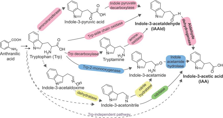
Bioscience Reports (2021) 41 BSR20210598
                                                                                                                                         https://doi.org/10.1042/BSR20210598

           of events, once E. huxleyi begins to senesce, it releases p-coumaric acid which is sensed by P. gallaeciensis. P. gallae-
           ciensis then synthesizes roseobacticides to kill the algae and scavenge nutrients from its victim for its growth [3]. In
           the natural world, there are several innovative means that pathogens use to gain entry into the host. Rabies virus, a
           member of the Rhabdoviridae family, enters into nerve ending-rich muscle and tissues through the bite of an animal
           and then proliferates and travels via nerve cells to the brain causing hydrophobia, hallucinations, and eventually death
           [4]. The blast disease of rice is caused by the fungus Magnaporthe oryzae which produces a flattened, hyphal cell
           from the germinating spore with an infection peg that penetrates the host causing brown diamond-shape lesions on
           the rice leaves [5]. Interestingly, many plant-associated microbes synthesize a plant hormone or its close mimic to
           gain entry and establish themselves by modulating the host plant’s metabolism as well as their own virulence genes
           [6–8]. An extensively studied example of hormone-mediated plant–microbe pathogenesis is the crown gall disease
           caused by the Gram-negative soil bacterium, Agrobacterium tumefaciens. The bacterium senses chemoattractants

                                                                                                                                                                                           Downloaded from http://portlandpress.com/bioscirep/article-pdf/41/8/BSR20210598/919617/bsr-2021-0598c.pdf by guest on 29 November 2021
           such as phenolic compounds released from wounded regions on the plants and initiates transfer of bacterial DNA to
           the host chromosome which hijacks the metabolism in the infected plant cells [9]. A. tumefaciens harbors a special-
           ized tumor-inducing (Ti) plasmid which encodes an array of genes that aid in its transfer, replication, and in overall
           plant–microbe pathogenesis [10]. The T-DNA region of the Ti plasmid is transferred to the plant nuclear DNA, which
           initiates the biosynthesis of plant hormones auxin and cytokinins via the iaaM and iaaH, and ipt pathways, respec-
           tively, which are absent from the host plant (Figure 1). This results in uncontrolled cell proliferation and subsequently
           crown gall tumors [10].
              Another intriguing example is that of Pseudomonas syringae, a bacterial plant pathogen that manipulates hor-
           mone signaling as a means of breaking the host plant defense [8,11]. It gains entry into the tomato plant by producing
           the phytotoxin coronatine, a molecular mimic of the plant hormone jasmonic acid-isoleucine, resulting in the bac-
           terial speck disease [7,12,13]. P. syringae also synthesizes the auxin indole-3-acetic acid (IAA), a common growth
           hormone synthesized by plants for cell enlargement, division, and differentiation and uses it to establish itself in its
           plant host [11,14]. IAA can be synthesized in bacteria from tryptophan as a precursor via five distinct routes, and also
           via tryptophan-independent pathways which mostly yet remain to be characterized in detail [15–18] (Figure 1).
              Three of the tryptophan-dependent pathways result in the formation of a common intermediate
           indole-3-acetaldehyde (IAAld) which is oxidized by an aldehyde dehydrogenase (ALDH) to IAA. ALDHs are
           housekeeping enzymes that play a major role in the detoxification of reactive aldehydes by converting them into their
           corresponding carboxylic acid [20–23]. ALDHs are known to participate in important functions such as polyamine
           catabolism [24], ethanol metabolism [25,26], xenobiotic metabolism [27–29], and plant cell wall ester synthesis
           [30,31], yet, many ALDH-catalyzed reactions remain to be characterized in detail in bacteria.
              In 2018, the Kunkel laboratory reported the identification of six putative ALDHs in the pathogenic P. syringae
           pv. tomato strain DC3000 [11]. Biochemical analysis of these six homologs revealed that AldA, AldB, and AldC were
           capable of producing IAA from IAAld. Further, a high-resolution crystal structure of wildtype AldA was obtained.
           Additionally, 42 classes of genes encoding putative ALDHs in various Pseudomonas species have been recently re-
           ported, expanding the scope of this family of enzymes in bacterial metabolism [23]. The role of AldA was established
           to be a nicotinamide adenine dinucleotide (NAD+ )-dependent IAAld dehydrogenase that produces IAA [11]. In 2020,
           biochemical and structural characterization of AldC by Lee et al. revealed it to be a long-chain aliphatic ALDH [32].
           IAA produced by P. syringae has been reported to promote the virulence in Arabidopsis thaliana by two different
           mechanisms—one, it up-regulates the virulence gene expression in the bacterium, and second, it suppresses salicylic
           acid-mediated plant defenses by activating auxin signaling in the host plant (via the TIR1/AFB auxin co-receptor
           system) [8,11]. Studies with tir1/afb mutants show accumulation of IAA in A. thaliana, resulting in the deregulation
           of plant host signaling. This results in increased host susceptibility to the pathogen’s entry [8] (Figure 2). Targeting
           the arsenal of enzymes that P. syringae uses for infection is one the strategies to suppress its pathogenicity. To this
           end, Zhang et al. have recently reported a detailed study of the enzyme AldA to provide a glimpse into its molecular
           mechanism of action [33].
              The reaction mechanism of AldA can be broadly described in two parts. First, an active site catalytic residue Cys302
           forms a thiohemiacetal intermediate with the IAAld which is followed by a hydride transfer from the aldehyde to the
           NAD+ cofactor resulting in a thioacyl-enzyme intermediate. Second, the active site residue Glu267 activates a water
           molecule for the hydrolysis of the intermediate resulting in the release of the carboxylic acid product and the reduced
           cofactor NADH [11]. Zhang et al. created mutants of these two catalytic residues and other substrate-binding site
           residues to study the mechanism in detail using kinetic and structural tools [33].
              The activity of the wildtype and mutants of AldA were tested with IAAld which is the physiological substrate.
           As expected, enzyme assays using the AldA catalytic residue Cys302 and Glu267 mutants (Cys302 Ala, Glu267 Gln,
           Glu267 Ala) showed no detectable activity. Noticeably, mutations in the aliphatic and aromatic residues lining the

2   © 2021 The Author(s). This is an open access article published by Portland Press Limited on behalf of the Biochemical Society and distributed under the Creative Commons Attribution
    License 4.0 (CC BY).
Bioscience Reports (2021) 41 BSR20210598
https://doi.org/10.1042/BSR20210598
Downloaded from http://portlandpress.com/bioscirep/article-pdf/41/8/BSR20210598/919617/bsr-2021-0598c.pdf by guest on 29 November 2021
Figure 1. Bacterial biosynthesis of auxin
Certain plant-associated bacteria can produce auxin indole-3-acetic acid (IAA) from tryptophan (Trp) as a precursor using one or
more than one of the routes shown here. Among the known Trp-dependent pathways, three routes involving (i) aminotransferase
and indole-3-pyruvate decarboxylase, (ii) Trp-side chain oxidase, and (iii) Trp deacarboxylase and an amine-oxidase converge at
indole-3-acetaldehyde (IAAld) which is then oxidized by aldehyde dehydrogenase (ALDH) to yield IAA (enzymes shown in pink).
Another prominent route involves the intermediate indole-3-acetamide which can be synthesized directly from tryptophan by tryp-
tophan 2-monooxygenase (iaaM), and then converted into IAA via the enzyme indole acetamide hydrolase (iaaH) (enzymes shown in
blue). Alternately, indole-3-acetamide is synthesized via a two-step reaction involving a predicted dehydratase and nitrile hydratase
(enzymes shown in yellow) followed by conversion into IAA as mentioned previously. Finally, some bacteria appear to use the lesser
studied Trp-independent routes, though the precursors appear to be derived from Trp biosynthesis intermediates [16,17]. For ex-
ample, indole-3-acetonitrile, derived from a precursor of Trp yield IAA in a single-step reaction catalyzed by the nitrilase enzyme
(enzyme shown in green). The reactions for which genetic and biochemical evidence of the enzymes involved are yet to discovered
are shown with dashed arrows. Pseudomonas syringae DC3000 genome possesses genes for amine oxidase, nitrilase, indole ac-
etamide hydrolase, ALDH, and a putative monooxygenase [19]. The enzymes shown with dashed arrows have some biochemical
evidence however, the encoding genes are yet unknown. Figure adapted from Spaepen and Vanderleyden (2011) [15] and Duca et
al. (2014) [18].
substrate-binding site of AldA were especially detrimental to the binding of IAA and lowered the catalytic efficiency
significantly. These mutants were also tested for activity with octanal, the preferred substrate of AldC. Interestingly,
the substitution of Phe169 with Trp (the corresponding residue found in AldC at that position) resulted in a higher
turnover with octanal as compared with IAAld, thus leading to a reversal in the substrate preference and catalytic
efficiency of the AldA Phe169 Trp mutant [33]. Such a shift in the preference for chemically distinct substrates arising
from a single point mutation indicates that a small number of precise changes in the active site are likely responsible
for the extensive biochemical scope of enzymes in the ALDH enzyme superfamily.
An interesting feature observed in the mechanism of ALDH enzymes is cofactor isomerization wherein after the
hydride transfer, the nicotinamide ring of the NAD(H) cofactor tilts away from the catalytic cysteine and glutamate
residues [35,36]. Zhang et al., captured the two orientations of the cofactor NAD+ in their crystal structure analysis
of AldA [33]. The ‘extended’ conformation where the nicotinamide half of the NAD+ cofactor is buried in the active
site, with the nicotinamide in close proximity to Cys302 and Glu267 , and the ribose part forming hydrogen bonds with
another active site residue Glu401 was found in the wildtype AldA enzyme. In contrast, a ‘contracted’ conformation
where the NAD+ nicotinamide ring is pulled away from the catalytic site and the nicotinamide-ribose orients towards
an active site residue Gln349 was found in the catalytically dead AldA Cys302 Ala mutant. This movement allows each
half-reaction to occur efficiently, the hydride transfer part followed by the final hydrolysis of the enzyme–product
© 2021 The Author(s). This is an open access article published by Portland Press Limited on behalf of the Biochemical Society and distributed under the Creative Commons Attribution 3
License 4.0 (CC BY).Bioscience Reports (2021) 41 BSR20210598
https://doi.org/10.1042/BSR20210598
Downloaded from http://portlandpress.com/bioscirep/article-pdf/41/8/BSR20210598/919617/bsr-2021-0598c.pdf by guest on 29 November 2021
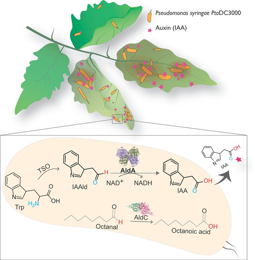
Figure 2. Plant pathogen P. syringae pv. tomato strain DC3000 synthesizes the auxin IAA, a plant growth hormone that
facilitates its entry into the growing plant, and causing wilting and discoloration of the infected parts
AldA is an IAAld dehydrogenase in P. syringae DC3000 which catalyzes the oxidation of IAAld to IAA coupled with reduction of
NAD+ cofactor. Please refer to Figure 1from Zhang et al. (2020) [33] for the detailed mechanism. AldC, another ALDH found in P.
syringae oxidizes a range of long-chain aliphatic aldehydes such as octanal to the corresponding acids. Such aliphatic molecules
have been hypothesized to assist microbes in gaining entry into the host cell and also to act as nutrients for their proliferation
[32,34].
complex which marks the completion of the reaction. A comparison of these two structures provides a glimpse into
the dynamic interactions of the AldA active site residues with NAD(H) through the course of reaction, and highlights
the molecular significance of these interactions in the mechanism of ALDH.
This series of investigations from the Kunkel and Jez labs on P. syringae ALDHs have opened several avenues in the
study of bacterial IAA biosynthesis and their role in plant–microbe interactions. For example, the three characterized
ALDH homologs AldA, AldB, and AldC were found via bioinformatics-based searches and share sufficient sequence
4 © 2021 The Author(s). This is an open access article published by Portland Press Limited on behalf of the Biochemical Society and distributed under the Creative Commons Attribution
License 4.0 (CC BY).Bioscience Reports (2021) 41 BSR20210598
https://doi.org/10.1042/BSR20210598
identity [11]. However, their biochemical characterization show that they have different substrate preferences which
would lead to them playing distinct functional roles in the cells. Obtaining a molecular level picture of the differences
between the ALDH homologs via detailed biochemical characterization with the enzymes and their mutants would
contribute to a bigger picture understanding of their function and physiological roles in P. syringae pathogenesis.
Further, in addition to ALDHs that synthesize IAA from IAAld, the P. synringae DC3000 genome also possesses
genes to convert tryptamine, indole-3-acetamide, and indole-3-acetonitrile into IAA [19] (Figure 1). Additionally, a
homolog of lysine monooxygenase has been hypothesized to synthesize indole-3-acetamide from tryptophan [11].
Functional characterization of these gene products are required to understand the flux of tryptophan to produce IAA
and validate all the operating pathways for auxin biosynthesis in the bacterium. Given the agro-biotechnological and
ecological importance of P. syringae [37,38], mechanistic enzymology studies such as these are important steps for
in-depth understanding of host–microbe interaction and to keep its pathogenicity in check.
Downloaded from http://portlandpress.com/bioscirep/article-pdf/41/8/BSR20210598/919617/bsr-2021-0598c.pdf by guest on 29 November 2021
Conclusion
IAA plays a vital role in plant growth, and doubles up as an agent that regulates bacterial virulence, thus aiding
the entry of P. syringae and other microbial plant pathogens into plants. Specifically inhibiting IAA biosynthesis in
pathogenic bacteria may therefore act as an effective strategy to reduce widespread microbial infections in plants.
Through the characterization and detailed mechanistic study of P. syringae AldA, the Jez and Kunkel laboratories
took a step in this direction [11,33]. The unique NAD(H) cofactor isomerization that occurs in ALDH enzymes
(and also seen in the P. syringae AldA structures in this work) is especially attractive—analyzing this step not only
broadens our basic understanding of how NAD(H) functions, but also provides a prospective target for inhibiting
the reaction at the halfway point and inactivating the enzyme. Finally, the ALDH family appears to accommodate
a variety of aldehyde substrates [21–23,25,39–41]. Analysis of the active site ‘aromatic box’ in AldA allowed for a
single amino acid substitution that altered the substrate preference from an indole-based substrate (aromatic) to an
octanal (aliphatic) with higher catalytic efficiency [33]. This opens up the possibility of directed evolution and enzyme
engineering of the ALDH superfamily members to design enzymes that can aid in the detoxification of a wide range
of aldehyde substrates in environmental, health, and industrial applications.
Competing Interests
The authors declare that there are no competing interests associated with the manuscript.
Funding
This work was supported by the Department of Biotechnology – Ramalingaswami Re-entry Fellowship [grant number
BT/RLF/Re-entry/12/2014 to Amrita B. Hazra)]; an Integrated-Ph.D. Program Institute Fellowship in Chemistry by IISER Pune [to
Ateek Shah]; and the Graduate Research Fellowship from the Council for Scientific and Industrial Research, India [grant number
09/936(0194)/2018-EMR-1 (to Yamini Mathur)].
Acknowledgements
The authors thank the Indian Institute of Science Education and Research (IISER) Pune for infrastructural support. We thank
Anand Krishnan, Anirban Hazra, and Rabin Patra for critical reading of the manuscript.
Abbreviations
ALDH, aldehyde dehydrogenase; IAA, indole-3-acetic acid; IAAld, indole-3-acetaldehyde; NAD+ , nicotinamide adenine dinu-
cleotide; T-DNA, transfer-DNA; Ti, tumor-inducing.
References
1 Tshikantwa, T.S., Ullah, M.W., He, F. and Yang, G. (2018) Current trends and potential applications of microbial interactions for human welfare. Front.
Microbiol. 9, 1156, https://doi.org/10.3389/fmicb.2018.01156
2 Braga, R.M., Dourado, M.N. and Araújo, W.L. (2016) Microbial interactions: ecology in a molecular perspective. Brazilian J. Microbiol. 47, 86–98,
https://doi.org/10.1016/j.bjm.2016.10.005
3 Seyedsayamdost, M.R., Case, R.J., Kolter, R. and Clardy, J. (2011) The Jekyll-and-Hyde chemistry of Phaeobacter gallaeciensis. Nat. Chem. 3,
331–335, https://doi.org/10.1038/nchem.1002
4 Yousaf, M.Z., Qasim, M., Zia, S., Khan, M.R., Ashfaq, U.A. and Khan, S. (2012) Rabies molecular virology, diagnosis, prevention and treatment. Virol. J.
9, 50, https://doi.org/10.1186/1743-422X-9-50
© 2021 The Author(s). This is an open access article published by Portland Press Limited on behalf of the Biochemical Society and distributed under the Creative Commons 5
Attribution License 4.0 (CC BY).Bioscience Reports (2021) 41 BSR20210598
https://doi.org/10.1042/BSR20210598
5 Shahriar, S.A., Imtiaz, A.A., Hossain, M.B., Husna, A. and Eaty, M.N.K. (2020) Review: Rice blast disease. Annu. Res. Rev. Biol. 35, 50–64,
https://doi.org/10.9734/arrb/2020/v35i130180
6 Jones, J.D.G. and Dangl, J.L. (2006) The plant immune system. Nature 444, 323–329, https://doi.org/10.1038/nature05286
7 Xin, X.F. and He, S.Y. (2013) Pseudomonas syringae pv. tomato DC3000: a model pathogen for probing disease susceptibility and hormone signaling in
plants. Annu. Rev. Phytopathol. 51, 473–498, https://doi.org/10.1146/annurev-phyto-082712-102321
8 Djami-Tchatchou, A.T., Harrison, G.A., Harper, C.P., Wang, R., Prigge, M.J., Estelle, M. et al. (2020) Dual role of auxin in regulating plant defense and
bacterial virulence gene expression during Pseudomonas syringae PtoDC3000 pathogenesis. Mol. Plant Microbe Interact. 33, 1059–1071,
https://doi.org/10.1094/MPMI-02-20-0047-R
9 Escobar, M.A. and Dandekar, A.M. (2003) Agrobacterium tumefaciens as an agent of disease. Trends Plant Sci. 8, 380–386,
https://doi.org/10.1016/S1360-1385(03)00162-6
10 Gordon, J.E. and Christie, P.J. (2014) The Agrobacterium Ti plasmids. Microbiol. Spect. 2, 1–18, PLAS-0010–2013,
https://doi.org/10.1128/microbiolspec.PLAS-0010-2013
Downloaded from http://portlandpress.com/bioscirep/article-pdf/41/8/BSR20210598/919617/bsr-2021-0598c.pdf by guest on 29 November 2021
11 McClerklin, S.A., Lee, S.G., Harper, C.P., Nwumeh, R., Jez, J.M. and Kunkel, B.N. (2018) Indole-3-acetaldehyde dehydrogenase-dependent auxin
synthesis contributes to virulence of Pseudomonas syringae strain DC3000. PLoS Pathog. 14, e1006811,
https://doi.org/10.1371/journal.ppat.1006811
12 Preston, G.M. (2000) Pseudomonas syringae pv. tomato: the right pathogen, of the right plant, at the right time. Mol. Plant Pathol. 1, 263–275,
https://doi.org/10.1046/j.1364-3703.2000.00036.x
13 Katsir, L., Schilmiller, A.L., Staswick, P.E., He, S.Y. and Howe, G.A. (2008) COI1 is a critical component of a receptor for jasmonate and the bacterial
virulence factor coronatine. Proc. Natl. Acad. Sci. U.S.A. 105, 7100–7105, https://doi.org/10.1073/pnas.0802332105
14 Casanova-Sáez, R., Mateo-Bonmatı́, E. and Ljung, K. (2021) Auxin metabolism in plants. Cold Spring Harb. Perspect. Biol. 13, a039867,
https://doi.org/10.1101/cshperspect.a039867
15 Spaepen, S. and Vanderleyden, J. (2011) Auxin and plant-microbe interactions. Cold Spring Harb. Perspect. Biol. 3, a001438,
https://doi.org/10.1101/cshperspect.a001438
16 Prinsen, E. (1993) Azospirillum brasilense indole-3-acetic acid biosynthesis: evidence for a non-tryptophan dependent pathway. Mol. Plant Microbe
Interact. 6, 609, https://doi.org/10.1094/MPMI-6-609
17 Ahmad, E., Sharma, S.K. and Sharma, P.K. (2021) Deciphering operation of tryptophan-independent pathway in high indole-3-acetic acid (IAA)
producing Micrococcus aloeverae DCB-20. FEMS Microbiol. Lett. 367, fnaa190, https://doi.org/10.1093/femsle/fnaa190
18 Duca, D., Lorv, J., Patten, C.L., Rose, D. and Glick, B.R. (2014) Indole-3-acetic acid in plant-microbe interactions. Antonie Van Leeuwenhoek 106,
85–125, https://doi.org/10.1007/s10482-013-0095-y
19 Kanehisa, M., Sato, Y., Kawashima, M., Furumichi, M. and Tanabe, M. (2016) KEGG as a reference resource for gene and protein annotation. Nucleic
Acids Res. 44, 457–462, https://doi.org/10.1093/nar/gkv1070
20 Yoshida, A., Rzhetsky, A., Hsu, L.C. and Chang, C. (1998) Human aldehyde dehydrogenase gene family. Eur. J. Biochem. 251, 549–557,
https://doi.org/10.1046/j.1432-1327.1998.2510549.x
21 Sophos, N.A. and Vasiliou, V. (2003) Aldehyde dehydrogenase gene superfamily: the 2002 update. Chem. Biol. Interact. 143–144, 5–22,
https://doi.org/10.1016/S0009-2797(02)00163-1
22 Brocker, C., Vasiliou, M., Carpenter, S., Carpenter, C., Zhang, Y., Wang, X. et al. (2013) Aldehyde dehydrogenase (ALDH) superfamily in plants: gene
nomenclature and comparative genomics. Planta 237, 189–210, https://doi.org/10.1007/s00425-012-1749-0
23 Riveros-Rosas, H., Julián-Sánchez, A., Moreno-Hagelsieb, G. and Muñoz-Clares, R.A. (2019) Aldehyde dehydrogenase diversity in bacteria of the
Pseudomonas genus. Chem. Biol. Interact. 304, 83–87, https://doi.org/10.1016/j.cbi.2019.03.006
24 Brocker, C., Lassen, N., Estey, T., Pappa, A., Cantore, M., Orlova, V.V. et al. (2010) Aldehyde dehydrogenase 7A1 (ALDH7A1) is a novel enzyme involved
in cellular defense against hyperosmotic stress. J. Biol. Chem. 285, 18452–18463, https://doi.org/10.1074/jbc.M109.077925
25 Klyosov, A.A. (1996) Kinetics and specificity of human liver aldehyde dehydrogenases toward aliphatic, aromatic, and fused polycyclic aldehydes†.
Biochemistry 35, 4457–4467, https://doi.org/10.1021/bi9521102
26 Steinmetz, C.G., Xie, P., Weiner, H. and Hurley, T.D. (1997) Structure of mitochondrial aldehyde dehydrogenase: the genetic component of ethanol
aversion. Structure 5, 701–711, https://doi.org/10.1016/S0969-2126(97)00224-4
27 Coitinho, J.B., Pereira, M.S., Costa, D.M.A., Guimarães, S.L., Araújo, S.S., Hengge, A.C. et al. (2016) Structural and kinetic properties of the aldehyde
dehydrogenase nahf, a broad substrate specificity enzyme for aldehyde oxidation. Biochemistry 55, 5453–5463,
https://doi.org/10.1021/acs.biochem.6b00614
28 Crabo, A.G., Singh, B., Nguyen, T., Emami, S., Gassner, G.T. and Sazinsky, M.H. (2017) Structure and biochemistry of phenylacetaldehyde
dehydrogenase from the Pseudomonas putida S12 styrene catabolic pathway. Arch. Biochem. Biophys. 616, 47–58,
https://doi.org/10.1016/j.abb.2017.01.011
29 Zahniser, M.P.D., Prasad, S., Kneen, M.M., Kreinbring, C.A., Petsko, G.A., Ringe, D. et al. (2017) Structure and mechanism of benzaldehyde
dehydrogenase from Pseudomonas putida ATCC 12633, a member of the Class 3 aldehyde dehydrogenase superfamily. Protein Eng. Des. Select. 30,
273–280, https://doi.org/10.1093/protein/gzx015
30 Nair, R.B., Bastress, K.L., Ruegger, M.O., Denault, J.W. and Chapple, C. (2004) The Arabidopsis thaliana REDUCED EPIDERMAL FLUORESCENCE1 gene
encodes an aldehyde dehydrogenase involved in ferulic acid and sinapic acid biosynthesis. Plant Cell 16, 544–554,
https://doi.org/10.1105/tpc.017509
31 Bosch, M., Mayer, C.D., Cookson, A. and Donnison, I.S. (2011) Identification of genes involved in cell wall biogenesis in grasses by differential gene
expression profiling of elongating and non-elongating maize internodes. J. Exp. Bot. 62, 3545–3561, https://doi.org/10.1093/jxb/err045
6 © 2021 The Author(s). This is an open access article published by Portland Press Limited on behalf of the Biochemical Society and distributed under the Creative Commons
Attribution License 4.0 (CC BY).Bioscience Reports (2021) 41 BSR20210598
https://doi.org/10.1042/BSR20210598
32 Lee, S.G., Harline, K., Abar, O., Akadri, S.O., Bastian, A.G., Chen, H.Y.S. et al. (2020) The plant pathogen enzyme AldC is a long-chain aliphatic aldehyde
dehydrogenase. J. Biol. Chem. 295, 13914–13926, https://doi.org/10.1074/jbc.RA120.014747
33 Zhang, K., Lee, J.S., Liu, R., Chan, Z.T., Dawson, T.J., de Togni, E.S. et al. (2020) Investigating the reaction and substrate preference of
indole-3-acetaldehyde dehydrogenase from the plant pathogen Pseudomonas syringae PtoDC3000. Biosci. Rep. 40, BSR20202959,
https://doi.org/10.1042/BSR20202959
34 Crouzet, J., Arguelles-Arias, A., Dhondt-Cordelier, S., Cordelier, S., Pršić, J., Hoff, G. et al. (2020) Biosurfactants in plant protection against diseases:
Rhamnolipids and Lipopeptides case study. Front. Bioeng. Biotechnol. 8, 1014, https://doi.org/10.3389/fbioe.2020.01014
35 Perez-Miller, S.J. and Hurley, T.D. (2003) Coenzyme isomerization is integral to catalysis in aldehyde dehydrogenase. Biochemistry 42, 7100–7109,
https://doi.org/10.1021/bi034182w
36 Talfournier, F., Pailot, A., Stinès-Chaumeil, C. and Branlant, G. (2009) Stabilization and conformational isomerization of the cofactor during the catalysis
in hydrolytic ALDHs. Chem. Biol. Interact. 178, 79–83, https://doi.org/10.1016/j.cbi.2008.10.045
37 Xin, X.F., Kvitko, B. and He, S.Y. (2018) Pseudomonas syringae: what it takes to be a pathogen. Nat. Rev. Microbiol. 16, 316–328,
Downloaded from http://portlandpress.com/bioscirep/article-pdf/41/8/BSR20210598/919617/bsr-2021-0598c.pdf by guest on 29 November 2021
https://doi.org/10.1038/nrmicro.2018.17
38 Morris, C.E., Monteil, C.L. and Berge, O. (2013) The life history of Pseudomonas syringae: linking agriculture to earth system processes. Annu. Rev.
Phytopathol. 51, 85–104, https://doi.org/10.1146/annurev-phyto-082712-102402
39 Wang, M.F., Han, C.L. and Yin, S.J. (2009) Substrate specificity of human and yeast aldehyde dehydrogenases. Chem. Biol. Interact. 178, 36–39,
https://doi.org/10.1016/j.cbi.2008.10.002
40 Pemberton, T.A. and Tanner, J.J. (2013) Structural basis of substrate selectivity of 1-pyrroline-5-carboxylate dehydrogenase (ALDH4A1):
semialdehyde chain length. Arch. Biochem. Biophys. 538, 34–40, https://doi.org/10.1016/j.abb.2013.07.024
41 Riveros-Rosas, H., González-Segura, L., Julián-Sánchez, A., Dı́az-Sánchez, Á.G. and Muñoz-Clares, R.A. (2013) Structural determinants of substrate
specificity in aldehyde dehydrogenases. Chem. Biol. Interact. 202, 51–61, https://doi.org/10.1016/j.cbi.2012.11.015
© 2021 The Author(s). This is an open access article published by Portland Press Limited on behalf of the Biochemical Society and distributed under the Creative Commons Attribution 7
License 4.0 (CC BY).You can also read