Divergent Mechanisms Activating RAS and Small GTPases Through Post-translational Modification
←
→
Page content transcription
If your browser does not render page correctly, please read the page content below

REVIEW
                                                                                                                                                     published: 08 July 2021
                                                                                                                                            doi: 10.3389/fmolb.2021.707439

                                           Divergent Mechanisms Activating RAS
                                           and Small GTPases Through
                                           Post-translational Modification
                                           Natsuki Osaka 1, Yoshihisa Hirota 2,3, Doshun Ito 4,5, Yoshiki Ikeda 6, Ryo Kamata 1, Yuki Fujii 2,7,
                                           Venkat R. Chirasani 8, Sharon L. Campbell 8, Koh Takeuchi 9, Toshiya Senda 4,10,11 and
                                           Atsuo T. Sasaki 1,2,12,13*
                                           1
                                             Institute for Advanced Biosciences, Keio University, Tsuruoka, Japan, 2Division of Hematology and Oncology, Department of
                                           Internal Medicine, University of Cincinnati College of Medicine, Cincinnati, OH, United States, 3Department of Bioscience and
                                           Engineering, College of Systems Engineering and Science, Shibaura Institute of Technology, Saitama, Japan, 4Structural Biology
                                           Research Center, Institute of Materials Structure Science, High Energy Accelerator Research Organization (KEK), Tsukuba,
                                           Japan, 5Faculty of Environment and Information Studies, Keio University, Fujisawa, Japan, 6Department of Molecular Genetics,
                                           Institute of Biomedical Science, Kansai Medical University, Osaka, Japan, 7Graduate School of Science, Osaka City University,
                                           Osaka, Japan, 8Department of Biochemistry and Biophysics and Lineberger Comprehensive Cancer Center, University of North
                                           Carolina at Chapel Hill, Chapel Hill, NC, United States, 9Cellular and Molecular Biotechnology Research Institute, National Institute
                                           of Advanced Science and Technology, Tokyo, Japan, 10Department of Accelerator Science, School of High Energy Accelerator
                                           Science, SOKENDAI (The Graduate University for Advanced Studies), Tsukuba, Japan, 11Faculty of Pure and Applied Sciences,
                                           University of Tsukuba, Tsukuba, Japan, 12Department of Cancer Biology, University of Cincinnati College of Medicine, Columbus,
                           Edited by:
                                           OH, United States, 13Department of Neurosurgery, Brain Tumor Center at UC Gardner Neuroscience Institute, Cincinnati, OH,
                      Kwang-jin Cho,
Wright State University, United States     United States

                       Reviewed by:
                        Katalin Szaszi,    RAS is a founding member of the RAS superfamily of GTPases. These small 21 kDa
       St. Michael’s Hospital, Canada
                                           proteins function as molecular switches to initialize signaling cascades involved in various
                          Yuqi Wang,
 Saint Louis University, United States     cellular processes, including gene expression, cell growth, and differentiation. RAS is
                     Daniel Dempsey,       activated by GTP loading and deactivated upon GTP hydrolysis to GDP. Guanine
             Harvard Medical School,
                         United States
                                           nucleotide exchange factors (GEFs) and GTPase-activating proteins (GAPs) accelerate
                  *Correspondence:
                                           GTP loading and hydrolysis, respectively. These accessory proteins play a fundamental
                      Atsuo T. Sasaki      role in regulating activities of RAS superfamily small GTPase via a conserved guanine
                 atsuo.sasaki@uc.edu
                                           binding (G)-domain, which consists of five G motifs. The Switch regions lie within or
                                           proximal to the G2 and G3 motifs, and undergo dynamic conformational changes between
                   Specialty section:
        This article was submitted to      the GDP-bound “OFF” state and GTP-bound “ON” state. They play an important role in the
                Cellular Biochemistry,     recognition of regulatory factors (GEFs and GAPs) and effectors. The G4 and G5 motifs are
               a section of the journal
   Frontiers in Molecular Biosciences      the focus of the present work and lie outside Switch regions. These motifs are responsible
            Received: 10 May 2021          for the recognition of the guanine moiety in GTP and GDP, and contain residues that
           Accepted: 22 June 2021          undergo post-translational modifications that underlie new mechanisms of RAS regulation.
           Published: 08 July 2021
                                           Post-translational modification within the G4 and G5 motifs activates RAS by populating
                            Citation:
                                           the GTP-bound “ON” state, either through enhancement of intrinsic guanine nucleotide
   Osaka N, Hirota Y, Ito D, Ikeda Y,
    Kamata R, Fujii Y, Chirasani VR,       exchange or impairing GAP-mediated down-regulation. Here, we provide a
Campbell SL, Takeuchi K, Senda T and       comprehensive review of post-translational modifications in the RAS G4 and G5
          Sasaki AT (2021) Divergent
Mechanisms Activating RAS and Small        motifs, and describe the role of these modifications in RAS activation as well as
             GTPases Through Post-         potential applications for cancer therapy.
           translational Modification.
       Front. Mol. Biosci. 8:707439.       Keywords: RAS, post-translational modification, G-domain, ubiquitylation (ubiquitination), lysine modification,
   doi: 10.3389/fmolb.2021.707439          cysteine oxydation, cancer, RAS superfamily GTPase

Frontiers in Molecular Biosciences | www.frontiersin.org                             1                                                 July 2021 | Volume 8 | Article 707439
Osaka et al.                                                                                                              RAS Activation by Post-Translational Modification

INTRODUCTION                                                                             post-translational modifications outside the canonical Switch
                                                                                         regions. We will also discuss potential applications for cancer
RAS superfamily small GTPases consist of more than 170                                   therapy.
members. They act as molecular switches cycling between
GTP-bound “ON”- and GDP-bound “OFF”-states and play a
crucial role in transducing signals that direct various cellular                         THE OVERVIEW OF RAS STRUCTURE AND
activities (Wennerberg et al., 2005). The RAS superfamily and                            REGULATION
other GTPase families (e.g., heterotrimeric G-proteins,
elongation factors) contain a core guanine binding (G)-                                  The Conserved G-Motif Is Required for
domain that possesses a Rossman fold. This structural unit                               High-Affinity GTP and GDP Binding of RAS
enables high-affinity binding to GTP and GDP, as well as the                              The core G-domain of RAS superfamily small GTPases consists
ability to hydrolyze GTP (Figure 1A). RAS proteins have been the                         of a six-stranded β-sheet and five α-helices, which contain five
subject of intense investigation, as they are the most prevalent                         functional motifs, G1-G5 motifs (Figures 1A,C; Wennerberg
oncoprotein in human cancer. In this review, we will focus on the                        et al., 2005; Wittinghofer and Vetter, 2011). The G1 motif is
RAS G-protein and introduce a new layer of the regulation by                             also referred to as P-loop or Walker A/phosphate-binding loop.

  FIGURE 1 | Overview of RAS structures and the guanine nucleotide-dependent interactions of G4 and G5 motifs. (A) A schematic diagram of the RAS G-domain.
  Upper: Multiple sequence alignment of the RAS isotype G4 and G5 motifs and representative RAS superfamily members are shown. Conserved residues are annotated
  by asterisks. Lower: the secondary structures and topology of RAS. α-helices and β-sheets are shown in rectangle and arrow shape, respectively. Color theme for each
  G motif (G1: cyan, G2: light green, G3: green, G4: coral pink, G5: magenta) are consistent throughout the figures. (B) Interaction of H-RAS G-motifs with GDP (PDB:
  4Q21) with hydrogen bonds. The plots were generated by LigPlot (Wallace et al., 1995) and the modified for clarity. The hydrogen bonds are shown in gray dotted lines
  with the distance between atoms. For amino acid residues, the main chains are shown in black, and the side chains are shown in green. Each atom is shown in a sphere
  and colored as follows: carbon, black; oxygen, red; nitrogen, blue; phosphorous, purple; magnesium, lime yellow. (C) The crystal structure of GDP-bound H-RAS (PDB:
  4Q21). The hydrogen bonds are shown as dotted lines and Mg2+ ion as a purple sphere. Protein is shown as gray helix and the interacting residues of G4 and G5 motifs
  with the guanine moiety are shown in licorice representation in the inset. (D) Interactions of Lys117 within the G4 motif (upper panels) and Lys147 within the G5 motif
  (lower panels) with GDP-bound K-RAS (PDB: 6MBT) (left panels) and GTP-bound K-RAS (PDB: 5VQ2) (right panels). Hydrogen bond interactions are shown as dotted
  lines. Protein is rendered as cartoon and residues interacting with Lys117 or Lys147 are shown in licorice representation.

Frontiers in Molecular Biosciences | www.frontiersin.org                             2                                                July 2021 | Volume 8 | Article 707439
Osaka et al.                                                                                                               RAS Activation by Post-Translational Modification

  FIGURE 2 | Wild type and oncogenic RAS regulation by GEFs and GAPs. (A) The RAS nucleotide cycling regulated by GEF and GAP. GEFs bind to RAS, inducing
  conformation changes that reduce the RAS affinity for guanine nucleotide ligands. This leads to the dissociation of GDP and the formation of the nucleotide-free apo-form
  of RAS from the GDP-bound “OFF” state. Stochastic GTP loading to the apo-form of RAS facilitating the GTP-bound “ON” state, due to the higher GTP/GDP ratios in the
  cell. GAPs bind to the GTP-bound RAS and increases its intrinsic GTPase activity for GTP hydrolysis. (B) Activation mechanism of oncogenic RAS mutant. Upper:
  The RAS G12V oncogenic mutant impairs both intrinsic GTPase activity and GAP-dependent GTP hydrolysis. Lower: the RAS K117R mutant maintains intrinsic GTPase
  activity and GAP-dependent GTP-hydrolysis, but decreases the nucleotide affinity, leading to an increased GTP/GDP exchange.

                                                                                          145
The G2 and G3 motifs contain regions termed Switch I and Switch II                           SAK147 in human RAS), which also interacts with the guanine
(collectively referred to as Switch regions). The P-loop and Switch                       moiety and is required for selective and high-affinity binding of RAS
regions form interactions with the β- and γ-phosphate groups of                           to guanine nucleotides (Figure 1B). The amino group of Ala146
GTP, GDP and Mg2+. The Switch regions differ in conformation                              forms a hydrogen bond with the O6 atom of the guanine ring, and
between the GDP-bound “OFF” state to the GTP-bound “ON” state                             the amino group of Lys147 forms a hydrogen bond with the N2
(Kinoshita et al., 1999; Wittinghofer and Vetter, 2011). The GTP-                         atom of the guanine ring (Figure 1D; Pai et al., 1989).
bound “ON” state is considered the active state as it adopts a
conformation that leads to increased affinity for downstream                               RAS Regulation by GEFs and GAPs
effectors (e.g., RAFs, class I PI3Ks), thereby transmitting signals.                      In mammalian cells, three families of GEFs and six families of
For example, the affinity of the GTP-bound RAS for RAF1 (CRAF)                             GAPs have been identified that act on RAS (Vigil et al., 2010;
is approximately 1000-fold higher than that of GDP-bound RAS                              Henning et al., 2015; Li et al., 2018; Gray et al., 2020; Stalnecker
(Herrmann et al., 1995; Kiel et al., 2009).                                               and Der., 2020). Similarly, there are multiple GEFs and GAPs
    The G4 and G5 motifs—the focus of this review—play a critical                         associated with other RAS superfamily small GTPases (Bos et al.,
role in the high-affinity binding of RAS to GTP and GDP through                            2007; Cherfils and Zeghouf, 2013). GEFs are regulated by kinase-
guanine base and ribose recognition (Vetter and Wittinghofer, 2001;                       mediated phosphorylation and interactions with second
Wittinghofer and Vetter, 2011). In fact, the substitution of Lys117 or                    messengers (e.g., Ca2+, diacylglycerol, cAMP), which is often
Asp119 in the G4 motif significantly reduces guanine ligand binding,                       coupled with changes in subcellular localization (Bos et al.,
leading to greatly enhanced guanine nucleotide dissociation (Feig                         2007; Vigil et al., 2010; Cherfils and Zeghouf, 2013). In
et al., 1986; Denayer et al., 2008; Baker et al., 2013b). In the RAS                      unstimulated cells, RAS exists predominately in the GDP-
superfamily, the G4 motif contains an “N-K-X-D” sequence (X                               bound “OFF” state. Once the GEF is activated or co-localized
denotes any amino acid, 116NKCD119 in human RAS) and is a major                           with RAS, the GEF binds to RAS and interferes with the RAS/
determinant of guanine nucleotide specificity. The amino acid                              guanine ligand. This leads to the dissociation of GDP from RAS.
residues in the G4 motif are strictly conserved, except for the                           As the affinity of RAS to GTP and GDP is similar (Feuerstein
third position (X). In the structure of the GDP-bound RAS,                                et al., 1987; John et al., 1993; Ford et al., 2009), the frequency of
Lys117 in the G4 motif interacts with Gly13 of the G1 motif and                           RAS activation reflects the intracellular GTP/GDP ratio (5∼80
the guanine nucleotide ribose sugar (Figures 1B,D). Since Lys117                          fold) in mammalian cells (Traut, 1994), to promote the
and Asp119 are highly conserved residues present in the guanine-                          population of RAS in the GTP-bound “ON” state via a
specificity region of all guanine-nucleotide-binding proteins,                             stochastic GTP loading (Figure 2A). RAS is deactivated upon
mutations at these residues significantly alter the nucleotide                             hydrolysis of the phosphate bond between the β- and
exchange rates. Mutations in Lys117 drastically reduce the                                γ-phosphate of GTP. Although the rate of intrinsic GTP
nucleotide-binding affinity and influence interactions with P-loop                          hydrolysis activity is slow, RAS GAPs bind to GTP-bound
residues. As Asp119 makes a key hydrogen bond interaction with the                        RAS and stimulate GTP hydrolysis. In the structure of RAS
guanine N1 atom (Figures 1B,D; Pai et al., 1989), mutations in                            GAPs (p120 RASGAP) and NF1-bound RAS, GAP binding
Asp119 will also influence nucleotide binding affinity (Cool et al.,                        stabilizes the active site and provides an arginine finger, which
1999). The influence of Asp119 mutations on nucleotide-binding                             directly interacts with the β- and γ-phosphate of GTP, to greatly
affinity is significantly lower than that of Lys117 mutations. The G5                       enhance the GTP hydrolysis rate of RAS (Figure 2A; Scheffzek
motif has an “S-A-X” sequence (X denotes any amino acid,                                  et al., 1997; Kötting et al., 2008).

Frontiers in Molecular Biosciences | www.frontiersin.org                              3                                               July 2021 | Volume 8 | Article 707439
Osaka et al.                                                                                                     RAS Activation by Post-Translational Modification

  FIGURE 3 | A chronicle of RAS-related discoveries highlighted in this review.

Oncogenic Mutation Within the G4 and G5                                               nucleotide exchange rate that renders the GTPase less sensitive to
Motifs                                                                                GEF-regulation (Denayer et al., 2008; Janakiraman et al., 2010;
In mammalian cells, there are three isotypes of RAS, named H-RAS,                     Figure 2B lower panel). As indicated in the previous section “The
K-RAS, and N-RAS. Single point mutations in RAS that promote                          Conserved G-Motif is Required for High-Affinity GTP and GDP
constitutive RAS activation and tumorigenesis (Bos, 1989;                             Binding of RAS,” a subset of amino acids in the G4 and G5 motifs
Downward, 2003; Malumbres and Barbacid, 2003; Karnoub and                             are highly conserved as they directly interact with the guanine
Weinberg, 2008; Pylayeva-Gupta et al., 2011; Ratner and Miller,                       ring and are important for the high affinity and specificity of the
2015) and developmental disorders (Tidyman and Rauen, 2009;                           guanine nucleotide. For example, even conservative mutations, such
Rauen, 2013; Borrie et al., 2017; Simanshu et al., 2017) were first                    as K117N, K117R, and K147R, can significantly increase nucleotide
identified in the early 1980s (Figure 3; Chang et al., 1982). These                    exchange rate and populate RAS in the GTP-bound “ON” state
were later found to be present in approximately 25% of human                          (Sasaki et al., 2011; Figure 2B lower panel). X-ray structural analysis
cancers (Forbes et al., 2010; Prior et al., 2012; Prior et al., 2020), and            indicates that the guanidium group of Arg117 associated with the
over 100 oncogenic mutations have since been identified in human                       K-RAS K117R mutant forms an additional interaction with the
RAS. Among them, the K-RAS G12C oncogenic mutation is present                         amide group of Asn85, resulting in destabilization of key
in about 3–14% of cancer patients (Prior et al., 2012; Prior et al.,                  nucleotide ligand interactions with the G4 motif (Lys117) and
2020; Nassar et al., 2021) and has been targeted for drug discovery                   P-loop (Gly13) (Denayer et al., 2008; Figure 1D). These
efforts (Ostrem et al., 2013; Lito et al., 2016; Janes et al., 2018; Hallin           observations suggest that the conserved amino acids in the G4
et al., 2020; Moore et al., 2020). However, the K-RAS G12C                            and G5 motifs are critical for guanine nucleotide-binding—i.e.,
inhibitors do not act on other oncogenic mutants as they lack the                     perturbations in these key residues may promote RAS activation.
reactive cysteine at position 12 needed for covalent ligation and
inhibition. Thus, further understanding of RAS regulatory
mechanisms is critical to developing new therapeutic approaches                       Post-translational Modifications Outside
for targeting RAS-driven cancers and developmental disorders.                         the Switch Regions
    Gly12 and Gly13 in the G1 motif and Gln61 in the G3 motif are                     While missense mutations within the key residues in G4 and G5
known as hot spots for RAS oncogenic mutations (Moore et al.,                         motifs can promote RAS activation, post-translational
2020; Prior et al., 2020). One common feature of these mutants is                     modification (PTM) of these residues is yet another mechanism
that they are impaired in GTP hydrolysis and thus populated in the                    that can alter guanine nucleotide interactions and RAS activity.
GTP-bound “ON” state (Figure 2B upper panel) (Gideon et al.,                          PTMs of proteins are key regulatory events in many cellular
1992). In addition to the impaired GTP hydrolysis, the G13D and                       processes. Eukaryotic cells possess a variety of enzymes
Q61L mutants are unique in that they also display enhanced                            responsible for PTMs, such as Ser/Thr/Tyr kinases,
intrinsic guanine nucleotide exchange (Smith et al., 2013). The                       methyltransferases, acetyltransferases, and ubiquitin ligases.
improvements in sequencing technology in the 2000s have                               PTMs by these enzymes are dynamic and, in most cases,
uncovered additional point mutations in the G4 (e.g., K117N)                          reversible. It is well-known that the G-domain and C-terminal
and G5 (e.g., A146T) motifs (Edkins et al., 2006; Denayer et al.,                     region of RAS is regulated by various PTMs (Ahearn et al., 2018).
2008; Wójcik et al., 2008; Smith et al., 2010; Gremer et al., 2011;                   Furthermore, accumulating evidence indicates that RAS undergoes
Niihori et al., 2011) that promote RAS activation.                                    S-nitrosylation of select cysteine residues, as well as acetylation,
    These oncogenic mutations in the G4 and G5 motifs of RAS                          methylation, and ubiquitylation of lysine residues within the G4
retain GTP hydrolytic activity but greatly accelerate the guanine                     and G5 motifs (Lander et al., 1995; Sasaki et al., 2011; Knyphausen

Frontiers in Molecular Biosciences | www.frontiersin.org                          4                                         July 2021 | Volume 8 | Article 707439
Osaka et al. RAS Activation by Post-Translational Modification
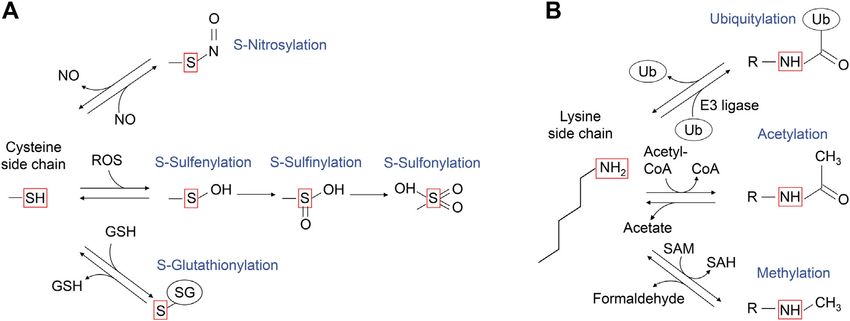
FIGURE 4 | A schematic diagram of the post-translational modifications of cysteine and lysine side chains. (A) The sulfur atom of cysteine side chain can undergo
several oxidative modifications, including those shown in the red box. S-nitrosylation can be generated upon reaction with nitric oxide (NO). Upon the reaction with
reactive oxygen species (ROS), the sulfur atom of cysteine side chain can undergo S-sulfenylation, and further oxidation to S-sulfinic and S-sulfonic states. The cysteine
side chain can also form mixed disulfides, including reaction with glutathione (GSH) to undergo reversible S-glutathionylation. (B) The ε-amino group of lysine side
chain can undergo several modifications as shown in the red box. The portion of modified lysine side chains is shown as “R-NH”. Ubiquitylation is mediated by ubiquitin
E3 ligase, while deubiquitylation is mediated by deubiquitylases. Lysine acetyltransferases use acetyl-CoA as the acetyl-donor for lysine acetylation, which can be
reversed by acetylated lysine deacetylases. Lysine methyltransferases use S-adenosylmethionine as a methyl donor for lysine methylation, which is reversed by
methylated lysine demethylase, coproducing formaldehyde.
et al., 2016; Yoshino et al., 2019; Figure 3). These PTMs can of the RAS cysteine oxidation research tracked back to 1995
upregulate RAS activity by increasing the guanine nucleotide (Figure 3).
exchange rate and/or inhibiting GAP-mediated GTP hydrolysis. Novogrodsky’s group at the Tel Aviv University found that
treatment of RAS with a variety of oxidative reagents, including
hydrogen peroxide (H2O2), hemin, Hg2+, and NO, increases
PTM WITHIN THE RAS G4 MOTIF (116NKCD119) cellular RAS activity (Lander et al., 1995). Further, Cys118 was
identified as the primary S-nitrosylation site in H-RAS. Cys118 is
S-Oxidation and S-Nitrosylation of Cys118 the most exposed solvent-accessible cysteine amongst three
in the G4 Motif cysteine residues within the G-domain (Lander et al., 1996).
Cells are often exposed to various stresses, such as increased Biochemical and structural studies of Cys118-nitrosylated
reactive oxygen species (ROS). ROS are continuously generated H-RAS and a redox insensitive H-RAS variant (C118S) revealed
through the mitochondrial electron transport chain, peroxidases, that neither nitrosylation at this solvent-exposed site or mutation
xanthine oxidase, lipoxygenase, NADPH oxidases, and heme- perturbs RAS structure, nucleotide cycling, or association with the
enzyme reactions. ROS can be generated by exogenous stimuli, RAS binding domain of CRAF (Mott et al., 1997; Williams et al.,
such as UV and ionizing radiation, ethanol intake, oxidized food, 2003). Subsequent functional analysis revealed that treatment with
metal ion overload (e.g., Fe and Cu), and smoking. Also, nitric S-nitrosocysteine (CysNO), an NO donor, increases the GDP
oxide (NO) is generated endogenously by nitric oxide synthases dissociation rate by ∼200-fold, resulting in the increased
(NOS) and exogenously by nitrogen oxides in air pollution (NOX) guanine nucleotide exchange rate, in the absence of a GEF
(e.g., car exhaust) and nitro compounds (Davies, 2016). (Williams et al., 2003; Heo and Campbell, 2004; Heo et al.,
Cysteine is a key amino acid in proteins for maintaining redox 2005; Figure 5). Biochemical analysis revealed that various
balance. Cysteine has a reactive thiol side chain (Cys-SH), which oxidants (e.g., superoxide, CysNO), but not H2O2, which
can undergo one- and two-electron oxidation reactions. Also, produce a Cys118 thiol radical intermediate, can cause
cysteine can undergo several reversible oxidative modifications, oxidation of the guanine nucleotide and destabilize guanine
including S-sulfenylation (Cys-SOH), S-nitrosylation (Cys-SNO), nucleotide-binding (Heo and Campbell, 2005), leading to
and S-glutathionylation (Cys-SSG) (Figure 4A; Paulsen and enhanced guanine nucleotide exchange.
Carroll, 2013). In addition, some cysteine residues in proteins
are more redox-sensitive than others because of changes in the side
chain orientation, charge, and altered exposure to ROS, affecting Conservation of Cys118 Within RAS
the efficiency of modification. For example, PTEN, a lipid Superfamily Members
phosphatase that antagonizes class I PI3K signaling by About 20% of small GTPases possess a cysteine residue at the
dephosphorylation of PI(3,4,5)P3, has a redox-sensitive cysteine position equivalent to Cys118 in the RAS superfamily. Within the
residue in its catalytic center, which undergoes S-sulfenylation, RAS and RAB sub-classes, 25 and 30% of these retain the Cys118
leading to PTEN inactivation and increased class I PI3K signaling (RAS isotypes numbering) (Figure 6; Wennerberg et al., 2005),
(Lee et al., 2002; Leslie et al., 2003; Zhang et al., 2017). The RAS respectively. Similar to H-RAS, a RAS sub-class member RAP1A and
GTPases are also regulated by cysteine oxidation, with the history a RAB sub-class member RAB3 undergo cysteine S-nitrosylation at
Frontiers in Molecular Biosciences | www.frontiersin.org 5 July 2021 | Volume 8 | Article 707439
Osaka et al.                                                                                                          RAS Activation by Post-Translational Modification

  FIGURE 5 | A schematic diagram highlighting the role of G4 and G5 post-translational modifications in RAS activation. Monoubiquitylation of RAS at Lys117, as well
  as S-nitrosylation of RAS at Cys118, increases GDP dissociation, leading to an increased GTP/GDP exchange rate. In contrast, monoubiquitylation of RAS at Lys147
  impedes GAP-mediated GTP hydrolysis, which populates the active RAS GTP-bound “ON” state.

  FIGURE 6 | The conservation of amino acids within the G4 and G5 motifs. Sequence alignment performed using Clustal Omega (https://www.ebi.ac.uk/Tools/
  msa/clustalo/). The amino acid sequence logo for the G4 and G5 motifs was created using WebLogo (https://weblogo.berkeley.edu/logo.cgi).

the cysteine residue in the G4 motif, leading to enhanced guanine                     Ubiquitylation of Lys117 in G4 Motif
nucleotide exchange resulting in elevated RAS activity (Heo and                       Lysine is a positively charged amino acid containing a long
Campbell, 2005; Heo et al., 2005). Thus, the role of Cys118 oxidation                 aliphatic sidechain and can undergo several post-translational
in regulation of GTPase activity appears to be conserved in several                   modifications, such as acetylation, methylation, and
RAS and RAB sub-class GTPases, and possibly in the other small                        ubiquitylation (Figure 4B). Ubiquitylation is a large lysine
GTPases with the cysteine residue equivalent to RAS Cys118 (Raines                    PTM, in which the 76 amino acid residue protein ubiquitin is
et al., 2007; Lim et al., 2008; Davis et al., 2011; Mitchell et al., 2013).           conjugated to the ε-amine of the lysine residue in the target

Frontiers in Molecular Biosciences | www.frontiersin.org                          6                                              July 2021 | Volume 8 | Article 707439
Osaka et al.                                                                                         RAS Activation by Post-Translational Modification

protein through an isopeptide bond formation to its carboxyl              Biochemical, NMR, and computational analyses indicated that
group of C-terminal glycine. The conjugated ubiquitin can be              ubiquitin makes dynamic non-specific contacts with RAS, yet
further polyubiquitylated. Lys48-linked polyubiquitylation                since the modification is large (∼8 kDa), it alters the
induces proteasome-dependent protein degradation (Heride                  conformation of Switch regions and dynamics of RAS
et al., 2014). This process typically requires four or more               structure (Baker et al., 2013a; Hobbs et al., 2013). This, in
polyubiquitin chains (Thrower et al., 2000; Miller and Gordon,            turn, alters recognition by GAP and effector proteins. In
2005). Protein monoubiquitylation, on the other hand, does not            particular, the Lys147 monoubiquitylation enhances the
promote protein degradation but regulates other cell functions            association with the specific K-RAS effectors: CRAF, BRAF,
such as endocytic trafficking (Haglund et al., 2003; Mosesson              and class I PI3K in HEK293T cells, while binding affinity
et al., 2003) and DNA damage response (Uckelmann and Sixma,               appears unaffected with other effectors, such as phospholipase
2017).                                                                    C (PLC) and calmodulin. These findings revealed a new function
    In 2011, RAS was identified as a target for monoubiquitylation         for ubiquitylation in modulating signaling through specific
(Figure 3; Sasaki et al., 2011). Cell biology experiments conducted       downstream pathways (Sasaki et al., 2011). While Lys147
in HEK293T cells determined that both H- and K-RAS are targets            monoubiquitylation of GDP-bound K-RAS significantly
for monoubiquitylation. Monoubiquitylation of H- and K-RAS                enhances the affinity to CRAF (more than 40-fold),
appeared to promote RAS activation, as the ubiquitylated RAS              monoubiquitylated GTP-bound K-RAS shows attenuated
were more populated in GTP-bound “ON” state and showed                    binding affinity for the RAS binding domain of certain RAS
enhanced association with RAS effectors compared to the non-              effectors (CRAF, RALGDS, and PI3Ks) (Thurman et al., 2017).
modified RAS. These findings suggest that the                               These results suggest that monoubiquitylation in K-RAS Lys147
monoubiquitylation of RAS is linked to RAS activation (Sasaki             facilitates RAF association and promotes signaling in a GTP-
et al., 2011). Tandem affinity purification of ubiquitylated H- and         independent manner. Also, further analysis showed that the
K-RAS4B (hereafter K-RAS) followed by mass spectrometry                   linker length (at least seven to eight residues) and protein
analysis identified Lys117 and Lys147 as major sites for                   ligation size of ubiquitin are critical for the GAP defect
monoubiquitylation, respectively. NMR analysis and cell                   (Hobbs et al., 2013).
biology experiments showed that monoubiquitylation of                         Consistent with these results, cell biological analysis indicated
Lys117 stimulates nucleotide exchange in the absence of RAS               that Lys147 monoubiquitylation promotes GTP loading of
GEF and thereby induces GTP loading and RAS activation (Baker             K-RAS. In mouse xenograft assays, a K-RAS G12V/K147L
et al., 2013b; Figure 5).                                                 double mutant that cannot be ubiquitylated showed
                                                                          significantly decreased tumor mass and volume, compared to
                                                                          oncogenic K-RAS G12V expressing isogenic control cells,
Conservation of Lys117 Within RAS                                         suggesting a critical role of Lys147 monoubiquitylation, or
Superfamily Members                                                       possibly through other modifications (e.g., acetylation,
The lysine residue within the “N-K-X-D” G4 motif is highly                methylation), in tumor progression (Sasaki et al., 2011).
conserved within the RAS superfamily (Figure 6). Within the
RAS, RAB, and ARF sub-classes, almost all of these retain Lys117          Acetylation of RAS Lys147 in the G5 Motif
(RAS isotypes numbering), while a few exceptions exist within the         Lysine acetylation is a prevalent post-translational modification
RHO sub-class GTPases (e.g., CDC42, TCL, RHOH)                            in eukaryotes and bacteria, and is mediated by the transfer of an
(Wennerberg et al., 2005). Furthermore, the lysine residue                acetyl CoA acetyl group by a cognate lysine acetyltransferase (Ali
within the G4 motif is also highly conserved within the other             et al., 2018; Nakayasu et al., 2017). Acetylation of lysine decreases
G-protein families (Dever et al., 1987). Hence, it is considered          the overall positive charge of lysine residues and can create a
that    the    GEF-independent       activation   via     Lys117          docking site for other proteins (Figure 4B). Beyond its well-
monoubiquitylation may be a fundamental mechanism to                      characterized role in regulating gene transcription through
regulate the activity of small GTPases and perhaps the other              histone modification, lysine acetylation regulates diverse
G-proteins as well.                                                       cellular processes through non-histone proteins (Ali et al., 2018).
                                                                              Recent studies have shown that Lys147 in K-RAS also
                                                                          undergoes acetylation (Knyphausen et al., 2016; Song et al.,
PTM WITHIN RAS G5 MOTIF (145SAK147)                                       2016). The K-RAS K147Q mutation, which was generated to
                                                                          mimic Lys147-acetylation, increased the rate of guanine
Ubiquitylation of RAS Lys147 in the G5 Motif                              nucleotide exchange approximately three-fold higher than
Lys147 monoubiquitylation upregulates RAS activity in a manner            wild-type K-RAS (Song et al., 2016), which implies that
distinct from Lys117 monoubiquitylation (Figure 5). Lys147 lies           acetylation of Lys147 in K-RAS may be involved in regulating
outside the Switch regions (Figures 1A,C). Using ubiquitin-               guanine nucleotide exchange. However, the K147Q mutant may
conjugated K-RAS, our group discovered that Lys147                        not mimic lysine acetylation as substitution of Lys147 with
monoubiquitylation alters conformational dynamics of the                  glutamine may disrupt a key interaction(s) important for
Switch I and II regions and interferes with association of and            guanine nucleotide-binding. Indeed, it has been shown that
downregulation by RAS GAPs while slightly altering GEF-                   Lys147 acetylation did not affect the intrinsic and the GEF-
dependent GDP/GTP exchange (Baker et al., 2013a; Figure 5).               dependent guanine nucleotide exchange (Knyphausen et al.,

Frontiers in Molecular Biosciences | www.frontiersin.org              7                                         July 2021 | Volume 8 | Article 707439
Osaka et al. RAS Activation by Post-Translational Modification
2016). Further studies are warranted to define the role of Lys147 to the functional difference of these RAS sub-classes remains
acetylation in K-RAS functions. unknown.
Methylation of RAS Lys147 in the G5 Motif
Protein methylation also occurs on side chain nitrogen atoms of POTENTIAL THERAPEUTIC APPLICATION
lysine, arginine, and histidine residues. In contrast to the long-
studied lysine acetylation, the roles of lysine-methylations beyond Oncogenic RAS Specific Inhibitors
chromatin regulation are less well characterized, despite its earlier Although RAS has been considered “undruggable” (Gysin et al.,
discovery in Salmonella typhimurium flagellin protein in 1959 2011; Samatar and Poulikakos, 2014; Stephen et al., 2014; Papke
(Ambler and Rees, 1959). Lysine modifications are more diverse and Der, 2017; Welsch et al., 2017), recent discoveries identified
than acetylation and can involve the transfer of one, two, or three covalent inhibitors that target Cys12 which is the reactive cysteine
methyl groups to the ε-amine of a lysine side chain through the within the K-RAS G12C oncogenic mutant by designed peptide
conjugation of a methyl group from S-adenosyl methionine mimetics (Ostrem et al., 2013; Yoo et al., 2020). These inhibitors
(SAM) by a lysine methyltransferase (Figure 4B). Unlike are shown to suppress tumor progression (Lito et al., 2016; Janes
ubiquitylation and acetylation, lysine methylation maintains its et al., 2018). Recently, Sotorasib, a K-RAS G12C inhibitor, has
overall positive charge. It is thus believed that the major function been granted accelerated approval by the Food and Drug
of lysine methylation is to provide a docking site for the proteins Administration (FDA) (Canon et al., 2019; Hong et al., 2020)
that recognize and bind methylated lysine (e.g., MBT and Tudor for the treatment of non-small-cell lung cancer (NSCLC). In
domains) (Lanouette et al., 2014; Teske and Hadden, 2017). addition, other K-RAS G12C inhibitors are now in multiple
In 2019, mass spectrometry analysis of the clinical trials, including phase II and phase III studies (Clinical
immunoprecipitated endogenous RAS identified dimethylation Trial number: NCT04613596; NCT04685135; NCT04793958;
at Lys5, adjacent to the G1 motif, as well as monomethylation at NCT04449874; NCT04699188) (Hallin et al., 2020). While
Lys147 in H-RAS (Figure 3) (Yoshino et al., 2019). While it is most K-RAS mutations occur at codon 12 (e.g., G12V, G12D),
currently unclear whether Lys5 dimethylation is specific for all G12C is only one of the mutations that can lead to oncogenic RAS
RAS isotypes, Lys147 is unique to the H-RAS. Given that activation at this position. Hence, there is a need to develop
substitutions at Lys147 to alanine, cysteine, or leucine do not therapeutics effective for other RAS mutant-driven cancers.
significantly alter RAS activity (Sasaki et al., 2011; Baker et al.,
2013a), it has been speculated that methylation of Lys147 does
not alter RAS structure and that methylation of Lys147 may affect Targeting the Enzymes Responsible for RAS
the H-RAS function by creating a docking site or blocking other PTMs
PTMs. It is worth noting that methylation can prevent protein Given that the post-translational modifications identified in the
degradation by antagonizing ubiquitylation at the same targeted G4 and G5 motifs are mediated by enzymes, we postulate that
lysine residue (Lanouette et al., 2014); in yeast, 43% of methylated further mechanistic understanding of RAS regulation by PTMs of
lysine residues are predicted to undergo ubiquitylation as well G4 and G5 motifs may unveil new approaches to suppress the
(Pang et al., 2010). Given that Lys147 in K-RAS undergoes RAS oncogenic activity that targets these modification enzymes
monoubiquitylation, Lys147 methylation may negatively (Figure 7). While the enzymes involved in RAS methylation
regulate RAS activation and monoubiquitylation-mediated remain unclear, several enzymes for RAS ubiquitylation and
effector switching. acetylation have been identified. Lysine deacetylases, HDAC6
and SIRT2, are suggested to negatively regulate K-RAS
acetylation in cancer cells (Yang et al., 2013; Knyphausen
Conservation of Lys147 Within RAS et al., 2016). RABEX5, an E3 Ubiquitin ligase, catalyzes mono-
Superfamily Members and di-ubiquitylation of H- and N-RAS, but not K-RAS, which
The lysine residue within the “S-A-K” G5 motif is conserved in downregulates RAS activity (Xu et al., 2010; Yan et al., 2010;
about 45% of RAS superfamily members (Figure 6; Wennerberg Washington et al., 2020). The ubiquitylation site(s) by RABEX5
et al., 2005). The adjacent serine and alanine residues within the remains unclear. A deubiquitinase OTUB1 has been identified as
G5 motif are also highly conserved in each sub-class (Figure 6). a negative regulator of RAS through a mammalian protein-
Thus, the PTM of Lys147 (RAS isotypes numbering) may not be protein interaction screening using H-RAS G12V mutant as
limited to RAS but present in other RAS superfamily GTPases. the bait (Baietti et al., 2016). As Lys117 or Lys147
The G5 motif within some of the RHO, RAB, and ARF sub-classes ubiquitylation upregulates RAS activity, it is unlikely that
contain “S-A-L,” “S-A-T,” “C-A-L,” and “C-A-T” sequences RABEX5 and OTUB1 modulate ubiquitylation of either Lys
(Figure 6), and may undergo different PTMs within the G5 117 or Lys147 in the G4 and G5 motifs. Hence, further studies
motif (e.g., phosphorylation at threonine residue of “S-A-T” exploring enzymes responsible for RAS ubiquitylation are
motif and S-oxidation or S-nitrosylation at cysteine residue of required.
“C-A-L” motif). Of note, the G5 motif is absent in several other A promising new strategy to antagonize aberrant RAS
G-proteins (e.g., heterotrimeric G-proteins and elongation signaling involves RAS degradation through ubiquitylation.
factors). Whether the diverse sequences associated with the G5 These proteolysis-targeting chimera (PROTAC) approaches have
motif in comparison to the more conserved G4 motif contribute proven to be an effective strategy for inhibiting specific protein
Frontiers in Molecular Biosciences | www.frontiersin.org 8 July 2021 | Volume 8 | Article 707439Osaka et al. RAS Activation by Post-Translational Modification
function of RAS proteins. Divergent mechanisms involved in RAS
activation through PTMs of the G4 and G5 motifs are likely to enable
RAS to function at the distinctive subcellular localization, timing,
and kinetics, apart from the canonical RAS regulatory pathway by
GEFs and GAPs. Thus, RAS PTMs may play an important role in
developing a new therapeutic approach for RAS-driven cancers. One
of the next important steps will be to identify enzymes responsible
for RAS PTMs as well as to clarify the physiological significance of
these modifications in developmental processes, homeostasis, and
disease states. PTMs associated with RAS G4 and G5 motifs may
represent novel “Achille’s heels” for new anti-RAS approaches.
Further understanding of these mechanisms might shed light on
the development of effective therapeutic approaches.
AUTHOR CONTRIBUTIONS
FIGURE 7 | A RAS activation model by monoubiquitylation at Lys117 NO and AS organized and wrote the manuscript. YH, DI, RK, and
and Lys147. For RAS Lys117 ubiquitylation, a current working model is that
Lys117 monoubiquitylation increase GTP-bound RAS by increased GTP/
VC projected to draw the illustration. NO, AS, YH, DI, YI, RK,
GDP exchange. The Lys147 monoubiquitylation inhibits the RAS GAP and YF conceptualized. NO, AS, TS, KT, and SC edited the
binding to RAS, which leads to the increased GTP-bound RAS. Hence, manuscript. All authors contributed to the article and approved
targeting the E3 ligase responsible for Lys117 and/or Lys147 the submitted version.
monoubiquitylation is expected to suppress the RAS activation and possibly
decrease tumorigenic activity or progression of cancer.
FUNDING
targets (Churcher, 2017; Coleman and Crews, 2018). PROTACs The work is supported in part by the MTP UC-Brain Tumor
induce proteolysis of a target protein by linking a target protein to the Center grant and NIH grants R21NS100077 and R01NS089815
specific E3 ubiquitin ligase via a chemical tag (Khan et al., 2020). to AS. Support is also provided by Project for Cancer Research
Importantly, PROTACs specifically targeting K-RAS or the K-RAS and Therapeutic Evolution (P-CREATE; JP20cm0106173 to AS,
G12C mutant have recently been developed (Bery et al., 2020; Bond TS, and KT) from Japan Agency for Medical Research and
et al., 2020). Identifying RAS E3 ligases could aid in the application of Development (AMED). NO is supported by the Japan Society
PROTAC approaches for therapeutic inhibition of RAS as RAS- for the Promotion of Science (JSPS), KAKENHI grant number
specific ligases may facilitate spatial/temporal localization needed for JP20H03165 and the Japan Science and Technology (JST) grant
efficient RAS degradation. Clarifying which enzymes are responsible number JP20356709. YH is supported, in part, by JSPS
for RAS acetylation and methylation may provide another indirect KAKENHI grant number JP18KK0455 and JP 21K11709. DI
way to suppress RAS activity by modulating these PTMs. is supported by JSPS KAKENHI grant number 21J00755. YI is
supported by JSPS KAKENHI grant number JP20K07624. RK is
supported by JSPS KAKENHI grant number 21K15019. SC is
CONCLUDING REMARKS supported in part by NIH grants 3R35GM134962 and
P01CA203567. TS is supported by the Platform Project for
Post-translational modifications contribute to the diversification of Supporting Drug Discovery and Life Science Research (Basis for
protein function as well as the robustness to intra- and extracellular Supporting Innovative Drug Discovery and Life Science
stress for maintaining cellular functions. Among the many post- Research (BINDS)) from AMEDunder Grant Numbers
translational modifications, S-oxygenation, S-nitrosylation, JP19am0101071. This work was also supported in part by
monoubiquitylation, acetylation, and methylation described in research funds from the Sumitomo Foundation, and the
this review reflect reversible modifications that can modulate the Yamagata prefectural government, and the City of Tsuruoka.
Ambler, R. P., and Rees, M. W. (1959). Ɛ-N-Methyl-lysine in Bacterial Flagellar
REFERENCES Protein. Nature 184, 56–57. doi:10.1038/184056b0
Baietti, M. F., Simicek, M., Abbasi Asbagh, L., Radaelli, E., Lievens, S., Crowther, J.,
Ahearn, I., Zhou, M., and Philips, M. R. (2018). Posttranslational Modifications of et al. (2016). OTUB 1 Triggers Lung Cancer Development by Inhibiting RAS
RAS Proteins. Cold Spring Harb. Perspect. Med. 8, a031484. doi:10.1101/ Monoubiquitination. EMBO Mol. Med. 8, 288–303. doi:10.15252/
cshperspect.a031484 emmm.201505972
Ali, I., Conrad, R. J., Verdin, E., and Ott, M. (2018). Lysine Acetylation Goes Baker, R., Lewis, S. M., Sasaki, A. T., Wilkerson, E. M., Locasale, J. W., Cantley, L. C., et al.
Global: From Epigenetics to Metabolism and Therapeutics. Chem. Rev. 118, (2013a). Site-specific Monoubiquitination Activates Ras by Impeding GTPase-
1216–1252. doi:10.1021/acs.chemrev.7b00181 Activating Protein Function. Nat. Struct. Mol. Biol. 20, 46–52. doi:10.1038/nsmb.2430
Frontiers in Molecular Biosciences | www.frontiersin.org 9 July 2021 | Volume 8 | Article 707439Osaka et al. RAS Activation by Post-Translational Modification Baker, R., Wilkerson, E. M., Sumita, K., Isom, D. G., Sasaki, A. T., Dohlman, H. G., et al. Gideon, P., John, J., Frech, M., Lautwein, A., Clark, R., Scheffler, J. E., et al. (1992). (2013b). Differences in the Regulation of K-Ras and H-Ras Isoforms by Mutational and Kinetic Analyses of the GTPase-Activating Protein (GAP)-p21 Monoubiquitination. J. Biol. Chem. 288, 36856–36862. doi:10.1074/jbc.C113.525691 Interaction: the C-Terminal Domain of GAP Is Not Sufficient for Full Activity. Bery, N., Miller, A., and Rabbitts, T. (2020). A Potent KRAS Macromolecule Mol. Cel. Biol. 12, 2050–2056. doi:10.1128/mcb.12.5.2050 Degrader Specifically Targeting Tumours with Mutant KRAS. Nat. Commun. Gray, J. L., Delft, F., and Brennan, P. E. (2020). Targeting the Small GTPase 11, 3233. doi:10.1038/s41467-020-17022-w Superfamily through Their Regulatory Proteins. Angew. Chem. Int. Ed. 59, Bond, M. J., Chu, L., Nalawansha, D. A., Li, K., and Crews, C. M. (2020). Targeted 6342–6366. doi:10.1002/anie.201900585 Degradation of Oncogenic KRASG12C by VHL-Recruiting PROTACs. ACS Gremer, L., Merbitz-Zahradnik, T., Dvorsky, R., Cirstea, I. C., Kratz, C. P., Zenker, Cent. Sci. 6, 1367–1375. doi:10.1021/acscentsci.0c00411 M., et al. (2011). Germline KRAS Mutations Cause Aberrant Biochemical and Borrie, S. C., Brems, H., Legius, E., and Bagni, C. (2017). Cognitive Dysfunctions in Physical Properties Leading to Developmental Disorders. Hum. Mutat. 32, Intellectual Disabilities: The Contributions of the Ras-MAPK and PI3K-AKT- 33–43. doi:10.1002/humu.21377 mTOR Pathways. Annu. Rev. Genom. Hum. Genet. 18, 115–142. doi:10.1146/ Gysin, S., Salt, M., Young, A., and McCormick, F. (2011). Therapeutic Strategies for annurev-genom-091416-035332 Targeting Ras Proteins. Genes & Cancer 2, 359–372. doi:10.1177/ Bos, J. L. (1989). Ras Oncogenes in Human Cancer: a Review. Cancer Res. 49, 1947601911412376 4682–4689. Haglund, K., Di Fiore, P. P., and Dikic, I. (2003). Distinct Monoubiquitin Signals in Bos, J. L., Rehmann, H., and Wittinghofer, A. (2007). GEFs and GAPs: Critical Receptor Endocytosis. Trends Biochem. Sci. 28, 598–604. doi:10.1016/ Elements in the Control of Small G Proteins. Cell 129, 865–877. doi:10.1016/ j.tibs.2003.09.005 j.cell.2007.05.018 Hallin, J., Engstrom, L. D., Hargis, L., Calinisan, A., Aranda, R., Briere, D. M., et al. Canon, J., Rex, K., Saiki, A. Y., Mohr, C., Cooke, K., Bagal, D., et al. (2019). The (2020). The KRAS G12CInhibitor MRTX849 Provides Insight toward Clinical KRAS(G12C) Inhibitor AMG 510 Drives Anti-tumour Immunity. Therapeutic Susceptibility of KRAS-Mutant Cancers in Mouse Models and Nature 575, 217–223. doi:10.1038/s41586-019-1694-1 Patients. Cancer Dis. 10, 54–71. doi:10.1158/2159-8290.CD-19-1167 Chang, E. H., Gonda, M. A., Ellis, R. W., Scolnick, E. M., and Lowy, D. R. (1982). Hennig, A., Markwart, R., Esparza-Franco, M. A., Ladds, G., and Rubio, I. (2015). Human Genome Contains Four Genes Homologous to Transforming Genes of Ras Activation Revisited: Role of GEF and GAP Systems. Biol. Chem. 396, Harvey and Kirsten Murine Sarcoma Viruses. Proc. Natl. Acad. Sci. 79, 831–848. doi:10.1515/hsz-2014-0257 4848–4852. doi:10.1073/pnas.79.16.4848 Heo, J., and Campbell, S. L. (2004). Mechanism of p21RasS-Nitrosylation and Cherfils, J., and Zeghouf, M. (2013). Regulation of Small GTPases by GEFs, GAPs, Kinetics of Nitric Oxide-Mediated Guanine Nucleotide Exchange†. and GDIs. Physiol. Rev. 93, 269–309. doi:10.1152/physrev.00003.2012 Biochemistry 43, 2314–2322. doi:10.1021/bi035275g Churcher, I. (2017). Protac-induced Protein Degradation in Drug Discovery: Heo, J., and Campbell, S. L. (2005). Superoxide Anion Radical Modulates the Breaking the Rules or Just Making New Ones? J. Med. Chem. 61, 444–452. Activity of Ras and Ras-Related GTPases by a Radical-Based Mechanism doi:10.1021/acs.jmedchem.7b01272 Similar to that of Nitric Oxide. J. Biol. Chem. 280, 12438–12445. Coleman, K. G., and Crews, C. M. (2018). Proteolysis-targeting Chimeras: doi:10.1074/jbc.M414282200 Harnessing the Ubiquitin-Proteasome System to Induce Degradation of Heo, J., Prutzman, K. C., Mocanu, V., and Campbell, S. L. (2005). Mechanism of Specific Target Proteins. Annu. Rev. Cancer Biol. 2, 41–58. doi:10.1146/ Free Radical Nitric Oxide-Mediated Ras Guanine Nucleotide Dissociation. annurev-cancerbio-030617-050430 J. Mol. Biol. 346, 1423–1440. doi:10.1016/j.jmb.2004.12.050 Cool, R. H., Schmidt, G., Lenzen, C. U., Prinz, H., Vogt, D., and Wittinghofer, A. Heride, C., Urbé, S., and Clague, M. J. (2014). Ubiquitin Code Assembly and (1999). The Ras Mutant D119N Is Both Dominant Negative and Activated. Disassembly. Curr. Biol. 24, R215–R220. doi:10.1016/j.cub.2014.02.002 Mol. Cel. Biol. 19, 6297–6305. doi:10.1128/mcb.19.9.6297 Herrmann, C., Martin, G. A., and Wittinghofer, A. (1995). Quantitative Analysis of Davies, M. J. (2016). Protein Oxidation and Peroxidation. Biochem. J. 473, the Complex between P21 and the Ras-Binding Domain of the Human Raf-1 805–825. doi:10.1042/BJ20151227 Protein Kinase. J. Biol. Chem. 270, 2901–2905. doi:10.1074/jbc.270.7.2901 Davis, M. F., Vigil, D., and Campbell, S. L. (2011). Regulation of Ras Proteins by Hobbs, G. A., Gunawardena, H. P., Baker, R., Chen, X., and Campbell, S. L. (2013). Reactive Nitrogen Species. Free Radic. Biol. Med. 51, 565–575. doi:10.1016/ Site-specific Monoubiquitination Activates Ras by Impeding GTPase- j.freeradbiomed.2011.05.003 Activating Protein Function. Small GTPases 4, 186–192. doi:10.4161/sgtp.26270 Denayer, E., Parret, A., Chmara, M., Schubbert, S., Vogels, A., Devriendt, K., et al. Hong, D. S., Fakih, M. G., Strickler, J. H., Desai, J., Durm, G. A., Shapiro, G. I., et al. (2008). Mutation Analysis in Costello Syndrome: Functional and Structural (2020). KRASG12C Inhibition with Sotorasib in Advanced Solid Tumors. N. Characterization of theHRASp.Lys117Arg Mutation. Hum. Mutat. 29, Engl. J. Med. 383, 1207–1217. doi:10.1056/NEJMoa1917239 232–239. doi:10.1002/humu.20616 Janakiraman, M., Vakiani, E., Zeng, Z., Pratilas, C. A., Taylor, B. S., Chitale, D., et al. Dever, T. E., Glynias, M. J., and Merrick, W. C. (1987). GTP-binding Domain: (2010). Genomic and Biological Characterization of Exon 4 KRAS Mutations in Three Consensus Sequence Elements with Distinct Spacing. Proc. Natl. Acad. Human Cancer. Cancer Res. 70, 5901–5911. doi:10.1158/0008-5472.CAN-10- Sci. 84, 1814–1818. doi:10.1073/pnas.84.7.1814 0192 Downward, J. (2003). Targeting RAS Signalling Pathways in Cancer Therapy. Nat. Janes, M. R., Zhang, J., Li, L.-S., Hansen, R., Peters, U., Guo, X., et al. (2018). Rev. Cancer 3, 11–22. doi:10.1038/nrc969 Targeting KRAS Mutant Cancers with a Covalent G12C-specific Inhibitor. Cell Edkins, S., O’Meara, S., Parker, A., Stevens, C., Reis, M., Jones, S., et al. (2006). 172, 578–589. doi:10.1016/j.cell.2018.01.006 Recurrent KRAS Codon 146 Mutations in Human Colorectal Cancer. Cancer. John, J., Rensland, H., Schlichting, I., Vetter, I., Borasio, G. D., Goody, R. S., et al. Biol. Ther. 5, 928–932. doi:10.4161/cbt.5.8.3251 (1993). Kinetic and Structural Analysis of the Mg(2+)-Binding Site of the Feig, L. A., Pan, B. T., Roberts, T. M., and Cooper, G. M. (1986). Isolation of Ras Guanine Nucleotide-Binding Protein p21H-Ras. J. Biol. Chem. 268, 923–929. GTP-Binding Mutants Using an In Situ colony-binding Assay. Proc. Natl. Acad. doi:10.1016/s0021-9258(18)54022-9 Sci. 83, 4607–4611. doi:10.1073/pnas.83.13.4607 Karnoub, A. E., and Weinberg, R. A. (2008). Ras Oncogenes: Split Personalities. Feuerstein, J., Goody, R. S., and Wittinghofer, A. (1987). Preparation and Nat. Rev. Mol. Cel Biol. 9, 517–531. doi:10.1038/nrm2438 Characterization of Nucleotide-free and Metal Ion-free P21 "apoprotein". Khan, S., He, Y., Zhang, X., Yuan, Y., Pu, S., Kong, Q., et al. (2020). PROteolysis J. Biol. Chem. 262, 8455–8458. doi:10.1016/s0021-9258(18)47433-9 TArgeting Chimeras (PROTACs) as Emerging Anticancer Therapeutics. Forbes, S. A., Bindal, N., Bamford, S., Cole, C., Kok, C. Y., Beare, D., et al. (2010). Oncogene 39, 4909–4924. doi:10.1038/s41388-020-1336-y COSMIC: Mining Complete Cancer Genomes in the Catalogue of Somatic Kiel, C., Filchtinski, D., Spoerner, M., Schreiber, G., Kalbitzer, H. R., and Mutations in Cancer. Nucleic Acids Res. 39, D945–D950. doi:10.1093/nar/ Herrmann, C. (2009). Improved Binding of Raf to Ras·GDP Is Correlated gkq929 with Biological Activity. J. Biol. Chem. 284, 31893–31902. doi:10.1074/ Ford, B., Boykevisch, S., Zhao, C., Kunzelmann, S., Bar-Sagi, D., Herrmann, C., jbc.M109.031153 et al. (2009). Characterization of a Ras Mutant with Identical GDP- and GTP- Kinoshita, K., Sadanami, K., Kidera, A., and Go, N. (1999). Structural Motif of Bound Structures,. Biochemistry 48, 11449–11457. doi:10.1021/bi901479b Phosphate-Binding Site Common to Various Protein Superfamilies: All- Frontiers in Molecular Biosciences | www.frontiersin.org 10 July 2021 | Volume 8 | Article 707439
Osaka et al.                                                                                                                   RAS Activation by Post-Translational Modification

    Against-All Structural Comparison of Protein-Mononucleotide Complexes.                   Pang, C., Gasteiger, E., and Wilkins, M. R. (2010). Identification of Arginine- and
    Protein Eng. 12, 11–14. doi:10.1093/protein/12.1.11                                          Lysine-Methylation in the Proteome of Saccharomyces cerevisiae and its
Knyphausen, P., Lang, F., Baldus, L., Extra, A., and Lammers, M. (2016). Insights                Functional Implications. BMC Genomics 11, 92. doi:10.1186/1471-2164-11-92
    into K-Ras 4B Regulation by post-translational Lysine Acetylation. Biol. Chem.           Papke, B., and Der, C. J. (2017). Drugging RAS: Know the Enemy. Science 355,
    397, 1071–1085. doi:10.1515/hsz-2016-0118                                                    1158–1163. doi:10.1126/science.aam7622
Kötting, C., Kallenbach, A., Suveyzdis, Y., Wittinghofer, A., and Gerwert, K. (2008).        Paulsen, C. E., and Carroll, K. S. (2013). Cysteine-Mediated Redox Signaling:
    The GAP Arginine finger Movement into the Catalytic Site of Ras Increases the                 Chemistry, Biology, and Tools for Discovery. Chem. Rev. 113, 4633–4679.
    Activation Entropy. Proc. Natl. Acad. Sci. 105, 6260–6265. doi:10.1073/                      doi:10.1021/cr300163e
    pnas.0712095105                                                                          Prior, I. A., Hood, F. E., and Hartley, J. L. (2020). The Frequency of Ras Mutations
Lander, H. M., Milbank, A. J., Tauras, J. M., Hajjar, D. P., Hempstead, B. L.,                   in Cancer. Cancer Res. 80, 2969–2974. doi:10.1158/0008-5472.CAN-19-3682
    Schwartz, G. D., et al. (1996). Redox Regulation of Cell Signalling. Nature 381,         Prior, I. A., Lewis, P. D., and Mattos, C. (2012). A Comprehensive Survey of Ras
    380–381. doi:10.1038/381380a0                                                                Mutations in Cancer. Cancer Res. 72, 2457–2467. doi:10.1158/0008-5472.CAN-
Lander, H. M., Ogiste, J. S., Teng, K. K., and Novogrodsky, A. (1995). p21 as a                  11-2612
    Common Signaling Target of Reactive Free Radicals and Cellular Redox Stress.             Pylayeva-Gupta, Y., Grabocka, E., and Bar-Sagi, D. (2011). RAS Oncogenes:
    J. Biol. Chem. 270, 21195–21198. doi:10.1074/jbc.270.36.21195                                Weaving a Tumorigenic Web. Nat. Rev. Cancer 11, 761–774. doi:10.1038/
Lanouette, S., Mongeon, V., Figeys, D., and Couture, J. F. (2014). The Functional                nrc3106
    Diversity of Protein Lysine Methylation. Mol. Syst. Biol. 10, 724. doi:10.1002/          Raines, K., Bonini, M., and Campbell, S. (2007). Nitric Oxide Cell Signaling:
    msb.134974                                                                                   S-Nitrosation of Ras Superfamily GTPases. Cardiovasc. Res. 75, 229–239.
Lee, S.-R., Yang, K.-S., Kwon, J., Lee, C., Jeong, W., and Rhee, S. G. (2002).                   doi:10.1016/j.cardiores.2007.04.013
    Reversible Inactivation of the Tumor Suppressor PTEN by H2O2. J. Biol. Chem.             Ratner, N., and Miller, S. J. (2015). A RASopathy Gene Commonly Mutated in
    277, 20336–20342. doi:10.1074/jbc.M111899200                                                 Cancer: the Neurofibromatosis Type 1 Tumour Suppressor. Nat. Rev. Cancer
Leslie, N. R., Bennett, D., Lindsay, Y. E., Stewart, H., Gray, A., and Downes, C. P.             15, 290–301. doi:10.1038/nrc3911
    (2003). Redox Regulation of PI 3-kinase Signalling via Inactivation of PTEN.             Rauen, K. A. (2013). The RASopathies. Annu. Rev. Genom. Hum. Genet. 14,
    EMBO J. 22, 5501–5510. doi:10.1093/emboj/cdg513                                              355–369. doi:10.1146/annurev-genom-091212-153523
Li, S., Balmain, A., and Counter, C. M. (2018). A Model for RAS Mutation Patterns            Samatar, A. A., and Poulikakos, P. I. (2014). Targeting RAS-ERK Signalling in
    in Cancers: Finding the Sweet Spot. Nat. Rev. Cancer 18, 767–777. doi:10.1038/               Cancer: Promises and Challenges. Nat. Rev. Drug Discov. 13, 928–942.
    s41568-018-0076-6                                                                            doi:10.1038/nrd4281
Lim, K.-H., Ancrile, B. B., Kashatus, D. F., and Counter, C. M. (2008). Tumour               Sasaki, A. T., Carracedo, A., Locasale, J. W., Anastasiou, D., Takeuchi, K., Kahoud,
    Maintenance Is Mediated by eNOS. Nature 452, 646–649. doi:10.1038/                           E. R., et al. (2011). Ubiquitination of K-Ras Enhances Activation and Facilitates
    nature06778                                                                                  Binding to Select Downstream Effectors. Sci. Signaling 4, ra13. doi:10.1126/
Lito, P., Solomon, M., Li, L.-S., Hansen, R., and Rosen, N. (2016). Allele-specific               scisignal.2001518
    Inhibitors Inactivate Mutant KRAS G12C by a Trapping Mechanism. Science                  Scheffzek, K., Ahmadian, M. R., Kabsch, W., Wiesmüller, L., Lautwein, A., Schmitz,
    351, 604–608. doi:10.1126/science.aad6204                                                    F., et al. (1997). The Ras-RasGAP Complex: Structural Basis for GTPase
Malumbres, M., and Barbacid, M. (2003). RAS Oncogenes: the First 30 Years. Nat.                  Activation and its Loss in Oncogenic Ras Mutants. Science 277, 333–338.
    Rev. Cancer 3, 459–465. doi:10.1038/nrc1097                                                  doi:10.1126/science.277.5324.333
Miller, J., and Gordon, C. (2005). The Regulation of Proteasome Degradation by               Simanshu, D. K., Nissley, D. V., and McCormick, F. (2017). RAS Proteins and
    Multi-Ubiquitin Chain Binding Proteins. FEBS Lett. 579, 3224–3230.                           Their Regulators in Human Disease. Cell 170, 17–33. doi:10.1016/
    doi:10.1016/j.febslet.2005.03.042                                                            j.cell.2017.06.009
Mitchell, L., Hobbs, G. A., Aghajanian, A., and Campbell, S. L. (2013). Redox                Smith, G., Bounds, R., Wolf, H., Steele, R. J. C., Carey, F. A., and Wolf, C. R. (2010).
    Regulation of Ras and Rho GTPases: Mechanism and Function. Antioxid.                         Activating K-Ras Mutations Outwith ’hotspot’ Codons in Sporadic Colorectal
    Redox Signaling 18, 250–258. doi:10.1089/ars.2012.4687                                       Tumours - Implications for Personalised Cancer Medicine. Br. J. Cancer 102,
Moore, A. R., Rosenberg, S. C., McCormick, F., and Malek, S. (2020). RAS-targeted                693–703. doi:10.1038/sj.bjc.6605534
    Therapies: Is the Undruggable Drugged?. Nat. Rev. Drug Discov. 19, 533–552.              Smith, M. J., Neel, B. G., and Ikura, M. (2013). NMR-based Functional Profiling of
    doi:10.1038/s41573-020-0068-6                                                                RASopathies and Oncogenic RAS Mutations. Proc. Natl. Acad. Sci. 110,
Mosesson, Y., Shtiegman, K., Katz, M., Zwang, Y., Vereb, G., Szollosi, J., et al.                4574–4579. doi:10.1073/pnas.1218173110
    (2003). Endocytosis of Receptor Tyrosine Kinases Is Driven by                            Song, H. Y., Biancucci, M., Kang, H.-J., O’Callaghan, C., Park, S.-H., Principe, D. R.,
    Monoubiquitylation, Not Polyubiquitylation. J. Biol. Chem. 278,                              et al. (2016). SIRT2 Deletion Enhances KRAS-Induced Tumorigenesis In Vivo
    21323–21326. doi:10.1074/jbc.C300096200                                                      by Regulating K147 Acetylation Status. Oncotarget 7, 80336–80349.
Mott, H. R., Carpenter, J. W., and Campbell, S. L. (1997). Structural and Functional             doi:10.18632/oncotarget.12015
    Analysis of a Mutant Ras Protein that Is Insensitive to Nitric Oxide Activation†.        Stalnecker, C. A., and Der, C. J. (2020). RAS, Wanted Dead or Alive: Advances in
    Biochemistry 36, 3640–3644. doi:10.1021/bi962790o                                            Targeting RAS Mutant Cancers. Sci. Signal. 13, eaay6013. doi:10.1126/
Nakayasu, E. S., Burnet, M. C., Walukiewicz, H. E., Wilkins, C. S., Shukla, A. K.,               scisignal.aay6013
    Brooks, S., et al. (2017). Ancient Regulatory Role of Lysine Acetylation in              Stephen, A. G., Esposito, D., Bagni, R. K., and McCormick, F. (2014). Dragging Ras
    Central Metabolism. mBio 8, 1395. doi:10.1128/mBio.01894-17                                  Back in the Ring. Cancer Cell 25, 272–281. doi:10.1016/j.ccr.2014.02.017
Nassar, A. H., Adib, E., and Kwiatkowski, D. J. (2021). Distribution of KRASG12C             Teske, K. A., and Hadden, M. K. (2017). Methyllysine Binding Domains: Structural
    Somatic Mutations across Race, Sex, and Cancer Type. N. Engl. J. Med. 384,                   Insight and Small Molecule Probe Development. Eur. J. Med. Chem. 136, 14–35.
    185–187. doi:10.1056/NEJMc2030638                                                            doi:10.1016/j.ejmech.2017.04.047
Niihori, T., Aoki, Y., Okamoto, N., Kurosawa, K., Ohashi, H., Mizuno, S., et al.             Thrower, J. S., Hoffman, L., Rechsteiner, M., and Pickart, C. M. (2000). Recognition
    (2011). HRAS Mutants Identified in Costello Syndrome Patients Can Induce                      of the Polyubiquitin Proteolytic Signal. EMBO J. 19, 94–102. doi:10.1093/
    Cellular Senescence: Possible Implications for the Pathogenesis of Costello                  emboj/19.1.94
    Syndrome. J. Hum. Genet. 56, 707–715. doi:10.1038/jhg.2011.85                            Thurman, R., Siraliev-Perez, E., and Campbell, S. L. (2020). RAS Ubiquitylation
Ostrem, J. M., Peters, U., Sos, M. L., Wells, J. A., and Shokat, K. M. (2013).                   Modulates Effector Interactions. Small GTPases 11, 180–185. doi:10.1080/
    K-Ras(G12C) Inhibitors Allosterically Control GTP Affinity and Effector                       21541248.2017.1371267
    Interactions. Nature 503, 548–551. doi:10.1038/nature12796                               Tidyman, W. E., and Rauen, K. A. (2009). The RASopathies: Developmental
Pai, E. F., Kabsch, W., Krengel, U., Holmes, K. C., John, J., and Wittinghofer, A.               Syndromes of Ras/MAPK Pathway Dysregulation. Curr. Opin. Genet. Develop.
    (1989). Structure of the Guanine-Nucleotide-Binding Domain of the Ha-Ras                     19, 230–236. doi:10.1016/j.gde.2009.04.001
    Oncogene Product P21 in the Triphosphate Conformation. Nature 341,                       Traut, T. W. (1994). Physiological Concentrations of Purines and Pyrimidines.
    209–214. doi:10.1038/341209a0                                                                Mol. Cel. Biochem. 140, 1–22. doi:10.1007/BF00928361

Frontiers in Molecular Biosciences | www.frontiersin.org                                11                                                 July 2021 | Volume 8 | Article 707439
You can also read