COVID-19 NC Collaboratory Projects
←
→
Page content transcription
If your browser does not render page correctly, please read the page content below
COVID-19 NC Collaboratory Projects
                                  Final Narrative
                                 February 12, 2021

Therapeutics I: Transitioning/Optimizing New Drugs For Pulmonary Delivery

                                 PI: Ric Boucher, MD
     Co-Is: Charles Esther, PhD, Ray Pickles, PhD, Brian Button, PhD, Alessandra
Livraghi-Butrico, PhD, Claire Doerschuk, MD, Matt Wolfgang, PhD, Alexander Kabanov,
                                 PhD, Gauri Rao, PhD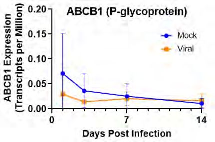
EXECUTIVE SUMMARY
A large number of drugs that have promising antiviral activity for COVID-19 in the laboratory have
proven ineffective clinically. For many of these agents, lack of clinical effectiveness reflects the challenges
of getting sufficient drug into the lung to have antiviral activity using intravenous or oral dosing regimens.
Our project explored the therapeutic potential of aerosolizing drugs directly to the airway cell
surface using a pharmacokinetic/pharmacodynamic (PK/PD) system in cell culture that mimics what can
be achieved using clinical nebulizers in patients. Our working hypothesis was that aerosolizing drugs
directly into the airway will improve the therapeutic index by delivering high concentrations directly to airway
epithelia (the site of SARS-CoV-2 infection) while minimizing systemic side effects.

Evaluations were performed on drugs that are active within the cell (remdesivir, hydroxychloroquine) as well
as those that are only active on cell surfaces (surfactants/baby shampoo, reducing agents). For both
remdesivir and hydroxychloroquine, we showed that a single aerosolized dose could generate intracellular
concentrations of active drug similar to, and in some cases greater than, what can be achieved by standard
intravenous or oral doses. For remdesivir, we demonstrated that aerosolized drug is metabolized to the
active metabolite at concentrations predicted to be antiviral, though gene expression studies suggested
that active SARS-CoV-2 infection could inhibit this conversion. For hydroxycholorquine, we showed that drug
accumulates intracellularly, undergoes minimal metabolism, and stays in the cell for several days after
dosing. For both drugs, we demonstrated that diffusion into the extracellular fluid is limited, suggesting that
the inhaled route will avoid many of the side effects of systemic dosing.

Our studies on cell surface active agents demonstrated challenges for this class of therapeutics. We showed
that while a gentle surfactant such as baby shampoo can readily kill SARS-CoV-2 and other viruses in a
test tube and is safe to apply to airway epithelia, application of a baby shampoo/saline mix widely used for
sinus disease did not have any impact on viral levels in infected cultured cells. These findings paralleled those
from a clinical study by colleagues at Vanderbilt in which nasal rinses with baby shampoo/saline did not alter
viral levels in the noses of COVID-19 patients. Mass spectrometric pharmacokinetics showed that baby
shampoo contains a mix of short and longer chain surfactants, and that the longer chain surfactants that have
antiviral activity are rapidly lost when delivered to airway epithelia, likely because they bind to and/or are
incorporated into epithelial cell membranes. We observed similar overall findings with reducing agents, which
had significant antiviral activity in test tube but limited duration of activity on cell surfaces.

In summary, we found that drugs that are active intracellularly, such as remdesivir and hydroxychloroquine, are
excellent candidates for aerosolization. Even brief aerosolized treatments generated high
intracellular concentrations similar or better that those achieved by systemic dosing to levels predicted to
have antiviral activity. Although further work needs to be done, our studies demonstrate that an aerosolized
approach not only improves the therapeutic index for these agents but also offers a non-invasive route of
delivery that would permit treatment earlier in the course of disease when antivirals are likely to be more
effective. Our studies also uncovered challenges to utilization of cell surface active agents. Though in each
case the challenges were unique to the drug, the results highlight the value of our PK/PD system to identify
such problems prior to clinical trials.

INTRODUCTION
The COVID-19 pandemic has led to an unprecedented
search for antiviral agents. While many drugs have promising
antiviral activity against SARS-CoV-2 in vitro, most have
failed to demonstrate clinical efficacy when tested in patients.
For many drugs, a significant limiting factor is the difficulty of
achieving sufficient concentrations at the site of pulmonary         Fig. 1. SARS-CoV-2 Infection of Airway Epithelia.
infection (the airway epithelia, Fig. 1)1 early enough in the        Scanning electron microscopy of cultured human
disease course to be effective while limiting systemic side          bronchial epithelial cells 96 hours after SARS-CoV-2
effects. A unique opportunity to enhance the efficacy of             infection [from Ehre et al, NEJM 2020].
pulmonary antiviral therapeutics exists because of the ability to deliver compounds via the aerosol route, which
improves their therapeutic index by increasing concentrations in the target tissue while limiting systemic
toxicities. Therefore, the goal of this project was to develop a world-class human respiratory epithelial testing
facility to characterize the pharmacokinetic (PK) and pharmacodynamic (PD) properties of antiviral drugs
delivered by aerosol vs systemic approaches in respiratory epithelia. The PK system employs novel aerosolized
delivery of test agents to human bronchial or nasal epithelial cells (HBE, HNE) and utilized mass spectrometric-
based analyses to assess test agent distribution between airway surface and intracellular compartments. The
PD system utilizes model coronavirus test systems (e.g., GFP – NL63) to provide a drug-screening platform to
directly assess antiviral activity. These PK and PD systems were enhanced through utilization of confocal
microscopy to assess impact of drugs on HBE and RNA sequencing technologies to assess expression of
transporters and other proteins that can influence drug concentrations.

The Therapeutics I Team. The team was led by Drs. Boucher and Esther. Dr. Boucher is an adult pulmonologist
and director of the Marsico Lung Institute (MLI) with extensive experience developing therapeutics for respiratory
disorders. Dr. Esther is a pediatric pulmonologist and director of the pharmacokinetics/pharmacodynamics core
for drug measurement within MLI. Other members of the team included Drs. Brain Button, Raymond Pickles,
and Guari Roa. Dr. Button has extensive experience in cultured human bronchial epithelial and designed an
aerosolized drug delivery device for cultured cells that mimics the properties of clinical nebulizers. Dr. Pickles is
an experienced virologist who tested the antiviral impacts of drugs, and Dr. Rao provided pharmacological
expertise to help guide the experiments. Drs. Esther and Pickles received direct support from this award, as did
two research specialists who performed the cell culture studies directed by the Button laboratory. All personnel
supported on this award were employees of the University of North Carolina at Chapel Hill.

The Therapeutics I team met (virtually) at regular intervals to plan experiments and discuss results. Given the
fairly short time frame of support available (6 months), the team elected to focus primary efforts on four agents:
the intracellularly active drugs hydroxychloroquine and remdesivir, a cell surface active surfactant (baby
shampoo), and novel cell surface active reducing agents. The team also completed pilot studies of other potential
antiviral agents such as ivermectin and camostat.

RESULTS
Remdesivir. The nucleoside inhibitor remdesivir is one of the few FDA approved drugs for COVID-19, and while
it demonstrated significant clinical benefit in SARS-CoV-2 infected patients in U.S. trials, subsequent
international trials failed to demonstrate any efficacy for this agent2. These findings suggest that improving
therapeutic index by aerosolized dosing may translate to better clinical outcomes. Furthermore, since remdesivir
can currently only be given intravenously, its current use is effectively limited to hospitalized patients. Like other
antiviral agents, it is likely that remdesivir would be most effective if given early in the course of disease. A non-
invasive, inhaled route of administration could facilitate drug delivery earlier after initial infection or even
prophylactically in exposed individuals.

To explore the aerosolized potential of remdesivir, we compared HBE treated with remdesivir given by
aerosolization (Aero) to cells treated with drug in basolateral media to mimic systemic dosing (Baso). For these
experiments, the basolateral treatment was 0.5 µM remdesivir in media, representing typical serum
concentrations of the drug when given intravenously. For aerosolization, we utilized 0.5 mM remdesivir
(maximum solubility in saline) given by aerosolization at doses mimicking a single nebulizer treatment for various
time intervals: a typical 10 min treatment as well as longer 20 min (2x) and 40 min (4x) inhaled treatments.
Concentrations of remdesivir were measured by mass spectrometry in the airway surface liquid (ASL), the cell
culture media (basolateral side), and in intracellular lysates 24 hours after dosing. Because remdesivir must be
converted to a triphosphate metabolite for antiviral activity, we also measured intracellular concentrations of the
triphosphate metabolite. Since ASL and intracellular contents are diluted during cell lysis and processing, in
parallel experiments we assessed the extent of this dilution by adding urea into the basolateral media and
allowed it to diffuse throughout all extra-and intracellular compartments prior to analysis, then measuring urea
concentrations by mass spectrometry [ref]. This allowed us to determine that ASL was diluted 10-fold and
intracellular contents were diluted 50-fold during processing, and measured values were corrected for this
dilution factor.
In these experiments, remdesivir was not detected in ASL when
given by basolateral dosing, but ASL remdesivir increased in a
dose dependent manner when drug was given by aerosolization
(Fig. 2A). Contrasting results were observed in media. In the
basolateral treatment group, substantial drug remained in media
(~25% of original concentrations), whereas drug concentrations
in media were substantially lower in HBE treated by
aerosolization (Fig. 2B). Intracellular remdesivir concentrations
were very low in basolaterally treated cells, suggesting extensive
metabolism, but were significantly higher in cells treated by
aerosolization (Fig. 2C). Interestingly, the intracellular
remdesivir concentrations were not dose dependent, suggesting
possible saturable compartmentalization of drug. Most
importantly, the concentrations of the active triphosphorylated
remdesivir metabolite exhibited dose dependent increases with
aerosolization, with concentrations at the highest aerosolized
dose exceeding that of drug given basolaterally (Fig. 2D).
                                                                          Fig. 2. PK of remdesivir. HBE were treated with
                                                                          remdesivir either basolaterally (Baso) or by
In summary, these experiments suggest that inhaled remdesivir             aerosolization (Aero) as described in the text.
has significant potential as a therapeutic for SARS-CoV-2.                Remdesivir concentrations (corrected for dilution
Reasonable aerosolization strategies can generate intracellular           during processing) are reported in A) ASL, B) media,
concentrations of the active metabolite that are higher than those        and C) intracellularly. D) Intracellular concentrations
expected from systemic dosing. Further studies are needed to              of the active remdesivir triphosphate metabolite.
identify the optimal dosing for antiviral efficacy and the duration of intracellular drug in HBE to guide dosing
frequency.

Hydroxychloroquine.        Another potential COVID-19 drug examined with the PK/PD system was
hydroxychloroquine (HCQ). This drug was felt to be an ideal candidate for aerosolized drug delivery since it has
1) proven activity against SARS-CoV-2 and several other viruses in vitro, 2) a PK profile suggesting slow,
suboptimal uptake into the lung with systemic dosing3, and 3) known cardiac toxicity that can limit systemic
dosing. To examine HCQ PK in airway epithelia, we treated HBE cells with 5 µM drug in media to mimic typical
serum concentrations achieved with systemic dosing in comparison to HBE treated with aerosolization at rates
mimicking a single nebulized dose at either
low (10 mM) or high (40 mM) concentrations.
Samples were collected 24 hours after
dosing, and concentrations of HCQ and its
metabolites were measured using mass
spectrometric methods developed for this
purpose and corrected for dilution. These
experiments confirmed that HCQ accumulate
intracellularly   to   high     concentration.
Intracellular HCQ concentrations from cells
treated with low aerosolized dose were
modestly lower than cells treated with
systemic dosing, whereas a high aerosolized
dose achieved levels higher than systemic
dosing (Fig. 3A). We detected minimal
metabolism of HCQ to its primary metabolites
desethylchloroquine         (DCQ)          and
didesethylchloroquine (DDCQ) with any Fig. 3. PK of HCQ. HBE were treated with HCQ either basolaterally (Baso)
treatment strategy (high dose aerosol shown or by aersoloization (Aero) as described in the text. HCQ concentrations
in Fig. 3B). To assess durability of treatment, are reported in A) ASL, B) media, and C) intracellularly. D) 24 hours after
we maintained cells treated with low dose       high dose (40 mM) aerosol treatment, minimal metabolism of HCQ to DCQ
                                                or DDCQ was observed. E) Cells treated with a single low dose (10 mM)
aerosolized HCQ for 96 hours after aerosolized HCQ were maintained for 96 hours post treatment, with media
treatment, changing media every 24 hours. changed every 24 hours. HCQ concentrations reported in both media and
These      experiments    determined       that intracellular compartments.
intracellular HCQ slowly diffused out into the cell culture media over time. However, this loss was modest, with
relatively low concentrations in media and preservation of intracellular drug concentrations even 96 hours post
a single aerosolized dose (Fig. 3C).
These experiments confirmed that aerosolization has the potential to deliver high, long lasting concentrations of
intracellular HCQ to airway epithelia with minimal systemic effects. Several clinical trials have shown that
systemically administered HCQ does not have significant clinical efficacy in COVID-194, but lack of efficacy could
reflect the known poor PK properties of systemically administered HCQ, with slow uptake into the lung coupled
with systemic side effects that limit dosing3. Our studies suggest that aerosolized HCQ could overcome these
limitations by delivering high, antiviral concentrations of drug directly to airway epithelia while minimizing drug
metabolism and systemic toxicity. The persistence of intracellular HCQ suggests that even intermittent
aerosolized dosing could provide persistent antiviral activity. The team is seeking support for further studies to
determine the potential of aerosolized HCQ as a therapeutic for COVID-19 or other viral respiratory diseases.
Cell surface surfactant (baby shampoo). Detergents and surfactants are well-established antivirals properties
since they readily disrupt the lipid membrane that surrounds SARS-CoV-, but most are too toxic to utilize directly
on airway epithelia. Baby shampoos are much less toxic than other detergents, and otolaryngologists have long
recommended dilute (0.5-1%) solutions of baby shampoo in sinus rinses to provide a gentle detergent effect5.
Therefore, topical application of dilute baby shampoo solutions holds promise as an antiviral strategy, particularly
in the nose as the initial portal of entry in SARS-CoV-26.
To explore the promise of topical surfactacts, we examined the safety and efficacy of dilute baby shampoo
(Johnson’s and Johnson’s baby shampoo) at concentrations (0.5%-1.0% in normal saline) similar to those
utilized in sinus rinses. Application of baby
shampoo did not have any apparent toxic A
effects on human nasal epithelia (HNE) or
impacts on cell composition as assessed
using confocal microscopy (Fig 4A).
Similarly, shampoo had only a mild and
transient effect on transepithelial airway
resistance as a measure of cellular integrity
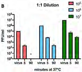
(Fig. 4B). To assess efficacy, SARS-CoV-2 B C
was mixed 1:1 with 0.5% baby shampoo (250 1.5
Relative Rt from baseline
µL shampoo in 24 mL normal saline) for 5 or
90 minutes, with titers of active virus 1.0
assessed using standard methods7.
Shampoo treatment decreased SARS-CoV-2 0.5
PBS
50μL/24mL
titers by ~2 orders of magnitude within 5 250μL/24mL
500μL/24mL
minutes of treatment, with further decreases 0.0
0 1 2 3 4 8 12 16 20 24
after 90 minutes (Fig. 4C). Even greater Hours
potency was observed with other viruses, D E
including the coronavirus NL63 (Fig. 4D) and
respiratory syncytial virus (RSV) (Fig. 4E).
For these experiments, we utilized
genetically modified NL63 coronavirus and
RSV that express Green Fluorescent Protein
(GFP), allowing the Tissue Culture Infectious
Dose (TCID50) to be easily tracked by the
intensity of the fluorescent signal8. This
approach allows us to rapidly test hypotheses Fig. 4. Baby Shampoo Safety and in vitro Efficacy. A) Confocal
without needing time in one of the few BSL-3 microscopy showing that application of baby shampoo to HNE did not result
in obvious evidence of cellular toxicity. B) Application of shampoo to HNE
facilities that are required for experiments resulted in short term decreases in transepithelial airway resistance (Rt)
with SARS-CoV-2. An additional benefit is only at higher concentrations, suggesting minimal toxicity. C) Treating
that data using NL63 is directly applicable to SARS-CoV-2 1:1 with shampoo reduced titers by two or more orders of
one of the viruses responsible for upper magnitude. D) Treating GFP-labeled NL-63 coronovirus 1:1 with shampoo
reduced tissue culture infectious dose (TCID) measured using fluorescence.
respiratory infections.Having established safety and antiviral activity,
we then explored efficacy of baby shampoo in an 10 8
Shampoo
7
PBS B
10
HNE model system of active viral infection. HNE 10 6
10 5
PFU/ml
were infected with SARS-CoV-2 at an MOI of 0.1, 10 4
and titers were obtained at 48 hours to verify 10 3
10 2
active infection. At 72 hours, cells were treated 10 1
with 200 µl of 0.5% shampoo or PBS as a control, 10
48
0
72 76 96
with the treatment removed by aspiration after 10 HPI
minutes to mimic typical mucociliary clearance D
time in the nose. Viral titers were then measured 80 Shampoo standard
four hours later (76 hours) and again at 96 hours. Shampoo wash 1 mi
As expected, viral titers were high at all 60 Shampoo wash 30 m
MS Peak Area
measured time points in the PBS (control) 40
treated cells (Fig. 5A). However, somewhat
surprisingly shampoo treatment had no 20
significant impact on viral titers at any timepoint. 0
Lack of efficacy in our HNE system parallels C8 C10 C12 C14 C16
CAPB Chain Length
C18
conclusions from a clinical trial by colleagues at Fig. 5. Baby Shampoo PK/PD. A) No differences in SARS-CoV-2
Vanderbilt, who found that nasal lavage of 0.5% titers were observed in HNE infected at day zero and treated with
baby shampoo had no significant impact on shampoo or PBS at 72 hours B) Baby shampoo nasal washes did not
nasal viral load in patients infected with SARS- affect SARS-CoV-2 viral shedding relative to control and saline alone,
as log10(change values from Ct value at first day to the max
CoV-2 (Fig. 5B). To better understand why baby measured
value of Ct)/days between two values) of the N1 nucleocapsid
shampoo did not exhibit antiviral activity on nasal expression. C). Full scan MS of baby shampoo dominated by six peaks
epithelia, we utilized MS to assess the PK that had masses and fragmentation patterns of various chain lengths
properties of shampoo when applied to HNE. of CAPB. D). Longer chain CAPB were depleted after baby shampoo
Baby shampoo is a proprietary mixture of was applied to then washed off HNE.
detergents and surfactants, but the dominant signal from full scan MS was found in six peaks with run times
between 7.6 and 10.1 that had masses and fragmentation patterns consistent with various chain lengths of
cocamidopropyl betaine (CAPB) (Fig. 5C). CAPB is the first listed surfactant in the baby shampoo ingredient list,
and the relative abundances of the different chain lengths, as assessed by peak area on chromatograms, was
similar to the known pattern of chain lengths in CAPB: mostly C12 with lesser quantities of lower and higher
chain lengths (Fig. 5D). We then evaluated samples in which shampoo was applied to HNE then washed after
1 or 30 minutes. We found that shorter chain lengths of CABP (C8 and C10) were readily recovered by HNE
wash, but that the concentrations of C12 were reduced significantly and the longest chain lengths (C14, C16,
C18) were below detection limits of the assay. These differences were observed in samples lavaged very quickly
(one minute) after application of baby shampoo to HNE and persisted in samples lavaged later (Fig. 5D).
Taken together, these findings indicated that while baby shampoo has antiviral properties and can be safely
applied to nasal epithelia, it does not have efficacy as a therapeutic. The most likely explanation is that the
antiviral activity of baby shampoo arises mainly from the longer chain surfactants, which are rapidly depleted
from baby shampoo applied to HNE by binding to or intercalating into epithelial membranes. These findings are
consistent with previous studies suggest that longer chain length surfactants are the most potent as antivirals
but bind readily to cells. Their rapid depletion on cell surfaces limits the clinical efficacy of baby shampoo and
likely other surfactant based therapies.
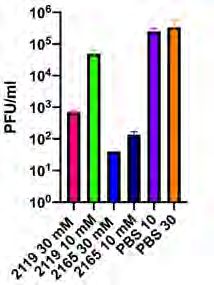
Reducing agents. Infection with SARS-CoV-2 depends upon an interaction between the viral spike protein and
a cellular receptor (ACE2). The viral spike protein has to have a specific configuration to bind well to the receptor,
which is maintained in part by the presence of disulfide bonds within the protein. Therefore reducing agents,
which break disulfide bonds, might change the configuration of the viral spike protein and reduce infectivity. We
tested this hypothesis through evaluation of two novel reducing agents from Parion Sciences, 2119 and 2165,
that are being developed as inhaled therapeutics for chronic lung diseases. Consistent with our hypothesis, both
2119 and 2165 were able to reduce the infectivity of SARS-CoV-2 treated prior to infection (Fig 6A). Similar
findings were seen with NL63 coronavirus and parainfluenza virus (PIV) (Fig 6B), both of which also rely on viral
protein-receptor interactions for infectivity. However, our previous studies have demonstrated that the inhaled
reducing agents are quickly oxidized on the cell surface (Fig 6C), suggesting a lack of durable antiviral action.Thus, we predict that the antiviral A B C
activity of reducing agents will be NL63 PIV
10 10 20
lost quickly after aerosolization. 2119-ox
10 8
Further development of newer 15 2119
Drug (µM)
TCID50
agents with prolonged cell surface 10 6
10
activity would be needed for 10 4
reducing agents to be effective as 5
10 2
clinical antiviral therapies.
10 0 0
Other agents. While the limited 0 2 4 6
19 B S
S
21 5 3 mM
10 M
M
21 5 3 mM
10 M
M
PB
65 0 m
m
65 0 m
m
Time (hours)
P
time of the award precluded
6 0
6 0
21 9 1
21 1
1
21
21
extensive investigation of other
agents, we did lay the groundwork Fig. 6. Reducing Agents. A) SARS-CoV-2 infectivity was significantly reduced by pre-
to examine other potential treatment with the reducing agents 2119 or 2165. B) Similar impact of reducing agents
was observed in NL63 and PIV. C) In bronchoalveolar lavage obtained from sheep at
therapies for COVID-19. various time intervals after treatment with nebulized 2119, very little native drug was
Camostat has potential antiviral detected. Oxidized 2119 (2119-ox) was present and cleared over time.
activity at the cell surface by
blocking cleavage of the viral spike protein needed for cell entry. Because the A
drug is rapidly metabolized, we developed methods to measure its primary
(and active) metabolite FOY-251 (Fig. 7A). We also developed methods to
measure ivermectin (Fig. 7B), an anti-parasitic drug that inhibits SARS-CoV-
replication in vitro but likely has poor distribution to the lungs with standard B
9
oral dosing . Development of these methods puts us in strong position for
future studies of these and other drugs that have antiviral activity.
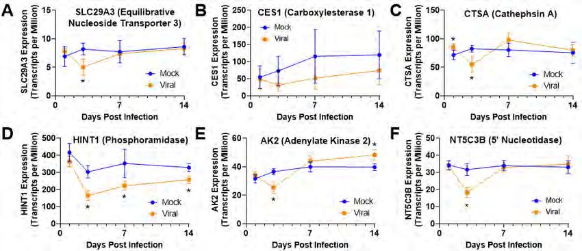
Viral effects on gene expression. Viral infection is known to affect gene Fig. 7. Other Agents. Mass
expression in airway epithelia, which could alter expression of genes involved spectrometric chromatograms of A)
in transport and metabolism of antiviral drugs. To assess potential impacts of FOY-251, the active metabolite of
viral infection on expression of relevant genes, we performed RNA camostat, and B) ivermectin
sequencing studies on cultured HBE infected with SARS-CoV-2. HBE were
obtained from
donors of a
variety of ages
from infants to
elderly, allowing
us to also assess
age related
changes in
expression. In
these
experiments,
gene expression
was measured at
day 1, 3, 7, and
14 post infection,
with viral titers Fig. 8. Gene Expression. HBE were infected with SARS-CoV-2 viral infected (orange) and mock infected
noted to be (blue), and expression over time was monitored for genes involved in remdesivir transport and metabolism
highest on day 3 including the nucleoside transporter SLC29A3 (A), the esterases CES1 (B) and CTSA (C), the
of infection. We phophoramidase HINT1 (D), the adenylate kinase AK2 (E), and the nucleotidase NT5C3B (F). Significant
decreases in expression in virally infected cells were noted for SLC29A3, CTSA, HIT1, AK2, and NT5C3B,
initially focused
on genes involved in transport and metabolism of remdesivir, since this drug relies on multiple transporters and
enzymes to be converted to its active, triphosphate form. Remdesivir is transported intracellularly via the
SLC29A3 nucleoside transporter and is then cleaved from its pro-drug form by carboxylesterase 1 (CES1) or
cathepsin A (CTSA). The drug is then converted into the nucleoside monophosphate by a phosphoramidase
(HINT1) and further phosphorylated to the active triphosphate by adenylate kinase (AK2). Active drug can be
desphosphorylated and inactivated by nucleotidases such as NTSC3B and NT5C2. In our gene expressionstudies, we observed that several of these genes were downregulated during active SARS-CoV-2 infection in
HBE, with the most significant changes observed at day 3 of active infection for SLC29A3, CTSA, HINT1, AK2,
and NTSC3B (Fig. 8A-F). These findings suggest that highly infected cells may have reduced ability to transport
remdesivir and convert it to its antiviral metabolite. We did not observe any significant impact of the age of the
donor on expression of these genes.
We performed a similar analysis for HCQ. Current literature suggests that
movement of HCQ into the cell is not highly dependent on transporters, as
the drug is sufficiently hydrophobic to readily cross lipid membranes.
Furthermore, intracellular accumulation is not transporter driven but results
from protonation of HCQ in acidic compartments such as lysosomes that
reduces its lipid permeability, effectively trapping it within the cell. However,
some studies have suggested that the efflux pump P-glycoprotein (ABCB1)
may influence intracellular HCQ concentrations, with evidence that HCQ
inhibits P-glycoprotein activity at high concentrations, and overexpression Fig. 9. Gene Expression. Gene
of P-glycoprotein reduces HCQ toxicity10. We do not believe this mechanism expression of P-glycoprotein (ABCB1)
would meaningfully impact HCQ concentrations in viral infection, since in was low and not influenced by SARS-
CoV-2 viral infection in HBE.
our RNA sequencing studies expression on ABCB1 in HBE was low and
not significantly influenced by viral infection (Fig. 9). We also do not believe viral infection would influence HCQ
metabolism, since the cytochrome P450 system likely involved in HCQ metabolism (CYP2C8, CYP3A4,
CYP2D6) are primarily expressed in the liver. Furthermore, we did not observe significant metabolism of HCQ
in HBE (see Fig 3). Therefore, unlike with remdesivir, we do not believe viral infection is likely to influence
intracellular HCQ concentrations in treated epithelia.
A B
Confocal Microscopy. Funding from this
proposal supported purchase of a new
confocal microscope that offers improved
resolution over our existing instruments.
Our primary purpose was to utilize this
instrument to assess for drug toxicities in
our PK/PD system, as we did for baby
shampoo (see Fig. 4A). This instrument Fig. 10. Confocal Microscopy. A) Confocal image of showing inflammasome
also proved useful in other studies of activation (PYCARD staining, green) in proximity to SARS-CoV-2 (red) in a
COVID-19, including evaluation of lung autopsy from a patient who died from COVID-19. Cell nuclei are stained
autopsied lungs from patients who died of blue with DAPI. B) Airway from a COVID-19 patient showing prominent
staining of human neutrophil elastase (ELANE, green) and naturally occurring
COVID-19. We were able to demonstrate protease inhibitor (SERPINA1, red).
increases in inflammasome activation
(Fig. 10A) as well as increased levels of neutrophil elastase and its naturally occurring inhibitor alpha-1
antitrypsin (Fig. 10B). These experiments highlight the role of inflammatory responses in COVID-19, which are
known to underlie much of the morbidity and mortality of this disease. They also identify potential targets for anti-
inflammatory treatments that are key aspects of COVID-19 treatment.
CONCLUSIONS
The goal of this project was to develop a respiratory epithelial PK/PD system to assess the therapeutic potential
of drugs for COVID-19, focusing on therapeutic agents suitable for aerosolization that were either active on the
cell surface and or required intracellular accumulation for efficacy. Our studies successfully demonstrated the
aerosolization potential of two intracellularly active drugs, remdesivir and HCQ. For both drugs, antiviral
intracellular concentrations could be achieved quickly using aerosolization strategies that equivalent to those
achievable with existing nebulizers. Although more work remains to be done, we anticipate that our findings can
be readily translated into clinical studies of aerosolized treatment early in disease when infection burden is less
or even prophylactically in exposed individuals. Our PK/PD system was also able to uncover challenges with cell
surface therapies that likely limit clinical utility. Although we have hypothesized that removal of drug by
mucociliary clearance would limit the potential of cell surface drugs, in fact lack of efficacy could be traced to
rapid inactivation of drug on cell surfaces. While disappointing, our system allows further testing of such agents
to identify strategies to overcome these challenges and increase the likelihood of success in future clinical trials.Indeed, funding from this proposal has left us well-positioned for future studies of aerosolized drug therapies for COVID-19 and other viral infections. References 1. Ehre C. SARS-CoV-2 Infection of Airway Cells. N Engl J Med 2020;383:969. 2. Cohen J, Kupferschmidt K. 'A very, very bad look' for remdesivir. Science 2020;370:642-3. 3. McChesney EW. Animal toxicity and pharmacokinetics of hydroxychloroquine sulfate. Am J Med 1983;75:11-8. 4. Maisonnasse P, Guedj J, Contreras V, et al. Hydroxychloroquine use against SARS-CoV-2 infection in non-human primates. Nature 2020;585:584-7. 5. Chiu AG, Palmer JN, Woodworth BA, et al. Baby shampoo nasal irrigations for the symptomatic post- functional endoscopic sinus surgery patient. Am J Rhinol 2008;22:34-7. 6. Hou YJ, Okuda K, Edwards CE, et al. SARS-CoV-2 Reverse Genetics Reveals a Variable Infection Gradient in the Respiratory Tract. Cell 2020;182:429-46 e14. 7. Sheahan TP, Sims AC, Zhou S, et al. An orally bioavailable broad-spectrum antiviral inhibits SARS-CoV- 2 in human airway epithelial cell cultures and multiple coronaviruses in mice. Sci Transl Med 2020. 8. Donaldson EF, Yount B, Sims AC, Burkett S, Pickles RJ, Baric RS. Systematic assembly of a full-length infectious clone of human coronavirus NL63. J Virol 2008;82:11948-57. 9. Chaccour C, Abizanda G, Irigoyen-Barrio A, et al. Nebulized ivermectin for COVID-19 and other respiratory diseases, a proof of concept, dose-ranging study in rats. Scientific reports 2020;10:17073. 10. Weiss J, Bajraktari-Sylejmani G, Haefeli WE. Interaction of Hydroxychloroquine with Pharmacokinetically Important Drug Transporters. Pharmaceutics 2020;12.
You can also read


