Circulating miR-145 as a Marker of Therapeutic Response to Anti- TNF Therapy in Patients With Ankylosing Spondylitis
←
→
Page content transcription
If your browser does not render page correctly, please read the page content below
Physiol. Res. 70: 255-264, 2021 https://doi.org/10.33549/physiolres.934542
Circulating miR-145 as a Marker of Therapeutic Response to Anti-
TNF Therapy in Patients With Ankylosing Spondylitis
Klára PRAJZLEROVÁ1,2, Martin KOMARC3, Šárka FOREJTOVÁ1,2, Karel PAVELKA1,2,
Jiří VENCOVSKÝ1,2, Ladislav ŠENOLT1,2, Mária FILKOVÁ1,2
1
Institute of Rheumatology, Prague, Czech Republic, 2Department of Rheumatology, First Faculty
of Medicine, Charles University, Prague, Czech Republic, 3Department of Anthropometrics and
Methodology, Faculty of Physical Education and Sport, Charles University, Prague, Czech
Republic
Received July 15, 2020
Accepted February 11, 2021
Epub Ahead of Print March 8, 2021
Summary Corresponding author
Circulating miRNAs appear promising therapeutic and prognostic K. Prajzlerová, Institute of Rheumatology, Na Slupi 4, 128 00
biomarkers. We aimed to investigate the predictive value of Prague, Czech Republic. Email: prajzlerova@revma.cz
circulating miRNAs on the disease outcome following anti-TNF
therapy in patients with ankylosing spondylitis (AS). Our study Introduction
included 19 AS patients assessed at baseline (M0), after three
(M3) and twelve months (M12) of therapy. Total RNA was
Ankylosing spondylitis (AS) is a chronic
isolated from plasma. A comprehensive analysis of 380 miRNAs
inflammatory disease that mainly affects the axial
using TaqMan Low Density Array (TLDA) was followed by a single
skeleton and typically presents with inflammatory back
assay validation of selected miRNAs. All AS patients had high
baseline disease activity and an excellent response to anti-TNF
pain (Taurog et al. 2016). Inflammatory back pain,
therapy at M3 and M12. TLDA analysis revealed the dysregulation reduced mobility of the spine, and radiographic evidence
of 17 circulating miRNAs, including miR-145. Single assay of sacroiliitis are all part of the modified New York
validation confirmed that miR-145 is significantly downregulated diagnostic criteria for AS (van der Linden et al. 1984).
at M3 compared to baseline. The decrease in the levels of The new ASAS (The Assessment of SpondyloArthritis
miR-145 from M0 to M3 negatively correlated with the change in International Society) classification criteria for axial
BASDAI from M0 to M3; and positively correlated with disease spondyloarthritis (AxSpA) use magnetic resonance
activity improvement from M3 to M12 as per BASDAI and ASDAS. imaging (MRI) of sacroiliac joints to detect spinal
The predictive value of the early change in miR-145 and levels of inflammation in patients with the non-radiographic stage
miR-145 at M3 were further validated by Receiver operating
of the disease and to enable earlier diagnosis (Rudwaleit
curves analysis. We show that the early change in circulating
et al. 2009). The most widely used tool for measuring
miR-145 may be a predictor for the future outcome of
disease activity of patients with AS is the BASDAI (Bath
AS patients treated with TNF inhibitors. Patients with a more
Ankylosing Spondylitis Disease Activity Index) that
significant decrease in miR-145 levels may show further
significant improvement of disease activity after 12 months.
reflects the disease activity from the patient´s perspective
Monitoring the expression of miR-145 in plasma in AS patients (Garrett et al. 1994). Later, the ASDAS (Ankylosing
may, therefore, influence our therapeutic decision-making. Spondylitis Disease Activity Score) for the assessment of
disease activity was developed (Lukas et al. 2009). This
Key words includes C-reactive protein (CRP) or erythrocyte
miRNA • Ankylosing spondylitis • anti-TNF therapy • Biomarker sedimentation rate (ESR) as laboratory markers of disease
activity. The worsening of spinal mobility is influenced
by inflammation in the early disease and structural
PHYSIOLOGICAL RESEARCH • ISSN 1802-9973 (online)
2021 Institute of Physiology of the Czech Academy of Sciences, Prague, Czech Republic
Fax +420 241 062 164, e-mail: physres@fgu.cas.cz, www.biomed.cas.cz/physiolres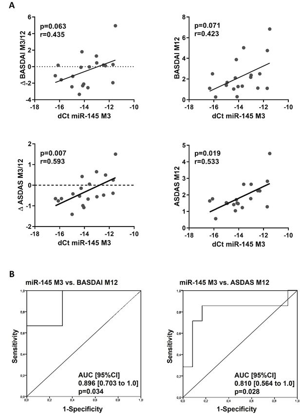
256   Prajzlerová et al.                                                                                         Vol. 70

damage in the radiographic phase (Machado et al. 2010).       Table 1. Patients were recruited from the outpatient clinic
The first evidence of good treatment response to anti-        of the Institute of Rheumatology in Prague. Written
TNF (tumor necrosis factor) agents in patients with AS        informed consent was obtained from all participants prior
was described almost 20 years ago (Brandt et al. 2000).       to enrolment. The study was approved by the local Ethics
Current data suggest that the early initiation of therapy     committee at the Institute of Rheumatology in Prague.
may reduce the radiographic progression in AS (Haroon
et al. 2013). Significant improvement of disease activity,    Samples and RNA isolation
functional outcomes and inhibition of radiographic                      Whole blood samples collected in EDTA tubes
progression have been shown in patients treated with          were obtained from all participants at baseline, at M3 and
anti-TNF biologic drugs (Taurog et al. 2016).                 M12 of therapy. Plasma was separated by centrifugation
          There are several biomarkers of activity or         within four hours of collection, ensuring constant pre-
treatment response being tested in AxSpA, but more            analytical conditions for all samples. All plasma samples
studies in large patient cohorts are needed for their         were stored at -80 °C, and no freeze-thaw cycles occurred
implementation in the clinical practice (Prajzlerova et al.   before use. Total RNA, including the miRNA fraction,
2016, Prajzlerova et al. 2017). CRP remains the best          was extracted from plasma samples (100μl) using
circulating marker for assessing disease activity and         miRNeasy Serum/Plasma Kit (Qiagen, Düsseldorf,
predicting treatment response and structural progression      Germany) according to manufacturer’s instructions.
(Prajzlerova et al. 2016). MicroRNA (miRNAs) are              Spike-in non-human synthetic miRNA (C. elegans cel-
small, non-coding RNAs important for post-                    miR-36, cel-miR-54, and cel-miR-238, Integrated DNA
transcriptional regulation of gene expression. Moreover,      Technologies, Coralville, IA, USA) were added to the
cell-free circulating miRNAs are released from the cells,     samples after the initial denaturation for further
are stable in body fluids, and may serve as promising         normalization. Total RNA was eluted in 24μl of RNase-
therapeutic and prognostic biomarkers, e.g., in               free water and stored at -80 °C. Total RNA concentration
rheumatoid arthritis, AxSpA, or other non-rheumatic           was measured using a NanoDrop 2000c spectropho-
diseases (Filkova et al. 2014, Hruskova et al. 2016,          tometer (Thermo Fisher Scientific, Waltham, MA, USA)
Prajzlerova et al. 2017, Dlouha and Hubacek 2017). We         in all samples.
have previously presented circulating miRNAs as
markers of spinal involvement and disease activity in         miRNA expression analysis
patients with AxSpA (Prajzlerova et al. 2017). The aim of              First, non-pooled individual plasma RNA
the current study was to investigate miRNAs as                samples (M0, M3, and M12 from randomly selected
predictors of treatment response to anti-TNF therapy in       3 patients) were used. Complementary DNA was
patients with AS.                                             obtained by reverse transcription using a TaqMan®
                                                              MicroRNA Reverse Transcription Kit with Megaplex RT
Methods                                                       Primers (Thermo Fisher Scientific) with equal RNA
                                                              input. cDNA was preamplified using 2x TaqMan®
Patients                                                      PreAmp Master Mix and Megaplex™ PreAmp Primers
          This study included 19 patients who fulfilled the   (all Thermo Fisher Scientific) on a PCR thermocycler
New York classification criteria for AS (van der Linden       (Bio-Rad Laboratories, Hercules, CA, USA). The
et al. 1984): 3 patients had stage I AS, 2 had stage II AS,   expression of 380 miRNAs was measured using Human
4 had stage III AS, 9 had stage IV AS, and 1 had a            Pool A TaqMan® Low Density Array (TLDA) platforms
bamboo spine – stage V AS (Braun et al. 2002). All            for microRNAs on QuantStudio 7Flex Real-Time PCR
patients commenced treatment with anti-TNF therapy: 6         System (Thermo Fisher Scientific). All steps were
received infliximab, 3 adalimumab, 5 golimumab, and 5         performed according to the manufacturer’s instructions.
were treated with etanercept. Clinical and laboratory         The dCt method was used for relative quantification as
parameters of disease activity were assessed using            follows: dCt=Ct(array average)-Ct(miRNA of interest),
BASDAI (Garrett et al. 1994), ASDAS-CRP (Lukas et             followed by x-fold change calculations.
al. 2009), ESR and CRP at baseline (M0) prior to the                   For further single assay validation, total RNA
start of treatment and after three (M3) and twelve months     from 19 non-pooled samples was reverse-transcribed
(M12) of therapy. Clinical characteristic is provided in      using TaqMan Real Time miRNA specific primers
2021 Circulating miR-145 in Ankylosing Spondylitis 257 Table 1. Clinical characteristics of patients with ankylosing spondylitis Variable M0 M3 M12 P value Gender; Female/Male 6/13 - - NA Age; years 38.16 [15.22] - - NA HLA-B27 positivity; n (%) 19 (100 %) - - NA CRP; mg/l 22.61 [29.47] 3.09 [8.01] 3.96 [5.70]
258   Prajzlerová et al.                                                                                                 Vol. 70

Fig. 1. Plasma expression of miR-145 at baseline, month 3, and month 12 of anti-TNF therapy. (A) Expression analyzed using
comprehensive TaqMan Low Density array and (B) single assay validation. Data expressed as median with interquartile range. NS, not
significant. * p≤0.05 dCt was calculated as follows: (A) dCt=Ct(array average)-Ct(miRNA of interest) and (B) Ct(spike-in average)-
Ct(miRNA of interest). Higher dCt values represent, therefore a higher expression of miR-145.

differences between M3 and M12 in any monitored
parameters of disease activity (p>0.05 for all
comparisons).

Analysis of miRNAs
         A comprehensive analysis of 380 circulating
miRNAs in 3 patients was performed using TLDA, as
described above. Out of the 380 miRNAs, 125 miRNAs
were detected in all samples, while 148 miRNAs were
detected at M0, 154 at M3, and 151 at M12. Only
miRNAs expressed consistently in all samples below
Ct cycle 30 and showing >1.5-fold change between at
least 2 groups in initial analysis were taken for further
validation (17 miRNAs in total). Single assay validation
confirmed any difference between groups in the                     Fig. 2. Negative correlation between the change in miR-145
                                                                   levels and BASDAI from month 0 to month 3. Δ dCt miR-145
expression of 9 miRNAs (Thermo Fisher Scientific Assay             M0/M3 is calculated as M3-M0 (numbers
2021 Circulating miR-145 in Ankylosing Spondylitis 259
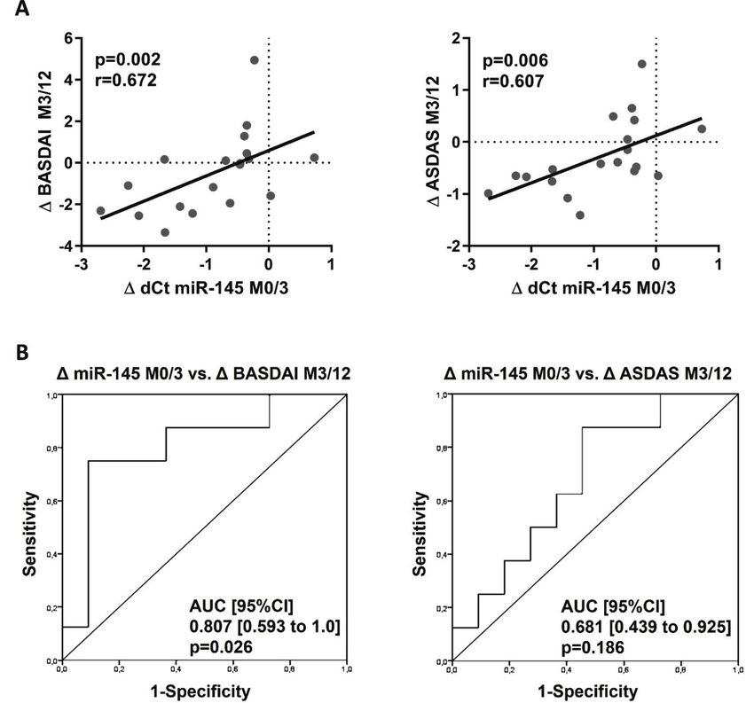
(p=0.002; r=0.672) and ASDAS (p=0.006; r=0.607) outcome at M12: the lower levels of miR-145 at M3, the
(Fig. 3A). Therefore, patients with a more significant more improvement of disease activity from M3 to M12
decrease in miR-145 levels associated with less initial and disease activity at M12 assessed by both BASDAI
improvement in BASDAI until M3 may show further (Δ BASDAI: p=0.063; r=0. 435 and BASDAI: p=0.071;
significant improvement of BASDAI (and ASDAS) at r=0.423) and ASDAS (Δ ASDAS: p=0.007; r=0.593 and
M12. Importantly, this was further confirmed by ROC ASDAS: p=0.019; r=0.533) (Fig. 4A). ROC analysis
analysis. The ROC curves showed that the decrease in the confirmed, that plasma levels of miR-145 at M3
miR-145 expression from M0 to M3 predicts the disease significantly predicted low disease activity at M12
activity improvement from M3 to M12 defined by defined by BASDAI (cut off 4) (AUC [95 % CI] 0.896
BASDAI (AUC [95 %CI] 0.807 [0.593 to 1.0], p=0.026), [0.703 to 1.0]; p=0.034) and ASDAS (cut of 2.1) (AUC
but not by ASDAS (AUC [95 %CI] 0.601 [0.439 to [95 % CI] 0.810 [0.564 to 1.0]; p=0.028) (Fig. 4B),
0.925], p=0.186) (Fig. 3B). nevertheless it did not predict remission defined by
Furthermore, there was a positive correlation ASDAS (cut off 1.3) (data not shown).
between the levels of miR-145 at M3 and disease
Fig. 3. Relationship between the early change in miR-145 expression from month 0 to month 3 and further clinical improvement from
month 3 to 12. (A) Correlation of decreasing in miR-145 expression from M0 to M3 with disease activity improvement from M3 to M12.
(B) ROC curves analysis of the change in miR-145 expression from M0 to M3 as a predictor of disease activity improvement from M3 to
M12 defined by BASDAI and ASDAS. Δ dCt miR-145 M0/M3 is calculated as M3-M0 (numbers260 Prajzlerová et al. Vol. 70 Fig. 4. Relationship between the expression of miR-145 in month 3 and disease activity outcome at month 12. (A) Correlation of expression of miR-145 at M3 with disease activity outcome at M12. (B) Receiver operator characteristic (ROC) curves analysis of expression of miR145 at M3 as a predictor for the low disease activity at M12 defined by BASDAI (
2021                                                                     Circulating miR-145 in Ankylosing Spondylitis   261

         Overall, as shown in Fig. 1, there was no          of therapeutic response to anti-TNF treatment in
significant change in miR-145 levels from M3 to M12.        RA patients (Castro-Villegas et al. 2015). Regarding
However, the plasma expression of miR-145 increased         individuals with AS treated with anti-TNF therapy,
from M3 to M12 in 10 patients (p0.05). In the first group with increasing     a minimum 1.5-fold change difference were selected for
levels of miR-145 from M3 to M12, 7 patients were           further validation. From selected miRNAs, only miR-145
treated with monoclonal antibodies (2 with adalimumab,      revealed a significant decrease from baseline to month
4 with golimumab, and 1 with infliximab) and the            three. There was still a significant decrease at month 12
remaining 3 with etanercept. In the second group,           compared to baseline, but there was no difference
7 patients were treated with monoclonal antibodies          compared to month three.
(1 with adalimumab, 1 with golimumab, and 5 with                      Next, we validated the pilot data and assessed
infliximab), and 2 were treated with etanercept.            the relationship between miR-145 and the disease activity
                                                            and treatment response in a larger patient cohort. We
VEGF and association with circulating miR-145 and           confirmed the decrease of miR-145 from baseline to M3
treatment response                                          but no change from M3 to M12. We found that the more
         As VEGF was shown as a direct target of            significant decrease in miR-145 expression correlated
miR-145 (Fan et al. 2012), we were interested in its        with poorer improvement of disease activity during the
association with circulating miR-145 in plasma. There       first 3 months. Importantly, our data showed that patients
was a trend towards the decrease in VEGF levels from        with a more significant reduction in miR-145 levels that
M0 to M3 (p=0.069), but there was no further change at      is associated with the little improvement in BASDAI
M12 compared to M3. Levels of VEGF do not correlate         until month 3 may develop further significant
with disease activity or its change at any time; however,   improvement and better disease outcomes after
the decrease in VEGF from M0 to M3 correlated with          12 months of therapy based on BASDAI and ASDAS.
decreased CRP (p=0.001; r=0.733) and ESR (p=0.023;          The assessment of treatment response to anti-TNF
r=0.517) from M0 to M3. There was no correlation            treatment after 3 months is essential for further treatment
between the levels of VEGF and miR-145 at any time          decision making: whether the switch is indicated or the
(p>0.1 for all comparisons) (data not shown).               patient remains on the same drug. Based on our miR-145
                                                            data, we propose that the therapeutic switch after
Discussion                                                  3 months would be premature as some patients will
                                                            improve significantly after 12 months on the same
         MicroRNAs are essential regulators of gene         therapy.
expression and therefore contribute to the physiology in              We hypothesized a potential link between
health as well as to the pathophysiology of diseases,       circulating miR-145 (and its reflection of disease
including inflammatory conditions (Filkova et al. 2012).    outcome) in our AS patients with its possible
Circulating miRNAs were shown to be very stable and         involvement in the pathogenesis of AS. MiR-145 is
easily accessible that makes them potential biomarkers      known mainly as a tumor suppressor that mediates cell
(Cortez et al. 2011). Circulating miRNAs miR-23a-3p         growth, invasion, and metastasis (Sachdeva and Mo,
and miR-223-3p were previously described as biomarkers      2010). VEGF was shown to be increased in patients with
262 Prajzlerová et al. Vol. 70
AS compared to healthy controls and decreased following Despite presenting miR-145 as a predictor of
anti-TNF therapy (Pedersen et al. 2010). VEGF is future response to anti-TNF therapy, our study has some
a crucial mediator in angiogenesis, is involved in limitations. Firstly, our data show the association of
osteogenesis, and, therefore, bone remodeling (Hu and miR-145 with a short-term disease outcome in advanced
Olsen 2016) and was previously described as a direct disease (most AS patients had cervical spine
target gene of miR-145 (Fan et al. 2012). Although we involvement). The predictive value of miR-145 for
proved the decrease in systemic VEGF after anti-TNF radiographic progression is missing and would need long-
therapy, as shown previously (Pedersen et al. 2010), we term follow up. Similarly, analysis of miR-145 in the
showed no associations between circulating miR-145 and non-radiographic disease would undoubtedly be of
VEGF. interest as well, although our previous study showed
The characteristic inflammation of no difference in miR-145 between healthy controls and
spondyloarthritis occurs at the interface between cartilage non-radiographic AxSpA. Our data also need validation
and bone in the sacroiliac joints, the spine, and the in larger patient cohorts. The data from published in vitro
entheses. The mechanisms of interaction between and in vivo studies show different roles of miR-145 in
inflammation, paradoxical bone destruction, and new inflammation or bone formation. Our data are descriptive
bone formation are still not completely understood. and show no association with selected mediators of
However, studies have suggested that the initial inflammation or tissue remodeling (e.g., CRP or VEGF).
inflammation (in which TNF is the principal cytokine We can only hypothesize the exact role of miR-145 in the
involved) causes erosions in cartilage and bone; these pathogenesis of AS and, especially, which
lesions are then filled in by fibrous tissue, that is later pathophysiologic aspect of TNF inhibition is reflected by
ossified, leading to abnormal bony growth changes in circulating miR-145 in patients with AS.
(syndesmophytes, bone bridges, and complete ankyloses) Although bone-remodeling appears suggestive, we are
(Sieper and Poddubnyy 2017, Watad et al. 2018, Cici unable to directly prove this as we didn´t look directly at
et al. 2019). Undoubtedly, the Wnt (Wingless) signaling the tissues of interest, and also, when circulating miRNAs
pathway is considered one of the primary regulators of are studied, they may originate from different cells or
bone metabolism. It is involved in osteoblast tissues and reflect various more or less specific tissue-
differentiation from mesenchymal precursors and related pathways.
osteoprogenitor cells, osteoblast regulation, proliferation, To conclude, we show that the early change in
and survival (Cici et al. 2019). MiR-145 was shown to circulating miR-145 may be a predictor for the future
negatively regulate chondrogenesis and bone formation outcome of AS patients treated with anti-TNF inhibitors.
(osteogenic differentiation, osteoblastogenesis or bone Patients with a more significant decrease in miR-145
regeneration) at multiple levels of Wnt signaling and levels may show further significant improvement of
other bone-homeostasis related mechanisms (Jia et al. disease activity after 12 months. Monitoring the
2013, Yang et al. 2011, Hao et al. 2018, Fukuda et al. expression of miR-145 in plasma in AS patients may,
2015) and to aggravate bone erosions by promoting therefore, influence our therapeutic decision-making.
osteoclastogenesis (Chen et al. 2018). We, therefore,
suggest that our data may reflect the involvement of Conflict of Interest
miR-145 in bone-remodeling in AS. In fact, in our There is no conflict of interest.
previous work, we showed lower levels of miR-145 in
plasma of AS patients than healthy controls and between Acknowledgements
non-radiographic AxSpA and advanced AS stages This work was supported by a grant 17-33127A of the
(Prajzlerova et al. 2017). We hypothesize that the Agency for Healthcare Research of the Czech Republic
decrease in miR-145 levels in the first 3 months of anti- and a project of the MHCR for conceptual research
TNF therapy can reflect the shift from the initial erosive development No. 023728.
process to the induction of reparative mechanisms There is no financial support or other benefits from
represented by new bone formation. In our study, these commercial sources for the work reported in the
could be reflected by further improvement in BASDAI manuscript.
score during follow-up.2021 Circulating miR-145 in Ankylosing Spondylitis 263
References
BRANDT J, HAIBEL H, CORNELY D, GOLDER W, GONZALEZ J, REDDIG J, THRIENE W, SIEPER J, BRAUN
J: Successful treatment of active ankylosing spondylitis with the anti-tumor necrosis factor alpha monoclonal
antibody infliximab. Arthritis Rheum 43: 1346-1352, 2000. https://doi.org/10.1002/1529-
0131(200006)43:63.0.CO;2-E
BRAUN J, VAN DER HEIJDE D, DOUGADOS M, EMERY P, KHAN MA, SIEPER J, VAN DER LINDEN S: Staging
of patients with ankylosing spondylitis: a preliminary proposal. Ann Rheum Dis 61 (Suppl 3): iii9-iii23, 2002.
https://doi.org/10.1136/ard.61.suppl_3.iii19
CASTRO-VILLEGAS C, PEREZ-SANCHEZ C, ESCUDERO A, FILIPESCU I, VERDU M, RUIZ-LIMON P,
AGUIRRE MA, JIMENEZ-GOMEZ Y, FONT P, RODRIGUEZ-ARIZA A, PEINADO JR, COLLANTES-
ESTEVEZ E, GONZALEZ-CONEJERO R, MARTINEZ C, BARBARROJA N, LOPEZ-PEDRERA C:
Circulating miRNAs as potential biomarkers of therapy effectiveness in rheumatoid arthritis patients treated
with anti-TNFalpha. Arthritis Res Ther 17: 49, 2015. https://doi.org/10.1186/s13075-015-0555-z
CICI D, CORRADO A, ROTONDO C, CANTATORE FP: Wnt Signaling and Biological Therapy in Rheumatoid
Arthritis and Spondyloarthritis. Int J Mol Sci 20: 5552 2019. https://doi.org/10.3390/ijms20225552
CIECHOMSKA M, BONEK K, MERDAS M, ZARECKI P, SWIERKOT J, GLUSZKO P, BOGUNIA-KUBIK K,
MASLINSKI W: Changes in MiRNA-5196 Expression as a potential biomarker of anti-TNF-alpha therapy in
rheumatoid arthritis and ankylosing spondylitis patients. Arch Immunol Ther Exp (Warsz) 66: 389-397, 2018.
https://doi.org/10.1007/s00005-018-0513-y
CORTEZ MA, BUESO-RAMOS C, FERDIN J, LOPEZ-BERESTEIN G, SOOD AK, CALIN GA: MicroRNAs in
body fluids--the mix of hormones and biomarkers. Nat Rev Clin Onco 8: 467-477, 2011.
https://doi.org/10.1038/nrclinonc.2011.76
DLOUHA D, HUBACEK JA: Regulatory RNAs and cardiovascular disease - with a special focus on circulating
microRNAs. Physiol Res 66: S21-S38, 2017. https://doi.org/10.33549/physiolres.933588
FAN L, WU Q, XING X, WEI Y, SHAO Z: MicroRNA-145 targets vascular endothelial growth factor and inhibits
invasion and metastasis of osteosarcoma cells. Acta Biochim Biophys Sin (Shanghai) 44: 407-414, 2012.
https://doi.org/10.1093/abbs/gms019
FILKOVA M, ARADI B, SENOLT L, OSPELT C, VETTORI S, MANN H, FILER A, RAZA K, BUCKLEY CD,
SNOW M, VENCOVSKY J, PAVELKA K, MICHEL BA, GAY RE, GAY S, JUNGEL A: Association of
circulating miR-223 and miR-16 with disease activity in patients with early rheumatoid arthritis. Ann Rheum
Dis 73: 1898-1904, 2014. https://doi.org/10.1136/annrheumdis-2012-202815
FILKOVA M, JUNGEL A, GAY RE, GAY S: MicroRNAs in rheumatoid arthritis: potential role in diagnosis and
therapy. BioDrugs 26: 131-141, 2012. https://doi.org/10.2165/11631480-000000000-00000
FUKUDA T, OCHI H, SUNAMURA S, HAIDEN A, BANDO W, INOSE H, OKAWA A, ASOU Y, TAKEDA S:
MicroRNA-145 regulates osteoblastic differentiation by targeting the transcription factor Cbfb. FEBS Lett589:
3302-3308, 2015. https://doi.org/10.1016/j.febslet.2015.09.024
GARRETT S, JENKINSON T, KENNEDY LG, WHITELOCK H, GAISFORD P, CALIN A: A new approach to
defining disease status in ankylosing spondylitis: the Bath Ankylosing Spondylitis Disease Activity Index.
J Rheumatol 21: 2286-2291, 1994.
HAO W, LIU H, ZHOU L, SUN Y, SU H, NI J, HE T, SHI P, WANG X: MiR-145 regulates osteogenic differentiation
of human adipose-derived mesenchymal stem cells through targeting FoxO1. Exp Biol Med (Maywood) 243:
386-393, 2018. https://doi.org/10.1177/1535370217746611
HAROON N, INMAN RD, LEARCH TJ, WEISMAN MH, LEE M, RAHBAR MH, WARD MM, REVEILLE JD,
GENSLER LS: The impact of tumor necrosis factor alpha inhibitors on radiographic progression in ankylosing
spondylitis. Arthritis Rheum 65: 2645-2654, 2013. https://doi.org/10.1002/art.38070
HRUSKOVA V, JANDOVA R, VERNEROVA L, MANN H, PECHA O, PRAJZLEROVA K, PAVELKA K,
VENCOVSKY J, FILKOVA M, SENOLT L: MicroRNA-125b: association with disease activity and the
treatment response of patients with early rheumatoid arthritis. Arthritis Res Ther 18: 124, 2016.
https://doi.org/10.1186/s13075-016-1023-0264 Prajzlerová et al. Vol. 70
HU K, OLSEN BR: The roles of vascular endothelial growth factor in bone repair and regeneration. Bone 91: 30-38,
2016. https://doi.org/10.1016/j.bone.2016.06.013
CHEN Y, WANG X, YANG M, RUAN W, WEI W, GU D, WANG J, GUO X, GUO L, YUAN Y: miR-145-5p
increases osteoclast numbers in vitro and aggravates bone erosion in collagen-induced arthritis by targeting
osteoprotegerin. Med Sci Monit 24: 5292-5300, 2018. https://doi.org/10.12659/MSM.908219
JIA J, TIAN Q, LING S, LIU Y, YANG S, SHAO Z: miR-145 suppresses osteogenic differentiation by targeting Sp7.
FEBS Lett 587: 3027-3031, 2013. https://doi.org/10.1016/j.febslet.2013.07.030
LUKAS C, LANDEWE R, SIEPER J, DOUGADOS M, DAVIS J, BRAUN J, VAN DER LINDEN S, VAN DER
HEIJDE D: Development of an ASAS-endorsed disease activity score (ASDAS) in patients with ankylosing
spondylitis. Ann Rheum Dis 68: 18-24, 2009. http://dx.doi.org/10.1136/ard.2008.094870
LV Q, LI Q, ZHANG P, JIANG Y, WANG X, WEI Q, CAO S, LIAO Z, LIN Z, PAN Y, HUANG J, LI T, JIN O,
WU Y, GU J: Disorders of microRNAs in peripheral blood mononuclear cells: as novel biomarkers of
ankylosing spondylitis and provocative therapeutic targets. Biomed Res Int 2015: 504208, 2015.
https://doi.org/10.1155/2015/504208
MACHADO P, LANDEWE R, BRAUN J, HERMANN KG, BAKER D, VAN DER HEIJDE D: Both structural damage
and inflammation of the spine contribute to impairment of spinal mobility in patients with ankylosing
spondylitis. Ann Rheum Dis 69: 1465-1470, 2010. https://doi.org/10.1136/ard.2009.124206
PEDERSEN SJ, HETLAND ML, SORENSEN IJ, OSTERGAARD M, NIELSEN HJ, JOHANSEN JS: Circulating
levels of interleukin-6, vascular endothelial growth factor, YKL-40, matrix metalloproteinase-3, and total
aggrecan in spondyloarthritis patients during 3 years of treatment with TNFalpha inhibitors. Clin Rheumatol
29: 1301-1309, 2010. https://doi.org/10.1007/s10067-010-1528-x
PRAJZLEROVA K, GROBELNA K, HUSAKOVA M, FOREJTOVA S, JUNGEL A, GAY S, VENCOVSKY J,
PAVELKA K, SENOLT L, FILKOVA M: Association between circulating miRNAs and spinal involvement in
patients with axial spondyloarthritis. PLoS One12: e0185323, 2017.
https://doi.org/10.1371/journal.pone.0185323
PRAJZLEROVA K, GROBELNA K, PAVELKA K, SENOLT L, FILKOVA M: An update on biomarkers in axial
spondyloarthritis. Autoimmun Rev 15: 501-509, 2016. https://doi.org/10.1016/j.autrev.2016.02.002
RUDWALEIT M, LANDEWE R, VAN DER HEIJDE D, LISTING J, BRANDT J, BRAUN J, BURGOS-VARGAS R,
COLLANTES-ESTEVEZ E, DAVIS J, DIJKMANS B, DOUGADOS M, EMERY P, VAN DER HORST-
BRUINSMA IE, INMAN R, KHAN MA, LEIRISALO-REPO M, VAN DER LINDEN S, MAKSYMOWYCH
WP, MIELANTS H, OLIVIERI I, STURROCK R, DE VLAM K, SIEPER, J: The development of Assessment
of SpondyloArthritis international Society classification criteria for axial spondyloarthritis (part I):
classification of paper patients by expert opinion including uncertainty appraisal. Ann Rheum Dis 68: 770-776,
2009. https://doi.org/10.1136/ard.2009.108217
SACHDEVA M, MO YY: miR-145-mediated suppression of cell growth, invasion and metastasis. Am J Transl Res 2:
170-180, 2010.
SIEPER J, PODDUBNYY D: Axial spondyloarthritis. Lancet 390: 73-84, 2017. https://doi.org/10.1016/S0140-
6736(16)31591-4
TAUROG JD, CHHABRA A, COLBERT RA: Ankylosing spondylitis and axial spondyloarthritis. N Engl J Med 375:
1303, 2016. https://doi.org/10.1056/NEJMc1609622
VAN DER LINDEN S, VALKENBURG HA, CATS A: Evaluation of diagnostic criteria for ankylosing spondylitis.
A proposal for modification of the New York criteria. Arthritis Rheum 27: 361-368, 1984.
https://doi.org/10.1002/art.1780270401
WATAD A, BRIDGEWOOD C, RUSSELL T, MARZO-ORTEGA H, CUTHBERT R, MCGONAGLE D: The early
phases of ankylosing spondylitis: emerging insights from clinical and basic science. Front Immunol 9: 2668,
2018. https://doi.org/10.3389/fimmu.2018.02668
YANG B, GUO H, ZHANG Y, CHEN L, YING D, DONG S: MicroRNA-145 regulates chondrogenic differentiation of
mesenchymal stem cells by targeting Sox9. PLoS One 6: e21679, 2011.
https://doi.org/10.1371/journal.pone.0021679You can also read