Case Report Perforated Meckel's Diverticulitis in a Patient with Unknown Intestinal Malrotation: Clinical Pitfall
←
→
Page content transcription
If your browser does not render page correctly, please read the page content below
Hindawi
Case Reports in Surgery
Volume 2021, Article ID 5595803, 3 pages
https://doi.org/10.1155/2021/5595803

Case Report
Perforated Meckel’s Diverticulitis in a Patient with Unknown
Intestinal Malrotation: Clinical Pitfall

          Marie Burgard , Floryn Cherbanyk , François Pugin , and Bernhard Egger
          Department of Surgery, HFR Fribourg-Cantonal Hospital Fribourg, Switzerland

          Correspondence should be addressed to Bernhard Egger; bernhard.egger@h-fr.ch

          Received 8 January 2021; Accepted 28 February 2021; Published 8 March 2021

          Academic Editor: Boris Kirshtein

          Copyright © 2021 Marie Burgard et al. This is an open access article distributed under the Creative Commons Attribution License,
          which permits unrestricted use, distribution, and reproduction in any medium, provided the original work is properly cited.

          Symptomatic Meckel’s diverticulum is rare in adults. The most frequent complications are intestinal obstruction and diverticulitis.
          Diagnosis of Meckel’s diverticulitis can be challenging due to nonspecific clinical manifestation of pain in the right lower abdominal
          quadrant, mimicking acute appendicitis. If associated with congenital malformation, such as intestinal malrotation, the anomalous
          anatomy makes the diagnosis even more challenging. In such cases, radiological imaging is essential to guide further management.
          We present a case of Meckel’s diverticulitis in which physicians were initially misguided because of the atypical clinical
          presentation. Yet, anamnestic details directed to a potential underlying malformation, leading to supplementary radiological
          examination and the final diagnosis.

1. Case Report                                                           quadrant. The clinical examination showed no abdominal
                                                                         tenderness, and the patient had normal blood values. The
Meckel’s diverticulum (MD) mostly remains asymptomatic                   patient was discharged with a diagnosis of mild sigmoid
with a lifetime onset of complications of 4% but decreasing              diverticulitis and treated with oral antibiotics. Twenty-four
with age [1]. Therefore, symptomatic MD in adults is rare.               hours later, he presented to our emergency department with
The most common complications are intestinal obstruction                 increasing abdominal pain, fever of 38.1°C, and nausea. A
and diverticulitis, which may lead to perforation, and gastroin-         detailed history revealed possible malrotation of the intestine.
testinal bleeding. Diverticulitis of such a diverticulum usually         Clinical examination revealed localized periumbilical and left
presents as acute right or diffuse abdominal pain with or with-           lower quadrant abdominal tenderness. Laboratory tests
out localized peritonitis. Preoperative diagnosis is achieved in         demonstrated a normal white blood cell count but increased
only 5-15% of cases, and inflamed MD is often misdiagnosed                C-reactive protein (52 mg/l).
as acute appendicitis [2]. Localized left-sided abdominal pain               Based on the clinical examination, the main diagnostic
as a manifestation of Meckel’s diverticulitis is highly unusual          hypothesis was sigmoid diverticulitis. Nevertheless, the
but can occur in patients with intestinal malrotation.                   possibility of a malrotated intestine led to the differential
     Acute diverticulitis of such a diverticulum may lead to             diagnosis of acute appendicitis. Abdominal CT revealed a
perforation, and treatment is emergency surgery. Emergency               complete intestinal malrotation and inflamed diverticulum
physicians should be aware of this pathology and keep it in              containing a calculus in the left upper quadrant without radio-
mind as a differential diagnosis of acute abdominal pain.                 logical signs of acute appendicitis (Figure 1(a)). The appen-
Our case illustrates Meckel’s diverticulitis in a patient                dix was localized in the left lower quadrant (Figure 2(a)).
presenting with left abdominal pain due to an undiagnosed                    The patient underwent exploratory laparoscopy, show-
intestinal malrotation.                                                  ing a perforated diverticulum at the antimesenteric border
     A 65-year-old male patient with a medical history of                of the ileum 50 cm from the ileocecal valve with local peri-
sigmoid diverticulosis presented to his general practitioner             tonitis (Figure 1(b)). A segmental intestinal resection with
with mild acute abdominal pain localized in the left lower               anastomosis and occasional appendectomy was performed.
2 Case Reports in Surgery
(a) (b)
Figure 1: (a) Coronal reformatted contrast-enhanced CT images thickened with maximal intensity projection demonstrate malrotation of the
small intestine with the jejunum and a large part of the ileum placed on the right side of the abdomen (black arrow). In the left hemiabdomen
is a blind-ending tubular structure arising from the distal small bowel (white arrow), which has a markedly thickened wall with extensive
surrounding fat stranding. Note the intraluminal calcified lesion (white arrowhead) contained in the saccular structure. (b) Laparoscopic
view of perforated Meckel diverticulitis.
⁎
⁎
(a) (b)
Figure 2: (a) Coronal reformatted contrast-enhanced CT image and (b) laparoscopic view demonstrate malrotation of the bowel with the
ileocaecal valve (white arrowhead), the caecum (asterisk), and the normal appendix (white arrow) placed on the left side of the abdomen.
Left hepatic lobe: black arrowhead.
Perioperative microbiological examination showed polymi-
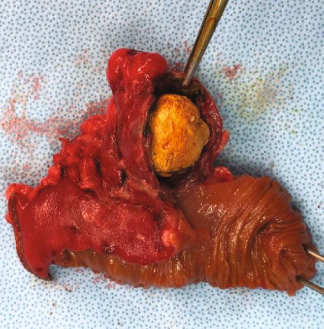
crobial intestinal flora. Examination of the 5 × 4:5 cm
diverticulum confirmed the intradiverticular voluminous
calculus (Figure 3). Definitive histology demonstrated
inflamed gastric mucosa in the diverticulum, confirming the
suspected diagnosis of a perforated MD. Due to the intestinal
malrotation, the MD and appendix were localized in the left
upper quadrant (Figure 2(b)). The postoperative outcome ⁎
was uneventful, and the patient was discharged home on
postoperative day 5 with oral antibiotics for another week.
The 6-week outpatient control was favorable.
MD is the most common malformation of the gastro-
intestinal tract and is found in 0.14-0.4% of autopsies [3].
It is a remnant of the vitelline duct and, in adults, is Figure 3: Surgical specimen: intestinal segment (asterisk), Meckel
usually located up to 100 cm from the ileocecal valve at diverticulum (white arrow), and intradiverticular calculus (white
the antimesenteric border of the ileum [4]. Anatomically, it arrowhead).
Case Reports in Surgery                                                                                                                           3

is a true diverticulum with all layers of the gastrointestinal                  The European Journal of Surgery, vol. 167, no. 7, pp. 518–524,
tract. Occasionally, it contains gastric or pancreatic                          2001.
mucosa [5].                                                               [3]   J. Dumper, S. Mackenzie, P. Mitchell, F. Sutherland, M. L.
    Most MD cases stay asymptomatic with a lifetime                             Quan, and D. Mew, “Complications of Meckel’s diverticula in
complication rate of only 4% [1]. These complications most                      adults,” Canadian Journal of Surgery, vol. 49, no. 5, p. 353, 2006.
often occur before the age of 8 years; thus, symptomatic                  [4]   K. Blouhos, K. A. Boulas, K. Tsalis et al., “Meckel's diverticulum
MD in adults is rare.                                                           in adults: surgical concerns,” Frontiers in Surgery, vol. 5, p. 55,
    As the diverticulitis can lead to perforation, the treatment                2018.
should be surgical. However, preoperative diagnosis can be                [5]   J. R. Robinson, H. Correa, A. S. Brinkman, and H. N. Lovvorn
difficult and is only achieved in 5-15% of cases, and inflamed                     III, “Optimizing surgical resection of the bleeding Meckel diver-
MD is often misdiagnosed as acute appendicitis [2]. Abdom-                      ticulum in children,” Journal of Pediatric Surgery, vol. 52, no. 10,
inal CT can help make the diagnosis; yet, most MD cases are                     pp. 1610–1615, 2017.
diagnosed intraoperatively during laparoscopy, especially if              [6]   J. J. Neville, J. Gallagher, A. Mitra, and H. Sheth, “Adult presen-
the diverticula do not contain a fecolith or foreign body. In                   tations of congenital Midgut malrotation: A Systematic
patients with unknown congenital malformation, clinical                         Review,” World Journal of Surgery, vol. 44, no. 6, pp. 1771–
diagnosis is even more difficult.                                                 1778, 2020.
    Intestinal malrotation is a congenital malformation due
to the incomplete rotation of the fetal intestine around the
superior mesenteric vessels. With an incidence of 0.2%, it is
diagnosed during the first year of life in 90% of cases [6]. Inci-
dental diagnosis in adult patients is rare. In patients with
unknown intestinal malrotation, the diagnostic and thera-
peutic delay of acute abdominal pain can be important, with
an increasing risk of morbidity and mortality. Physicians
should keep in mind the differential diagnosis of acute
appendicitis or Meckel’s diverticulitis in a patient presenting
with acute left abdominal pain, especially if there are anam-
nestic hints of intestinal malrotation. Abdominal CT may
help make the diagnosis; if the CT scan is not conclusive,
diagnostic laparoscopy may help and is mandatory in the
presence of peritonitis.
    Because of the possible diagnostic and therapeutic delay
in patients with intestinal malrotation, occasional resection
of noninflamed MD and the appendix should be considered
when performing laparoscopy for other reasons.

Additional Points
Highlights. (i) Meckel’s diverticulum is often misdiagnosed as
acute appendicitis. (ii) Intestinal malrotation can delay the
diagnosis of abdominal pain. (iii) Appendicitis or Meckel’s
diverticulitis can be responsible for left abdominal pain.

Conflicts of Interest
The authors declare that they have no conflicts of interest.

Authors’ Contributions
The first two authors, Marie Burgard and Floryn Cherbanyk,
contributed equally to this work.

References
[1] A. Parvanescu, M. Bruzzi, T. Voron et al., “Complicated
    Meckelʼs diverticulum,” Medicine, vol. 97, no. 38, article
    e12457, 2018.
[2] Y. Groebli, D. Bertin, and P. Mo, “Meckel's diverticulum in
    adults: retrospective analysis of 119 cases and historical review,”
You can also read


