Carotid intima-media thickness progression as surrogate marker for cardiovascular risk: Meta-analysis of 119 clinical trials involving 100,667 ...
←
→
Page content transcription
If your browser does not render page correctly, please read the page content below
Carotid intima-media thickness progression as surrogate
marker for cardiovascular risk: Meta-analysis of 119
clinical trials involving 100,667 patients

Short Title: cIMT progression as surrogate marker for CVD risk

Peter Willeit, MD, MPhil, PhD*; Lena Tschiderer, DI, BSc, PhD* et al.

*Authors contributed equally to this article.

The full list of authors is provided on page 14.

Corresponding author:

Peter Willeit MD MPhil PhD
Medical University of Innsbruck
Anichstraße 35, 6020 Innsbruck, Austria
Tel: +43 512 504-83493
Fax: +43 50 504-23852
Email: peter.willeit@i-med.ac.at

                                                -1-
Abstract

Background: To quantify the association between effects of interventions on carotid intima-
media thickness (cIMT) progression and their effects on cardiovascular disease (CVD) risk.

Methods: We systematically collated data from randomized controlled trials. cIMT was
assessed as the mean value at the common-carotid-artery; if unavailable, the maximum value
at the common-carotid-artery or other cIMT measures were utilized. The primary outcome was
a combined CVD endpoint defined as myocardial infarction, stroke, revascularization
procedures, or fatal CVD. We estimated intervention effects on cIMT progression and incident
CVD for each trial, before relating the two using a Bayesian meta-regression approach.

Results: We analyzed data of 119 randomized controlled trials involving 100,667 patients
(mean age 62 years, 42% female). Over an average follow-up of 3.7 years, 12,038 patients
developed the combined CVD endpoint. Across all interventions, each 10 μm/year reduction
of cIMT progression resulted in a relative risk for CVD of 0.91 (95% credible interval 0.87-
0.94), with an additional relative risk for CVD of 0.92 (0.87-0.97) being achieved independent
of cIMT progression. Taken together, we estimated that interventions reducing cIMT
progression by 10, 20, 30, or 40 μm/year would yield relative risks of 0.84 (0.75-0.93), 0.76
(0.67-0.85), 0.69 (0.59-0.79), or 0.63 (0.52-0.74). Results were similar when grouping trials by
type of intervention, time of conduct, time to ultrasound follow-up, availability of individual-
participant data, primary vs. secondary prevention trials, type of cIMT measurement, and
proportion of female patients.

Conclusions: The extent of intervention effects on cIMT progression predicted the degree of
CVD risk reduction. This provides a missing link supporting the usefulness of cIMT
progression as a surrogate marker for CVD risk in clinical trials.

Key Words: Intima-media thickness ■ Cardiovascular disease ■ Surrogate marker ■ Clinical
trials ■ Meta-analysis.

                                              -2-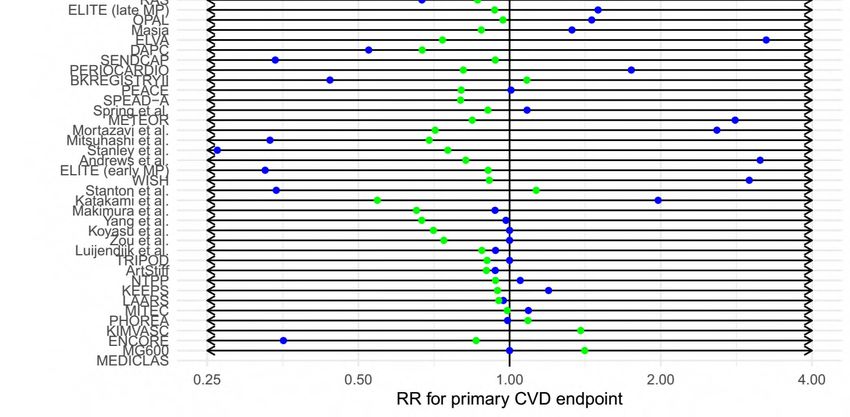
Non-standard Abbreviations and Acronyms

CI credible interval

cIMT Carotid intima-media thickness

CVD cardiovascular disease

RCT randomized controlled trial

RR relative risk

                                      -3-
Clinical Perspective
What Is New?
We analyzed data of 119 randomized controlled trials that involved 100,667 patients
and 12,038 incident cardiovascular disease events.
We used a Bayesian meta-regression approach to evaluate progression of carotid
intima-media thickness as a surrogate marker for cardiovascular events.
Our analysis revealed a statistically significant association between treatment effects
on progression of carotid intima-media thickness and treatment effects on
cardiovascular disease risk.
What Are the Clinical Implications?
Our paper provides the key missing link supporting the usefulness of carotid intima-
media thickness progression as a surrogate marker for cardiovascular disease risk in
clinical trials.
Using progression of carotid intima-media thickness as a surrogate endpoint in future
randomized controlled trials may facilitate and speed up the development and licensing
of new therapies.
-4-
Introduction

Carotid intima-media thickness (cIMT), the thickness of the intimal and medial layer of the
carotid artery wall, can be measured non-invasively using ultrasound imaging and is considered
a marker for the early stage of atherosclerosis.1 Mean values of cIMT in adults range around
650-900 µm and increase – on average – at a rate of 0-40 µm/year.2,3 A large number of
randomized controlled trials (RCTs) have demonstrated that therapeutic interventions may
slow progression of cIMT. However, it is uncertain whether effects on cIMT progression
translate into reduced risk of cardiovascular disease (CVD) events, that is whether cIMT
progression is a valid surrogate marker for CVD.

In 2005, Espeland et al. first proposed cIMT progression as a surrogate marker for CVD risk
based on findings in seven statin trials,4 but their arguments were based on limited data and
most researchers were reluctant to rely on cIMT results alone.5 In 2009, ARBITER-6 HALTS
was the first RCT to be terminated early based on findings for cIMT progression, showing
superiority of extended-release niacin over ezetimibe.6 This decision was controversial due to
the uncertain validity of the rate of progression of cIMT as a surrogate marker for clinical
endpoints.7,8 Two subsequent literature-based meta-regression analyses on this topic have
yielded conflicting results: Goldberger et al.9 observed an association of effects on cIMT
progression and risk of myocardial infarction, whereas Costanzo et al.10 found no statistically
significant association of changes in mean or maximal cIMT with risk of myocardial infarction
or stroke. Both of these meta-analyses have been criticized because of methodological flaws.11

To address this uncertainty, we conducted a comprehensive analysis of 119 RCTs involving a
total of 100,667 patients. Our aims were to: (i) quantify the reduction in CVD risk associated
with reducing cIMT progression by therapeutic intervention; (ii) explore cIMT progression as
a surrogate marker for different types of CVD endpoints as well as all-cause mortality; and (iii)
investigate differences according to the intervention type, method of cIMT assessment, and
other trial characteristics.

                                              -5-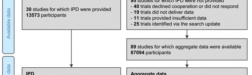
Methods

The datasets supporting the conclusions of this article are not made publicly available due to
legal restrictions arising from the data distribution policy of the PROG-IMT/Proof-ATHERO
collaborations and from the bilateral agreements between the consortium’s coordinating center
and participating studies, but they may be requested directly from individual study
investigators. Studies that shared individual-participant data have obtained informed consent
of the study participants and ethical approval by their respective institutional review boards.

The report of the results of our study adhere to the PRISMA-IPD guidelines (Table I in the
Supplement); the objectives and statistical methods in this paper have been described
previously12. We identified relevant RCTs published before 3 February 2020 through
systematic searches of ten medical knowledge databases, six clinical trial registries, and
reference lists of relevant publications and reviews (Table II in the Supplement). Trials were
eligible for inclusion if they: (1) had assigned patients randomly to two or more arms; (2) had
applied well-defined inclusion criteria; (3) had measured cIMT at trial baseline and at one or
more follow-up visits; and (4) had recorded incident CVD outcomes. We requested
anonymized patient-level data from these trials, performed comprehensive plausibility checks,
and were able to resolve any data-related queries through direct correspondence with trial
investigators. For trials for which patient-level data was unavailable, four authors (PW, LT,
EA, MWL) independently extracted the relevant data from the published literature and resolved
any discrepancies by consensus.

As a measure of cIMT, we gave preference to assessments of mean values at the common-
carotid-artery. If unavailable, we used maximum values at the common-carotid-artery or cIMT
at other sections of the carotid artery instead. In trials quantifying cIMT values at different sites
(i.e. left or right side, near or far vessel wall, or at different insonation angles), the arithmetic
mean of these measurements was used. The primary outcome was a combined CVD endpoint
defined as myocardial infarction, stroke, revascularization procedures (e.g. coronary or carotid
revascularization), or fatal CVD. For trials without data on cause-specific death, all-cause
mortality was included in the primary outcome instead. Table III in the Supplement provides
details on the assessment of cIMT progression and primary outcome definition in each trial.

                                                -6-
Statistical analysis

We conducted analyses according to a pre-specified analysis plan. For factorial trials, we
analyzed the intervention contrast anticipated to have the greatest effect on CVD risk. For trials
with more than two trial arms, we compared the arm that was – based on prior trials –
anticipated to have the greatest effect to the arm anticipated to have the least effect (or no effect
in case of placebo). For all trials, the latter group was used as reference.

The principal analysis consisted of three steps. First, we quantified intervention effects on
cIMT progression. For each trial for which patient-level data was available, we used a linear
mixed model to estimate the difference in yearly cIMT progression between trial arms. The
model included fixed effects for assigned treatment, time in study, and the interaction of the
two, plus an intercept and time variable allowed to vary randomly at the patient level. For each
trial for which literature-based data was available (i.e. tabular data extracted from the trials’
publications), we annualized differences in cIMT progression and calculated standard errors
from P values, if necessary.

Second, we quantified intervention effects on the CVD outcome. For each trial with patient-
level data, we fitted a Cox proportional-hazards model to estimate the log hazard ratio and its
standard error comparing the trial arms. If estimates were inestimable due to a low event
number, we applied an augmentation procedure to allow incorporation of the trial in the meta-
analysis.13 For each trial with literature-based data, we calculated the log risk ratio and its
standard error based on the number of events and patients in each trial arm. For trials in which
one arm had zero events, the number of events and non-events were each augmented by +0.5
in both trial arms. Hazard ratios and risk ratios are collectively described as measures of relative
risk (RR).

Third, to test whether effects on CVD risk depended on effects on cIMT progression, we used
a Bayesian meta-regression approach that models both effects simultaneously, while taking
into account the estimated precisions in these two effects.14 The principal analysis involved (i)
a model with an intercept of zero (i.e. forcing the regression line through the origin and thereby
assuming that all the effects on CVD risk operate through cIMT progression) and (ii) a model
with a non-zero intercept (i.e. allowing for an effect on CVD risk independent of cIMT
progression). The meta-regression also took into account the within-study correlation of the
                                           -7-
two effects, which was estimated using bootstrapping in the trials with patient-level data and
>30 events.15 For other trials, an overall correlation coefficient pooled using random-effects
meta-analysis was used instead. Further details on methods for assessing surrogacy are
provided in the Methods in the Supplement.
Subsidiary analyses evaluated surrogacy for individual disease endpoints and in trials grouped
by intervention type, time of conduct, time to ultrasound follow-up, availability of individual-
participant data, primary vs. secondary prevention trials, type of cIMT measure, and proportion
of female patients. A Bayesian approach was taken for estimation of the meta-regression model
parameters and for prediction (for details, see the Methods in the Supplement). Analyses were
performed using Stata 15, R 2.5.1 and JAGS 4.3.0. PW had full access to all the data in the
study and takes responsibility for the integrity of the data and the accuracy of the data analysis.
Results
Among 10,260 articles screened, we identified 119 trials involving 100,667 patients that met
the pre-specified inclusion criteria (Figure I in the Supplement). 103 trials (87%) had two
arms, seven had three arms, one had four arms, seven had a 2x2 factorial design, and one had
a 3x2 factorial design (Table 1). The trials employed antidiabetic (18 trials), antihypertensive
(19 trials), dietary/vitamin (20 trials), lipid-lowering (33 trials), and/or other interventions (37
trials). Mean age at baseline was 62 years (standard deviation 8); 42% were female. Over an
average follow-up duration of 3.7 years, 12,038 patients developed the primary CVD endpoint.
The median proportion of patients with repeat cIMT measurements across trials was 90%.
Seven large cardiovascular outcome trials had measured cIMT only in a subset of patients
(Table 1). Mean cIMT measured at the common-carotid-artery was available in 91 trials,
maximum cIMT at the common-carotid-artery in 49 trials, and other cIMT measures in 11
trials. Across contributing trials, the mean rate of cIMT progression was +9.1 µm/year (95%
confidence interval: 7.1 to 11.1) in control arms and +1.0 µm/year (-0.6 to 2.7) in interventions
arms. Across all contributing trials, the RR for CVD with intervention was 0.88 (0.83-0.92).
Results of the principal analysis are provided in Figure 1. Across all interventions, in the model
assuming an intercept of zero, each 10 μm/year reduction of cIMT progression was associated
with a RR for CVD of 0.88 (95% credible interval [CI] 0.85-0.91). In the model allowing for
-8-a non-zero intercept, the RR for CVD was 0.91 (0.87-0.94) per 10 μm/year slower cIMT
progression, with a further RR of 0.92 (0.87-0.97) achieved independent of cIMT progression.
Based on the non-zero intercept model, the proportion of variance in the CVD outcome
explained by cIMT progression was 98% albeit with a wide 95% CI (71-100%). Taken
together, we estimated that interventions that reduce cIMT progression by 10, 20, 30, or 40
µm/year would yield RRs of 0.84 (0.75-0.93), 0.76 (0.67-0.85), 0.69 (0.59-0.79), or 0.63 (0.52-
0.74).
Due to presence of effects on CVD risk unexplained by cIMT progression, subsequent analyses
focused on the non-zero intercept model. In outcome-specific analyses (Figure 2), RRs per 10
µm/year slower cIMT progression were 0.88 (0.82-0.94) for myocardial infarction, 0.92 (0.86-
1.00) for stroke, 0.90 (0.83-0.98) for revascularization procedures, 0.91 (0.83-1.01) for fatal
CVD, and 0.96 (0.89-1.04) for all-cause mortality. There was no evidence for differences in
the RR for CVD associated with slower cIMT progression nor in the intercept across trials
grouped by intervention type (Figure 3 and Figure 4). Similarly, there was no evidence for
differences in these RRs in trials grouped by time of conduct, time to ultrasound follow-up,
availability of individual-participant data, primary vs. secondary prevention trials, type of
cIMT measurements, and proportion of female patients (Figure 4, P values for heterogeneity
>0.05). In a sensitivity analysis that omitted trials with extreme effect sizes (i.e. cIMT
progression changes >80 µm/year or RR for CVD 4.0), the RR for CVD per 10
µm/year slower cIMT progression was 0.91 (0.87-0.95). Results were also highly robust across
leave-one-out cross-validation analyses (Figure II in the Supplement). Trial-specific
estimates are provided in Table IV in the Supplement.
Discussion
In this large-scale meta-analysis involving data from 119 RCTs and 100,667 patients, we
showed that interventions reducing cIMT progression are also likely to reduce CVD event rates
(summarized in Figure 5). Specifically, a 10 µm/year slower cIMT progression was associated
with a RR of 0.91 (95% CI 0.87-0.94) for the principal outcome of CVD, with the differences
in RR for CVD largely explained by the differences in cIMT progression. The same model also
indicated a non-zero intercept, overall and for different types of interventions, highlighting that
-9-a small but significant proportion of the intervention effect acted independently of cIMT
progression. By estimating CVD risk reductions according to specific reductions in cIMT
progression, we provide guidance to future trials in the cardiovascular field.5 Results were
robust for a range of disease endpoints and across clinically important trial characteristics,
including type of intervention or type of cIMT measurement.
Exploring the association between cIMT and CVD risk has some history. cIMT measured at a
single time-point is associated with incident CVD and provides incremental predictive value
over and beyond conventional CVD risk factors.190-192 For cIMT progression over time, our
earlier analyses of observational studies within the PROG-IMT collaboration indicated no
statistically significant association with subsequent CVD risk in individuals of the general
population,2 patients with diabetes mellitus,193 or patients at high CVD risk194. This null
association could be explained by the challenges of precisely estimating cIMT progression in
individuals over time. In contrast, our present report focuses on groups of patients in RCTs and
is therefore better suited to provide answers about the surrogate value of cIMT progression:
averaging across patients improves the signal-to-noise ratio, confounders are expected to be
balanced due to randomization, trial cohorts might be more homogeneous, and cIMT protocols
may be of higher quality in clinical trial settings.
Prior RCT data on cIMT progression as a surrogate marker for CVD risk are limited. Because
most RCTs reporting both cIMT and endpoints (with few exceptions63,70,97,127,170) have not been
designed as CVD outcome trials and as a range of intervention effect sizes is needed for
meaningful results, meta-analysis is the method of choice to investigate this question.195 Three
such pooled analyses had been undertaken before. Espeland et al. demonstrated that statin
treatment reduced cIMT progression and CVD risk in a concordant manner.4 In a meta-analysis
involving 28 RCTs of different intervention types, Goldberger et al. observed an association
between reduced cIMT progression and lower risk for non-fatal myocardial infarction, but
noted marked between-trials heterogeneity.9 A meta-analysis by Costanzo et al. involving 41
RCTs demonstrated no statistically significant relationship between slower cIMT progression
and risk of cardiovascular outcomes.10 Compared to these earlier reports, our meta-analysis
stands out by (i) exclusively conducting within-trial comparison (thereby upholding the
principle of randomization); (ii) increasing statistical power by involving >5 times as many
patients as the previously largest report10; (iii) enhancing validity by accessing patient-level
- 10 -data of 28 trials; and (iv) using modern statistical methods that incorporate uncertainties both
around the intervention effects on cIMT progression and CVD risk as well as their within-trial
correlation.
What do we know about the suitability of cIMT progression as a surrogate marker for CVD
risk? Ultrasound-based cIMT measurement fulfills several requirements of a surrogate
marker,196 including (i) high correlation with thickness of the vessel wall measured in
histological samples197; (ii) acceptable reproducibility198, which was further enhanced by clear
recommendations for measurement and technical improvements199; (iii) close correlation with
risk factors and prevalent CVD190-192; (iv) established correlation with atherosclerosis in other
vascular beds196; (v) association with occurrence of clinical events190-192; (vi) the ability to
change over time2,193; and (vii) the possibility to influence cIMT with interventions200. In the
present analysis, we have provided evidence for the last missing requirement not credibly
proven by earlier studies, namely that a change in cIMT progression is related to the change in
risk of CVD events.
Importantly, using cIMT progression as a surrogate endpoint in future RCTs may facilitate and
speed up development and licensing of new therapies. To illustrate this point, we conducted a
sample size calculation for a hypothetical future trial. For this calculation, we assumed 80%
power, several parameters similar to our individual-participant data (i.e. 2-year cumulative
incidence of CVD 6.57%, a standard deviation of cIMT 178 µm, and a correlation between
baseline and follow-up cIMT 0.79), no losses to follow-up, and a perfect relationship between
treatment effects on cIMT progression and those on the CVD outcome. To have 80% power to
detect a hazard ratio of 0.84, a future 2-year CVD outcome trial would require 8,600 patients
in each trial arm. In comparison, a future 2-year cIMT progression trial would require 470
patients per trial arm to detect a 10 µm/year reduction in cIMT progression (corresponding to
the above hazard ratio) at 2-years, also with a power of 80%. Consequently, a cIMT trial would
only require 5.5% of the sample size of a comparable CVD endpoint trial.
In addition to demonstrating the association between intervention effects on cIMT and
intervention effects on CVD risk, we found that the regression line had a small but significant
non-zero intercept, in the overall analysis and in all subgroups of trials investigated. The non-
zero intercept – which indicates that a small proportion of the intervention effect on CVD risk
- 11 -bypasses cIMT – may be explained by “pleiotropic” effects; meaning that the intervention
influences the clinical endpoint via multiple pathways. While effects of interventions on the
extent of atherosclerosis may be captured by cIMT progression, any effects on other
pathophysiological mechanisms related to CVD events, such as endogenous thrombogenesis
and fibrinolysis,1 may bypass cIMT progression and thereby lead to a non-zero intercept.
Alternative pathways have been described for many major cardiovascular substance groups,
including lipid-lowering medications (e.g. statins,1,201,202 fibrates,203 niacin,204 resins,205 and
omega-3 fatty acids206), antidiabetic medications (e.g. AMPK activators,207
thiazolidinediones,207 DPP-4 inhibitors,207,208 GLP-1 receptor agonists,207,208 SGLT-2
inhibitors208), or antihypertensive medications (e.g. beta-blockers,209 calcium channel-
inhibitors,210,211 angiotensin-II antagonists,212 ACE inhibitors212). Nevertheless, this finding
does not negate the main result that an intervention effect on cIMT predicts the effect on CVD
risk.
A major strength of our study is that we systematically collated and analyzed worldwide data
on cIMT progression and CVD outcomes published up to February 2020. Access to patient-
level data allowed us to include hitherto unpublished data and thereby reduce publication bias.
Supplementing our analysis with published data enhanced generalizability and statistical
power. Strengths of our meta-regression analysis include that it upholds randomization within
trials, allows for between-trials heterogeneity, makes no distributional assumption about the
true intervention effects on cIMT progression across trials (unlike standard bivariate random-
effects meta-analysis), and improved precision by incorporating within-trial correlations of
intervention effects on cIMT progression and CVD risk.
Our analysis also has limitations. First, our principal analysis combined trials of varying types
of interventions. While we conducted a sensitivity analysis by medication class, further
research is required to precisely quantify the differences in the surrogate value of cIMT by
intervention type. Second, our analysis involved a broad range of types of trial populations.
While sensitivity analysis revealed no evidence for differential effects in the setting of primary
vs. secondary prevention trials, further study is needed on specific trial populations, such as
patients with diabetes or chronic kidney disease. Third, the definition of the primary combined
CVD endpoint varied across the included trials. However, the differences were relatively minor
(see Table III in the Supplement), so we are confident that this does not constitute a major
- 12 -source of systematic bias. Finally, while ultrasound scanning protocols may have differed
across contributing trials – in particular before consensus guidelines were available213, there
was no evidence for effect modification by type of cIMT measure or baseline years of the trials.
Conclusions
In conclusion, effects of interventions on cIMT progression and on CVD risk are associated,
endorsing the usefulness of cIMT progression as a surrogate marker in clinical trials. Using
cIMT progression as a surrogate marker may be a useful tool to guide future development for
cardiovascular drugs.
Funding Sources
This work was supported by the Austrian Science Fund (FWF) [P 32488]; the Dr.-Johannes-
and-Hertha-Tuba Foundation; the German Research Foundation [DFG Lo 1569/2-1 and DFG
Lo 1569/2-3]; and the excellence initiative “Competence Centers for Excellent Technologies”
(COMET) of the Austrian Research Promotion Agency (FFG) “Research Center of Excellence
in Vascular Ageing: Tyrol, VASCage” [K-Project No. 843536], funded by Bundesministerium
für Verkehr, Innovation und Technologie (BMVIT), Bundesministerium für Bildung,
Wissenschaft und Forschung (BMWFW), Wirtschaftsagentur Wien, and Standortagentur Tirol.
Conflict of Interest Disclosures
P. Willeit reports grants from the German Research Foundation DFG, the Austrian Science
Fund FWF, the Austrian Research Promotion Agency FFG and the Dr.-Johannes-and-Hertha-
Tuba Foundation during the conduct of the study. L. Tschiderer reports grants from the Dr.-
Johannes-and-Hertha-Tuba Foundation during the conduct of the study and non-financial
support from Sanofi outside the submitted work. E. Allara was supported by a National Institute
for Health Research PhD studentship (NIHR BTRU-2014-10024) during the conduction of this
study and reports support from EU/EFPIA Innovative Medicines Initiative Joint Undertaking
BigData@Heart grant n° 116074 outside the submitted work. L. Seekircher reports non-
financial support from Sanofi outside the submitted work. H.C. Gerstein reports grants from
Sanofi, Eli Lilly, Astra Zeneca, Boehringer Ingelheim, Novo Nordisk, Merck, and Abbott, and
- 13 -personal fees from Sanofi, Eli Lilly, Astra Zeneca, Boehringer Ingelheim, Abbott, Novo
Nordisk, Merck, Jannsen, Kowa Research Institute, and Cirius outside the submitted work. E.
Stroes reports Lecturing/ad-boards fees paid to institution by Amgen, Sanofi-Regeneron,
Novartis, Athera, Mylan unrelated to the present work. K. Kapellas reports grants from the
National Health and Medical Research Council during the conduct of the study. M. Skilton
reports grants from the National Health and Medical Research Council of Australia during the
conduct of the study. M.G.A. van Vonderen reports grants from Abbott International and
Boehringer Ingelheim during the conduct of the study. S. Kiechl reports grants from the
Austrian Promotion Agency FFG outside the submitted work. G. Klingenschmid reports non-
financial support from Sanofi and Pfizer outside the submitted work. S.E. Kjeldsen reports
personal fees from Bayer, Merck KGaA, MSD, Sanofi, and Takeda outside the submitted work.
M.H. Olsen reports grants from the Novo Nordic Foundation outside the submitted work. N.
Sattar reports personal fees from Amgen, AstraZeneca, Boehringer Ingelheim, Eli Lilly,
Janssen, NAPP Pharmaceuticals, Novo Nordisk, and Sanofi, and grants from Boehringer
Ingelheim outside the submitted work. M.P.C. Grooteman reports grants from the Dutch
Kidney Foundation, Fresenius Medical Care Netherlands BV, Gambro Sweden, the Twiss
Fund, and ZON MW during the conduct of the study. P.J. Blankestijn reports grants from the
European Commission and other financial activities from Medtronic, Baxter, and Braun
outside the submitted work. M.L. Bots reports grants from AstraZeneca outside the submitted
work. M.J. Sweeting reports grants from the German Research Foundation during the conduct
of the study. S.G. Thompson reports grants from the UK Medical Research Council, the British
Heart Foundation, and the German Research Foundation DFG during the conduct of the study.
M.W. Lorenz reports grants from the German Research Foundation DFG during the conduct
of the study. Other authors have no conflicts of interests.
Authors
Peter Willeit, MD, MPhil, PhD1,2*; Lena Tschiderer, DI, BSc, PhD1*; Elias Allara, MD2,3;
Kathrin Reuber, MSc4; Lisa Seekircher, DI, Mag., BSc1; Lu Gao, BSc, MSc5; Ximing Liao,
BSc, MSc, PhD4; Eva Lonn, MD, MSc, FRCPC, FACC6,7; Hertzel C. Gerstein, MD, MSc,
FRCPC6,7; Salim Yusuf, MD, DPhil, MRCP6,7; Frank P. Brouwers, MD, PhD8; Folkert W.
Asselbergs, MD, PhD9; Wiek van Gilst, PhD10; Sigmund A. Anderssen, PhD11; Diederick E.
Grobbee, MD, PhD12; John J. P. Kastelein, MD, PhD, FESC13; Frank L. J. Visseren, MD14;
- 14 -George Ntaios, MD, MSc, PhD, FESO15; Apostolos I. Hatzitolios, MD, PhD, FESH16; Christos
Savopoulos, MD, PhD16; Pythia T. Nieuwkerk, PhD17; Erik Stroes, MD, PhD13; Matthew
Walters, MD, FRCP18; Peter Higgins, MD, MRCP19; Jesse Dawson, MD, FRCP, FESO19;
Paolo Gresele, MD, PhD20; Giuseppe Guglielmini, PhD20; Rino Migliacci, MD, PhD21; Marat
Ezhov, MD, PhD22; Maya Safarova, MD23; Tatyana Balakhonova, MD, PhD24; Eiichi Sato,
MD25; Mayuko Amaha, MD25; Tsukasa Nakamura, MD, PhD25; Kostas Kapellas, PhD26; Lisa
M. Jamieson, PhD26; Michael Skilton, PhD27; James A. Blumenthal, PhD28; Alan Hinderliter,
MD29; Andrew Sherwood, PhD28; Patrick J. Smith, PhD, MPH28; Michiel A. van Agtmael,
MD, PhD30; Peter Reiss, MD, PhD31,32; Marit G. A. van Vonderen, MD, PhD33; Stefan Kiechl,
MD1,34; Gerhard Klingenschmid, MD1; Matthias Sitzer, MD4,35; Coen D. A. Stehouwer, MD,
PhD, FESC36; Heiko Uthoff, MD, PD37; Zhi-Yong Zou, MD38; Ana R. Cunha, PhD39; Mario
F. Neves, MD, PhD39; Miles D. Witham, BMBCh, PhD40; Hyun-Woong Park, MD41; Moo-Sik
Lee, MD, PhD41,42; Jang-Ho Bae, MD, FACC43,44; Enrique Bernal, MD, PhD45; Kristian
Wachtell, MD, PhD, DrMedSci46; Sverre E. Kjeldsen, MD, PhD46; Michael H. Olsen, MD,
PhD, DMSc47; David Preiss, PhD, FRCPath, MRCP48; Naveed Sattar, MD, PhD49; Edith
Beishuizen, MD50; Menno V. Huisman, MD, PhD51; Mark A. Espeland, PhD52; Caroline
Schmidt, PhD53; Stefan Agewall, MD, PhD54; Ercan Ok, MD55; Gülay Aşçi, MD55; Eric de
Groot, MD, PhD56; Muriel P. C. Grooteman, MD, PhD57; Peter J. Blankestijn, MD58; Michiel
L. Bots, MD, PhD12; Michael J. Sweeting, PhD2,59†; Simon G. Thompson, DSc2†; Matthias W.
Lorenz, MD, PD4†; on behalf of the PROG-IMT and the Proof-ATHERO Study Groups.
*Authors contributed equally to this article.
†Authors contributed equally to this article.
1
Department of Neurology, Medical University of Innsbruck, Innsbruck, Austria; 2Department
of Public Health and Primary Care, University of Cambridge, Cambridge, UK; 3National
Institute for Health Research Blood and Transplant Research Unit in Donor Health and
Genomics, University of Cambridge, Cambridge, UK; 4Department of Neurology, Goethe
University, Frankfurt am Main, Germany; 5MRC Biostatistics Unit, University of Cambridge,
Cambridge, UK; 6Department of Medicine and Population Health Research Institute,
McMaster University, Hamilton, Ontario, Canada; 7Hamilton General Hospital, Hamilton,
- 15 -Ontario, Canada; 8Department of Cardiology, Haga Teaching Hospital, the Hague, the
Netherlands; 9Department of Cardiology, University Medical Center Utrecht, Utrecht, the
10
Netherlands; Department of Experimental Cardiology, University Medical Center
Groningen, Groningen, the Netherlands; 11Department of Sports Medicine, Norwegian School
12
of Sports Sciences, Oslo, Norway; Julius Center for Health Sciences and Primary Care,
13
University Medical Center Utrecht, Utrecht, the Netherlands; Department of Vascular
Medicine, Academic Medical Centre, University of Amsterdam, Amsterdam, the Netherlands;
14
Department of Vascular Medicine, University Medical Center Utrecht, Utrecht, the
15 16
Netherlands; Department of Medicine, University of Thessaly, Larissa, Greece; 1st
Propedeutic Department of Internal Medicine, Aristotle University of Thessaloniki,
17
Thessaloniki, Greece; Department of Medical Psychology, Amsterdam UMC- Location
AMC, Amsterdam, the Netherlands; 18School of Medicine, Dentistry and Nursing, University
of Glasgow, Glasgow, UK; 19Institute of Cardiovascular and Medical Sciences, University of
Glasgow, Glasgow, UK; 20Division of Internal and Cardiovascular Medicine, Department of
21
Medicine, University of Perugia, Perugia, Italy; Division of Internal Medicine, Cortona
22
Hospital, Cortona, Italy; Laboratory of Lipid Disorders, National Medical Research Center
23
of Cardiology, Moscow, Russia; Atherosclerosis Department, National Medical Research
Center of Cardiology, Moscow, Russia; 24Ultrasound Vascular Laboratory, National Medical
25
Research Center of Cardiology, Moscow, Russia; Division of Nephrology, Shinmatsudo
26
Central General Hospital, Chiba, Japan; Australian Research Centre for Population Oral
27
Health, University of Adelaide, Adelaide, SA, Australia; Boden Institute of Obesity,
Nutrition, Exercise and Eating Disorders, University of Sydney, Sydney, NSW, Australia;
28
Department of Psychiatry and Behavioral Sciences, Duke University Medical Center,
Durham, NC, USA; 29Department of Medicine, University of North Carolina, Chapel Hill, NC,
USA; 30Department of Internal Medicine, Amsterdam UMC, Vrije Universiteit, Amsterdam,
31
the Netherlands; Department of Global Health, Amsterdam UMC- Location AMC,
32
Amsterdam, the Netherlands; Amsterdam Institute for Global Health and Development,
33
University of Amsterdam, Amsterdam, the Netherlands; Department of Internal Medicine,
34
Medical Center Leeuwarden, Leeuwarden, the Netherlands; VASCage GmbH, Research
35
Centre on Vascular Ageing and Stroke, Innsbruck, Austria; Department of Neurology,
Klinikum Herford, Herford, Germany; 36Department of Internal Medicine and Cardiovascular
Research Institute Maastricht (CARIM), Maastricht University Medical Centre, Maastricht, the
- 16 -37
Netherlands; Department of Angiology, University Hospital Basel, Basel, Switzerland;
38
Institute of Child and Adolescent Health, School of Public Health, Peking University,
Beijing, China; 39Department of Clinical Medicine, State University of Rio de Janeiro, Rio de
40
Janeiro, Brazil; AGE Research Group, NIHR Newcastle Biomedical Research Centre,
Newcastle University and Newcastle-upon-Tyne Hospitals Trust, Newcastle, UK;
41
Department of Internal Medicine, Gyeongsang National University Hospital, Daejeon, South
Korea; 42Department of Preventive Medicine, Konyang University, Jinju, South Korea; 43Heart
44
Center, Konyang University Hospital, Daejeon, South Korea; Department of Cardiology,
Konyang University College of Medicine, Daejeon, South Korea; 45Infectious Diseases Unit,
Reina Sofia Hospital, Murcia, Spain; 46Department of Cardiology, Oslo University Hospital,
Oslo, Norway; 47Department of Internal Medicine, Holbaek Hospital, University of Southern
Denmark, Odense, Denmark; 48MRC Population Health Research Unit, Clinical Trial Service
49
Unit, Nuffield Department of Population Health, University of Oxford, Oxford, UK; BHF
Glasgow Cardiovascular Research Centre, University of Glasgow, Glasgow, UK;
50
Department of Internal Medicine, HMC+ (Bronovo), the Hague, the Netherlands;
51
Department of Thrombosis and Hemostasis, Leiden University Medical Center, Leiden, the
52
Netherlands; Department of Biostatistical Sciences, Wake Forest School of Medicine,
Winston-Salem, NC, USA; 53Wallenberg Laboratory for Cardiovascular Research, University
54
of Gothenburg, Gothenburg, Sweden; Oslo University Hospital Ullevål and Institute of
55
Clinical Sciences, University of Oslo, Oslo, Norway; Nephrology Department, Ege
University School of Medicine, Bornova-Izmir, Turkey; 56Imagelabonline & Cardiovascular,
57
Eindhoven and Lunteren, the Netherlands; Department of Nephrology, Amsterdam UMC,
Amsterdam, the Netherlands; 58Department of Nephrology, University Medical Center Utrecht,
Utrecht, the Netherlands; 59Department of Health Sciences, University of Leicester, Leicester,
UK;
Supplemental Material
Supplemental Methods
Supplemental Tables I-V
Supplemental Figures I-II
Full list of the PROG-IMT and the Proof-ATHERO study groups and their affiliations
- 17 -References
1. Libby P, Ridker PM, Hansson GK. Progress and challenges in translating the biology of
atherosclerosis. Nature. 2011;473:317–325. doi: 10.1038/nature10146.
2. Lorenz MW, Polak JF, Kavousi M, Mathiesen EB, Völzke H, Tuomainen T-P, Sander D,
Plichart M, Catapano AL, Robertson CM, et al. Carotid intima-media thickness
progression to predict cardiovascular events in the general population (the PROG-IMT
collaborative project): a meta-analysis of individual participant data. Lancet.
2012;379:2053–2062. doi: 10.1016/S0140-6736(12)60441-3.
3. Willeit P, Thompson SG, Agewall S, Bergström G, Bickel H, Catapano AL, Chien K-L,
de Groot E, Empana J-P, Etgen T, et al. Inflammatory markers and extent and progression
of early atherosclerosis: Meta-analysis of individual-participant-data from 20 prospective
studies of the PROG-IMT collaboration. Eur J Cardiovasc Prev Rehabil. 2016;23:194–
205. doi: 10.1177/2047487314560664.
4. Espeland MA, O'Leary DH, Terry JG, Morgan T, Evans G, Mudra H. Carotid intimal-
media thickness as a surrogate for cardiovascular disease events in trials of HMG-CoA
reductase inhibitors. Curr Control Trials Cardiovasc Med. 2005;6:3. doi: 10.1186/1468-
6708-6-3.
5. Peters SAE, den Ruijter HM, Grobbee DE, Bots ML. Results from a carotid intima-media
thickness trial as a decision tool for launching a large-scale morbidity and mortality trial.
Circ Cardiovasc Imaging. 2013;6:20–25. doi: 10.1161/CIRCIMAGING.112.978114.
6. Taylor AJ, Villines TC, Stanek EJ, Devine PJ, Griffen L, Miller M, Weissman NJ, Turco
M. Extended-release niacin or ezetimibe and carotid intima-media thickness. N Engl J
Med. 2009;361:2113–2122. doi: 10.1056/NEJMoa0907569.
7. Blumenthal RS, Michos ED. The HALTS trial--halting atherosclerosis or halted too early?
N Engl J Med. 2009;361:2178–2180. doi: 10.1056/NEJMe0908838.
8. Kastelein JJP, Bots ML. Statin therapy with ezetimibe or niacin in high-risk patients. N
Engl J Med. 2009;361:2180–2183. doi: 10.1056/NEJMe0908841.
9. Goldberger ZD, Valle JA, Dandekar VK, Chan PS, Ko DT, Nallamothu BK. Are changes
in carotid intima-media thickness related to risk of nonfatal myocardial infarction? A
critical review and meta-regression analysis. Am Heart J. 2010;160:701–714. doi:
10.1016/j.ahj.2010.06.029.
10. Costanzo P, Perrone-Filardi P, Vassallo E, Paolillo S, Cesarano P, Brevetti G, Chiariello
M. Does carotid intima-media thickness regression predict reduction of cardiovascular
events? A meta-analysis of 41 randomized trials. J Am Coll Cardiol. 2010;56:2006–2020.
doi: 10.1016/j.jacc.2010.05.059.
11. Bots ML, Taylor AJ, Kastelein JJP, Peters SAE, den Ruijter HM, Tegeler CH, Baldassarre
D, Stein JH, O'Leary DH, Revkin JH, et al. Rate of change in carotid intima-media
- 18 -thickness and vascular events: meta-analyses can not solve all the issues. A point of view.
J Hypertens. 2012;30:1690–1696. doi: 10.1097/HJH.0b013e32835644dc.
12. Lorenz MW, Bickel H, Bots ML, Breteler MMB, Catapano AL, Desvarieux M, Hedblad
B, Iglseder B, Johnsen SH, Juraska M, et al. Individual progression of carotid intima media
thickness as a surrogate for vascular risk (PROG-IMT): Rationale and design of a meta-
analysis project. Am Heart J. 2010;159:730‐736.e2. doi: 10.1016/j.ahj.2010.02.008.
13. White IR. Multivariate random-effects meta-analysis. Stata Journal. 2009;9:40–56. doi:
10.1177/1536867X0900900103.
14. Daniels MJ, Hughes MD. Meta-analysis for the evaluation of potential surrogate markers.
Stat Med. 1997;16:1965–1982. doi: 10.1002/(SICI)1097-
0258(19970915)16:173.0.CO;2-M.
15. Riley RD, Price MJ, Jackson D, Wardle M, Gueyffier F, Wang J, Staessen JA, White IR.
Multivariate meta-analysis using individual participant data. Res Synth Methods.
2015;6:157–174. doi: 10.1002/jrsm.1129.
16. The ACAPS Group. Rationale and design for the Asymptomatic Carotid Artery Plaque
Study (ACAPS). Control Clin Trials. 1992;13:293–314. doi: 10.1016/0197-
2456(92)90012-o.
17. Furberg CD, Adams HP, Applegate WB, Byington RP, Espeland MA, Hartwell T,
Hunninghake DB, Lefkowitz DS, Probstfield J, Riley WA. Effect of lovastatin on early
carotid atherosclerosis and cardiovascular events. Asymptomatic Carotid Artery
Progression Study (ACAPS) Research Group. Circulation. 1994;90:1679–1687. doi:
10.1161/01.CIR.90.4.1679.
18. DeFronzo RA, Tripathy D, Schwenke DC, Banerji M, Bray GA, Buchanan TA, Clement
SC, Henry RR, Hodis HN, Kitabchi AE, et al. Pioglitazone for diabetes prevention in
impaired glucose tolerance. N Engl J Med. 2011;364:1104–1115. doi:
10.1056/NEJMoa1010949.
19. Saremi A, Schwenke DC, Buchanan TA, Hodis HN, Mack WJ, Banerji M, Bray GA,
Clement SC, Henry RR, Kitabchi AE, et al. Pioglitazone slows progression of
atherosclerosis in prediabetes independent of changes in cardiovascular risk factors.
Arterioscler Thromb Vasc Biol. 2013;33:393–399. doi: 10.1161/ATVBAHA.112.300346.
20. Higgins P, Walters MR, Murray HM, McArthur K, McConnachie A, Lees KR, Dawson J.
Allopurinol reduces brachial and central blood pressure, and carotid intima-media
thickness progression after ischaemic stroke and transient ischaemic attack: a randomised
controlled trial. Heart. 2014;100:1085–1092. doi: 10.1136/heartjnl-2014-305683.
21. Orekhov AN, Sobenin IA, Korneev NV, Kirichenko TV, Myasoedova VA, Melnichenko
AA, Balcells M, Edelman ER, Bobryshev YV. Anti-atherosclerotic therapy based on
botanicals. Recent Pat Cardiovasc Drug Discov. 2013;8:56–66. doi:
10.2174/18722083113079990008.
- 19 -22. Taylor AJ, Kent SM, Flaherty PJ, Coyle LC, Markwood TT, Vernalis MN. ARBITER:
Arterial Biology for the Investigation of the Treatment Effects of Reducing Cholesterol: a
randomized trial comparing the effects of atorvastatin and pravastatin on carotid intima
medial thickness. Circulation. 2002;106:2055–2060. doi:
10.1161/01.CIR.0000034508.55617.65.
23. Taylor AJ, Sullenberger LE, Lee HJ, Lee JK, Grace KA. Arterial Biology for the
Investigation of the Treatment Effects of Reducing Cholesterol (ARBITER) 2: a double-
blind, placebo-controlled study of extended-release niacin on atherosclerosis progression
in secondary prevention patients treated with statins. Circulation. 2004;110:3512–3517.
doi: 10.1161/01.CIR.0000148955.19792.8D.
24. Devine PJ, Turco MA, Taylor AJ. Design and rationale of the ARBITER 6 trial (Arterial
Biology for the Investigation of the Treatment Effects of Reducing Cholesterol)-6-HDL
and LDL Treatment Strategies in Atherosclerosis (HALTS). Cardiovasc Drugs Ther.
2007;21:221–225. doi: 10.1007/s10557-007-6020-8.
25. Villines TC, Stanek EJ, Devine PJ, Turco M, Miller M, Weissman NJ, Griffen L, Taylor
AJ. The ARBITER 6-HALTS Trial (Arterial Biology for the Investigation of the
Treatment Effects of Reducing Cholesterol 6-HDL and LDL Treatment Strategies in
Atherosclerosis): final results and the impact of medication adherence, dose, and treatment
duration. J Am Coll Cardiol. 2010;55:2721–2726. doi: 10.1016/j.jacc.2010.03.017.
26. Laurent S, Boutouyrie P. Dose-dependent arterial destiffening and inward remodeling
after olmesartan in hypertensives with metabolic syndrome. Hypertension. 2014;64:709–
716. doi: 10.1161/HYPERTENSIONAHA.114.03282.
27. Salonen JT, Nyyssönen K, Salonen R, Lakka HM, Kaikkonen J, Porkkala-Sarataho E,
Voutilainen S, Lakka TA, Rissanen T, Leskinen L, et al. Antioxidant Supplementation in
Atherosclerosis Prevention (ASAP) study: a randomized trial of the effect of vitamins E
and C on 3-year progression of carotid atherosclerosis. J Intern Med. 2000;248:377–386.
doi: 10.1046/j.1365-2796.2000.00752.x.
28. Rissanen T, Voutilainen S, Nyyssönen K, Salonen R, Salonen JT. Low plasma lycopene
concentration is associated with increased intima-media thickness of the carotid artery
wall. Arterioscler Thromb Vasc Biol. 2000;20:2677–2681. doi:
10.1161/01.ATV.20.12.2677.
29. Salonen RM, Nyyssönen K, Kaikkonen J, Porkkala-Sarataho E, Voutilainen S, Rissanen
TH, Tuomainen T-P, Valkonen V-P, Ristonmaa U, Lakka H-M, et al. Six-year effect of
combined vitamin C and E supplementation on atherosclerotic progression: the
Antioxidant Supplementation in Atherosclerosis Prevention (ASAP) Study. Circulation.
2003;107:947–953. doi: 10.1161/01.CIR.0000050626.25057.51.
30. Smilde TJ, Trip MD, Wollersheim H, van Wissen S, Kastelein JJ, Stalenhoef AF.
Rationale, Design and Baseline Characteristics of a Clinical Trial Comparing the Effects
of Robust vs Conventional Cholesterol Lowering and Intima Media Thickness in Patients
with Familial Hypercholesterolaemia : The Atorvastatin versus Simvastatin on
- 20 -Atherosclerosis Progression (ASAP) Study. Clin Drug Investig. 2000;20:67–79. doi:
10.2165/00044011-200020020-00001.
31. Smilde TJ, van Wissen S, Wollersheim H, Trip MD, Kastelein JJ, Stalenhoef AF. Effect
of aggressive versus conventional lipid lowering on atherosclerosis progression in familial
hypercholesterolaemia (ASAP): a prospective, randomised, double-blind trial. Lancet.
2001;357:577–581. doi: 10.1016/S0140-6736(00)04053-8.
32. Zoungas S, McGrath BP, Branley P, Kerr PG, Muske C, Wolfe R, Atkins RC, Nicholls K,
Fraenkel M, Hutchison BG, et al. Cardiovascular morbidity and mortality in the
Atherosclerosis and Folic Acid Supplementation Trial (ASFAST) in chronic renal failure:
a multicenter, randomized, controlled trial. J Am Coll Cardiol. 2006;47:1108–1116. doi:
10.1016/j.jacc.2005.10.064.
33. Nanayakkara PWB, van Guldener C, ter Wee PM, Scheffer PG, van Ittersum FJ, Twisk
JW, Teerlink T, van Dorp W, Stehouwer CDA. Effect of a treatment strategy consisting
of pravastatin, vitamin E, and homocysteine lowering on carotid intima-media thickness,
endothelial function, and renal function in patients with mild to moderate chronic kidney
disease: results from the Anti-Oxidant Therapy in Chronic Renal Insufficiency (ATIC)
Study. Arch Intern Med. 2007;167:1262–1270. doi: 10.1001/archinte.167.12.1262.
34. Nanayakkara PWB, Teerlink T, Stehouwer CDA, Allajar D, Spijkerman A, Schalkwijk C,
ter Wee PM, van Guldener C. Plasma asymmetric dimethylarginine (ADMA)
concentration is independently associated with carotid intima-media thickness and plasma
soluble vascular cell adhesion molecule-1 (sVCAM-1) concentration in patients with
mild-to-moderate renal failure. Kidney Int. 2005;68:2230–2236. doi: 10.1111/j.1523-
1755.2005.00680.x.
35. Ahn CM, Hong SJ, Park JH, Kim JS, Lim D-S. Cilostazol reduces the progression of
carotid intima-media thickness without increasing the risk of bleeding in patients with
acute coronary syndrome during a 2-year follow-up. Heart Vessels. 2011;26:502–510. doi:
10.1007/s00380-010-0093-1.
36. Jalal DI, Decker E, Perrenoud L, Nowak KL, Bispham N, Mehta T, Smits G, You Z, Seals
D, Chonchol M, et al. Vascular Function and Uric Acid-Lowering in Stage 3 CKD. J Am
Soc Nephrol. 2017;28:943–952. doi: 10.1681/ASN.2016050521.
37. Andrews ES, Perrenoud L, Nowak KL, You Z, Pasch A, Chonchol M, Kendrick J, Jalal
D. Examining the effects of uric acid-lowering on markers vascular of calcification and
CKD-MBD; A post-hoc analysis of a randomized clinical trial. PLoS ONE.
2018;13:e0205831. doi: 10.1371/journal.pone.0205831.
38. Hedblad B, Wikstrand J, Janzon L, Wedel H, Berglund G. Low-dose metoprolol CR/XL
and fluvastatin slow progression of carotid intima-media thickness: Main results from the
Beta-Blocker Cholesterol-Lowering Asymptomatic Plaque Study (BCAPS). Circulation.
2001;103:1721–1726. doi: 10.1161/01.CIR.103.13.1721.
39. Bae J-H, Bassenge E, Kim K-Y, Synn Y-C, Park K-R, Schwemmer M. Effects of low-
dose atorvastatin on vascular responses in patients undergoing percutaneous coronary
- 21 -intervention with stenting. J Cardiovasc Pharmacol Ther. 2004;9:185–192. doi:
10.1177/107424840400900306.
40. Hodis HN, Mack WJ, Dustin L, Mahrer PR, Azen SP, Detrano R, Selhub J, Alaupovic P,
Liu C-r, Liu C-h, et al. High-dose B vitamin supplementation and progression of
subclinical atherosclerosis: a randomized controlled trial. Stroke. 2009;40:730–736. doi:
10.1161/STROKEAHA.108.526798.
41. Mercuri M, Bond MG, Sirtori CR, Veglia F, Crepaldi G, Feruglio FS, Descovich G, Ricci
G, Rubba P, Mancini M, et al. Pravastatin reduces carotid intima-media thickness
progression in an asymptomatic hypercholesterolemic mediterranean population: the
Carotid Atherosclerosis Italian Ultrasound Study. Am J Med. 1996;101:627–634. doi:
10.1016/S0002-9343(96)00333-6.
42. Preiss D, Lloyd SM, Ford I, McMurray JJ, Holman RR, Welsh P, Fisher M, Packard CJ,
Sattar N. Metformin for non-diabetic patients with coronary heart disease (the CAMERA
study): a randomised controlled trial. Lancet Diabetes Endocrinol. 2014;2:116–124. doi:
10.1016/S2213-8587(13)70152-9.
43. Hong S, Nam M, Little BB, Paik S, Lee K, Woo J, Kim D, Kang J, Chun M, Park Y.
Randomized control trial comparing the effect of cilostazol and aspirin on changes in
carotid intima-medial thickness. Heart Vessels. 2019;34:1758–1768. doi:
10.1007/s00380-019-01421-1.
44. Meuwese MC, de Groot E, Duivenvoorden R, Trip MD, Ose L, Maritz FJ, Basart DCG,
Kastelein JJP, Habib R, Davidson MH, et al. ACAT inhibition and progression of carotid
atherosclerosis in patients with familial hypercholesterolemia: the CAPTIVATE
randomized trial. JAMA. 2009;301:1131–1139. doi: 10.1001/jama.301.11.1131.
45. Beishuizen ED, van de Ree MA, Jukema JW, Tamsma JT, van der Vijver JCM, Meinders
AE, Putter H, Huisman MV. Two-year statin therapy does not alter the progression of
intima-media thickness in patients with type 2 diabetes without manifest cardiovascular
disease. Diabetes Care. 2004;27:2887–2892. doi: 10.2337/diacare.27.12.2887.
46. Mazzone T, Meyer PM, Feinstein SB, Davidson MH, Kondos GT, D’Agostino RB, Perez
A, Provost J-C, Haffner SM. Effect of pioglitazone compared with glimepiride on carotid
intima-media thickness in type 2 diabetes: a randomized trial. JAMA. 2006;296:2572–
2581. doi: 10.1001/jama.296.21.joc60158.
47. Lundby-Christensen L, Tarnow L, Boesgaard TW, Lund SS, Wiinberg N, Perrild H,
Krarup T, Snorgaard O, Gade-Rasmussen B, Thorsteinsson B, et al. Metformin versus
placebo in combination with insulin analogues in patients with type 2 diabetes mellitus-
the randomised, blinded Copenhagen Insulin and Metformin Therapy (CIMT) trial. BMJ
Open. 2016;6:e008376. doi: 10.1136/bmjopen-2015-008376.
48. Lundby-Christensen L, Almdal T, Boesgaard T, Breum L, Dunn E, Gade-Rasmussen B,
Gluud C, Hedetoft C, Jarloev A, Jensen T, et al. Study rationale and design of the CIMT
trial: the Copenhagen Insulin and Metformin Therapy trial. Diabetes Obes Metab.
2009;11:315–322. doi: 10.1111/j.1463-1326.2008.00959.x.
- 22 -49. Blankenhorn DH, Selzer RH, Crawford DW, Barth JD, Liu CR, Liu CH, Mack WJ,
Alaupovic P. Beneficial effects of colestipol-niacin therapy on the common carotid artery.
Two- and four-year reduction of intima-media thickness measured by ultrasound.
Circulation. 1993;88:20–28. doi: 10.1161/01.CIR.88.1.20.
50. Azen SP, Mack WJ, Cashin-Hemphill L, LaBree L, Shircore AM, Selzer RH, Blankenhorn
DH, Hodis HN. Progression of coronary artery disease predicts clinical coronary events.
Long-term follow-up from the Cholesterol Lowering Atherosclerosis Study. Circulation.
1996;93:34–41. doi: 10.1161/01.CIR.93.1.34.
51. Cashin-Hemphill L, Mack WJ, Pogoda JM, Sanmarco ME, Azen SP, Blankenhorn DH.
Beneficial effects of colestipol-niacin on coronary atherosclerosis. A 4-year follow-up.
JAMA. 1990;264:3013–3017. doi: 10.1001/jama.1990.03450230049028.
52. Penne EL, Blankestijn PJ, Bots ML, van den Dorpel MA, Grooteman MP, Nubé MJ, van
der Tweel I, ter Wee PM. Effect of increased convective clearance by on-line
hemodiafiltration on all cause and cardiovascular mortality in chronic hemodialysis
patients - the Dutch CONvective TRAnsport STudy (CONTRAST): rationale and design
of a randomised controlled trial ISRCTN38365125. Curr Control Trials Cardiovasc Med.
2005;6:8. doi: 10.1186/1468-6708-6-8.
53. Grooteman MPC, van den Dorpel MA, Bots ML, Penne EL, van der Weerd NC, Mazairac
AHA, den Hoedt CH, van der Tweel I, Lévesque R, Nubé MJ, et al. Effect of online
hemodiafiltration on all-cause mortality and cardiovascular outcomes. J Am Soc Nephrol.
2012;23:1087–1096. doi: 10.1681/ASN.2011121140.
54. Cao A-H, Wang J, Gao H-Q, Zhang P, Qiu J. Beneficial clinical effects of grape seed
proanthocyanidin extract on the progression of carotid atherosclerotic plaques. J Geriatr
Cardiol. 2015;12:417–423. doi: 10.11909/j.issn.1671-5411.2015.04.014.
55. Yamasaki Y, Kim Y-S, Kawamori R. Rationale and protocol of a trial for prevention of
diabetic atherosclerosis by using antiplatelet drugs: study of Diabetic Atherosclerosis
Prevention by Cilostazol (DAPC study). Cardiovasc Diabetol. 2006;5:16. doi:
10.1186/1475-2840-5-16.
56. Katakami N, Kim Y-S, Kawamori R, Yamasaki Y. The phosphodiesterase inhibitor
cilostazol induces regression of carotid atherosclerosis in subjects with type 2 diabetes
mellitus: principal results of the Diabetic Atherosclerosis Prevention by Cilostazol
(DAPC) study: a randomized trial. Circulation. 2010;121:2584–2591. doi:
10.1161/CIRCULATIONAHA.109.892414.
57. Hoogerbrugge N, de Groot E, de Heide LHM, de Ridder MAJ, Birkenhägeri JC, Stijnen
T, Jansen H. Doxazosin and hydrochlorothiazide equally affect arterial wall thickness in
hypertensive males with hypercholesterolaemia (the DAPHNE study). Doxazosin
Atherosclerosis Progression Study in Hypertensives in the Netherlands. Neth J Med.
2002;60:354–361.
- 23 -58. Ellingsen I, Seljeflot I, Arnesen H, Tonstad S. Vitamin C consumption is associated with
less progression in carotid intima media thickness in elderly men: A 3-year intervention
study. Nutr Metab Cardiovasc Dis. 2009;19:8–14. doi: 10.1016/j.numecd.2008.01.006.
59. Asci G, Tz H, Ozkahya M, Duman S, Demirci MS, Cirit M, Sipahi S, Dheir H, Bozkurt
D, Kircelli F, et al. The impact of membrane permeability and dialysate purity on
cardiovascular outcomes. J Am Soc Nephrol. 2013;24:1014–1023. doi:
10.1681/ASN.2012090908.
60. Ok ES, Asci G, Toz H, Ritz E, Kircelli F, Sever MS, Ozkahya M, Sipahi S, Dheir H,
Bozkurt D, et al. Glycated hemoglobin predicts overall and cardiovascular mortality in
non-diabetic hemodialysis patients. Clin Nephrol. 2014;82:173–180. doi:
10.5414/CN108251.
61. Hodis HN, Mack WJ, Shoupe D, Azen SP, Stanczyk FZ, Hwang-Levine J, Budoff MJ,
Henderson VW. Methods and baseline cardiovascular data from the Early versus Late
Intervention Trial with Estradiol testing the menopausal hormone timing hypothesis.
Menopause. 2015;22:391–401. doi: 10.1097/GME.0000000000000343.
62. Hodis HN, Mack WJ, Henderson VW, Shoupe D, Budoff MJ, Hwang-Levine J, Li Y,
Feng M, Dustin L, Kono N, et al. Vascular Effects of Early versus Late Postmenopausal
Treatment with Estradiol. N Engl J Med. 2016;374:1221–1231. doi:
10.1056/NEJMoa1505241.
63. Zanchetti A, Bond MG, Hennig M, Neiss A, Mancia G, Dal Palù C, Hansson L, Magnani
B, Rahn K-H, Reid JL, et al. Calcium antagonist lacidipine slows down progression of
asymptomatic carotid atherosclerosis: principal results of the European Lacidipine Study
on Atherosclerosis (ELSA), a randomized, double-blind, long-term trial. Circulation.
2002;106:2422–2427. doi: 10.1161/01.CIR.0000039288.86470.DD.
64. Wiklund O, Hulthe J, Wikstrand J, Schmidt C, Olofsson S-O, Bondjers G. Effect of
controlled release/extended release metoprolol on carotid intima-media thickness in
patients with hypercholesterolemia: a 3-year randomized study. Stroke. 2002;33:572–577.
doi: 10.1161/hs0202.102332.
65. Blumenthal JA, Babyak MA, Hinderliter A, Watkins LL, Craighead L, Lin P-H, Caccia
C, Johnson J, Waugh R, Sherwood A. Effects of the DASH diet alone and in combination
with exercise and weight loss on blood pressure and cardiovascular biomarkers in men
and women with high blood pressure: the ENCORE study. Arch Intern Med.
2010;170:126–135. doi: 10.1001/archinternmed.2009.470.
66. Smith PJ, Blumenthal JA, Babyak MA, Craighead L, Welsh-Bohmer KA, Browndyke JN,
Strauman TA, Sherwood A. Effects of the dietary approaches to stop hypertension diet,
exercise, and caloric restriction on neurocognition in overweight adults with high blood
pressure. Hypertension. 2010;55:1331–1338. doi:
10.1161/HYPERTENSIONAHA.109.146795.
67. Kastelein JJP, Akdim F, Stroes ESG, Zwinderman AH, Bots ML, Stalenhoef AFH,
Visseren FLJ, Sijbrands EJG, Trip MD, Stein EA, et al. Simvastatin with or without
- 24 -You can also read