B cell-activating factor modulates the factor VIII immune response in hemophilia A
←
→
Page content transcription
If your browser does not render page correctly, please read the page content below
The Journal of Clinical Investigation                                                                                                                                RESEARCH ARTICLE

B cell–activating factor modulates the factor VIII
immune response in hemophilia A
Bhavya S. Doshi,1,2 Jyoti Rana,3 Giancarlo Castaman,4 Mostafa A. Shaheen,2 Radoslaw Kaczmarek,3 John S.S. Butterfield,3
Shannon L. Meeks,5 Cindy Leissinger,6 Moanaro Biswas,3 and Valder R. Arruda1,2,7
Department of Pediatrics, Perelman School of Medicine, University of Pennsylvania, Philadelphia, Pennsylvania, USA. 2Divison of Hematology, Children’s Hospital of Philadelphia, Philadelphia,
1

Pennsylvania, USA. 3Herman B. Wells Center for Pediatric Research, Indiana University School of Medicine, Indianapolis, Indiana, USA. 4Center for Bleeding Disorders and Coagulation, Careggi University
Hospital, Florence, Italy. 5Department of Pediatrics, Aflac Cancer Center and Blood Disorders Center at Children’s Healthcare of Atlanta, Emory University School of Medicine, Atlanta, Georgia, USA. 6Section
of Hematology/Oncology, Tulane University School of Medicine, New Orleans, Louisiana, USA. 7Raymond G. Perelman Center for Cellular and Molecular Therapies, Children’s Hospital of Philadelphia,
Philadelphia, Pennsylvania, USA.

             Inhibitors of factor VIII (FVIII) remain the most challenging complication of FVIII protein replacement therapy in hemophilia
             A (HA). Understanding the mechanisms that guide FVIII-specific B cell development could help identify therapeutic targets.
             The B cell–activating factor (BAFF) cytokine family is a key regulator of B cell differentiation in normal homeostasis and
             immune disorders. Thus, we used patient samples and mouse models to investigate the potential role of BAFF in modulating
             FVIII inhibitors. BAFF levels were elevated in pediatric and adult HA inhibitor patients and decreased to levels similar to
             those of noninhibitor controls after successful immune tolerance induction (ITI). Moreover, elevations in BAFF levels were
             seen in patients who failed to achieve FVIII tolerance with anti-CD20 antibody–mediated B cell depletion. In naive HA mice,
             prophylactic anti-BAFF antibody therapy prior to FVIII immunization prevented inhibitor formation and this tolerance was
             maintained despite FVIII exposure after immune reconstitution. In preimmunized HA mice, combination therapy with anti-
             CD20 and anti-BAFF antibodies dramatically reduced FVIII inhibitors via inhibition of FVIII-specific plasma cells. Our data
             suggest that BAFF may regulate the generation and maintenance of FVIII inhibitors and/or anti-FVIII B cells. Finally, anti-
             CD20/anti-BAFF combination therapy may be clinically useful for ITI.

Introduction                                                                                               ing agents when inhibitors are present for breakthrough bleeds
One of the most immunogenic protein-based therapies is coag-                                               and/or surgery (10–12). Thus, the prevention and/or eradication
ulation factor VIII (FVIII) (1), which is missing or defective in the                                      of inhibitors of FVIII is of fundamental interest in the optimal
X-linked congenital bleeding disorder hemophilia A (HA). HA                                                management of HA (13). Several genetic and environmental risk
results from mutations in the F8 gene and affects 1 in 5000 males                                          factors have been implicated in inhibitor formation (14), including
born worldwide (2). Prophylactic FVIII replacement therapy pre-                                            F8 mutations (15); however, there is no clear predictor for which
vents bleeding-related morbidity and mortality (3), but the for-                                           patient will go on with certainty to develop an antibody response.
mation of anti-FVIII (α-FVIII) neutralizing alloantibodies, termed                                              A protracted course of high-dose FVIII infusions, termed the
inhibitors, represents the most significant therapeutic complication.                                      immune tolerance induction (ITI) protocol, is the only widely
    High-titer inhibitors occur in approximately 20% to 30% of                                             accepted method for establishing FVIII immunologic tolerance
patients with severe HA ( 5) render                                           there is a 5% to 35% recurrence risk (19), with higher rates seen in
replacement therapy ineffective and result in increased morbidity                                          patients who required addition of immunosuppressive agents after
and mortality (6–9). Although the bispecific antibody emicizum-                                            failing initial ITI (20). A variety of immunomodulatory drugs have
ab, which mimics FVIII activity, is useful for prophylaxis against                                         been tried in inhibitor patients with varying success rates and tox-
bleeding, patients still require additional treatment with bypass-                                         icities (21–24). Rituximab, a chimeric α-CD20 mAb that depletes
                                                                                                           mature B cells, has been tried for inhibitor eradication in ITI-re-
                                                                                                           fractory patients. However, both a prospective trial of rituximab
Conflict of interest: The authors have declared that no conflict of interest exists.                       alone and a retrospective review of rituximab use with FVIII ITI in
Copyright: © 2021, Doshi et al. This is an open access article published under the
                                                                                                           HA patients demonstrated limited success at tolerance induction
terms of the Creative Commons Attribution 4.0 International License.
Submitted: July 31, 2020; Accepted: February 23, 2021; Published: April 15, 2021.
                                                                                                           (25, 26). The reason for this modest effect remains unclear, and
Reference information: J Clin Invest. 2021;131(8):e142906.                                                 optimization of this therapeutic strategy with adjuncts to ritux-
https://doi.org/10.1172/JCI142906.                                                                         imab have not been pursued so far.

                                                                                                                                                                                                         1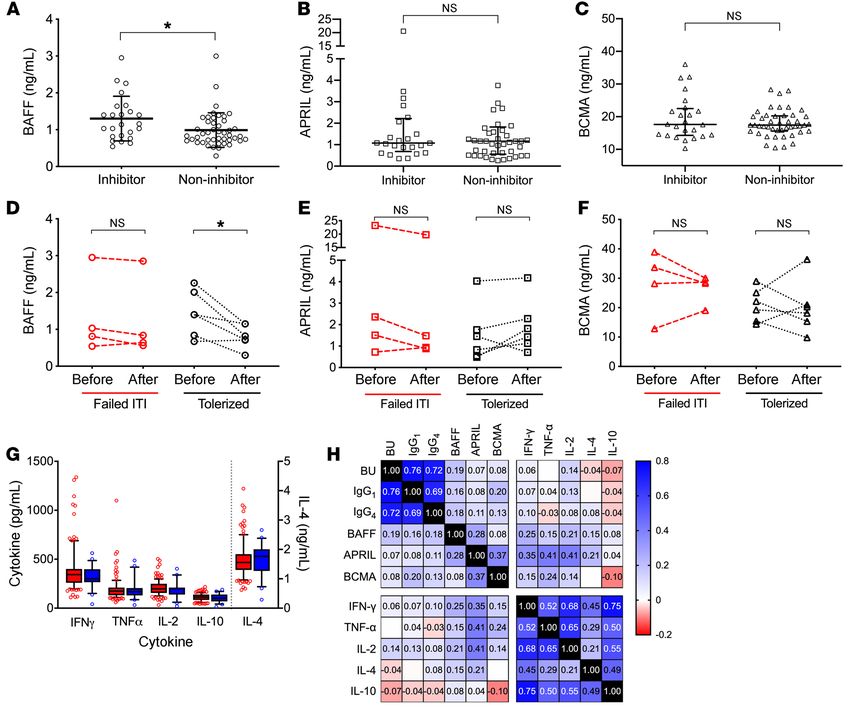
RESEARCH ARTICLE                                                                                                  The Journal of Clinical Investigation

                                                                                                         tional (TR), MZ, and other peripheral B cell subsets
Table 1. Characteristics of hemophilia A patients                                                        (40). APRIL binds to TACI and BCMA to promote
                                      Pediatric cohort                           Adult cohort            PC differentiation and survival. Expression of both
                         Inhibitor       Noninhibitor        P       Inhibitor     Noninhibitor   P      BAFF and APRIL is known to be increased under
n (%)                    24 (34.8)          45 (65.2)                22 (47.8)      24 (52.2)            proinflammatory conditions (43), thus contributing
                                                                                                         to pathogen responses (44).
Age (IQR)              3.41 (1.79–7.70) 8.08 (1.79–13.50)    NS     54 (18–63)     44 (28–57)     NS
                                                                                                              Elevations in plasma BAFF levels have been
Race                                                        0.029                                 NS
                                                                                                         implicated in several autoimmune disease pro-
  Caucasian              11 (45.8)          30 (66.7)                22 (47.8)      24 (52.2)
                                                                                                         cesses, leading to the development of a clinical-
  Black                  5 (20.8)           10 (22.2)                  0 (0)          0 (0)
                                                                                                         ly approved α-BAFF mAb, belimumab (45–48).
  Hispanic                2 (8.3)            4 (8.9)                   0 (0)          0 (0)
  Asian                   1 (4.2)            1 (2.2)                   0 (0)          0 (0)              Recent studies in allograft transplant recipients
  Other                  5 (20.8)             0 (0)                    0 (0)          0 (0)              demonstrate that high BAFF levels prior to trans-
Mutation                                                    0.016                                 NS     plant are associated with antibody-mediated rejec-
  Missense                2 (8.3)           19 (42.2)                  1 (4.5)       5 (20.8)            tion and elevated levels following α-CD20 therapy
  Nonsense               5 (20.8)           10 (22.2)                 3 (13.6)        2 (8.3)            may also contribute to the failure to induce humoral
  Inversion/Deletion     14 (58.3)          12 (26.7)                16 (72.7)      12 (50.0)            tolerance to the graft (49–51). Using both adult and
  Other                   3 (12.5)           4 (8.9)                   2 (9.1)       7 (15.2)            pediatric HA patient samples and HA mouse mod-
Severity                                                     NS                                   NS     els, here we investigate the hypothesis that BAFF
  Severe                 21 (87.5)          31 (68.9)                21 (95.5)       24 (100)            may play a role in the generation and sustenance
  Moderate                1 (4.2)            5 (11.1)                 1 (4.5)         0 (0)              of α-FVIII antibodies, especially in the context of
  Mild                    2 (8.3)           9 (20.0)                   0 (0)          0 (0)              α-CD20 therapy, which may influence therapeutic
                                                                                                         efficacy. These findings may offer additional thera-
                                                                                                         peutic targets for FVIII inhibitors in HA.

     The basic mechanisms driving FVIII inhibitor development                            Results
remain incompletely understood. Current evidence, predominantly
from animal studies, suggests that exogenous FVIII is taken up in the                    Analysis of samples from HA patient cohorts of distinct ages and
spleen by marginal zone (MZ) B cells, MZ macrophages, or delivered                       geographic backgrounds
to other antigen-presenting cells (APCs) and presented via major                         BAFF levels are higher in pediatric HA patients with persistent
histocompatibility (MHC) type II molecules to CD4+ T cells (27–31).                      FVIII inhibitors and correlate with α-FVIII antibody titers. Plasma
Under conditions of costimulation, CD4+ T cells, in particular T fol-                    samples from 69 patients followed at the Children’s Hospital
licular helper (Tfh) cells, activate cognate B cells to mature and prolif-               of Philadelphia (CHOP) Hemophilia Treatment Center (HTC)
erate within germinal centers (GCs) into antibody-secreting plasma                       were collected longitudinally. Demographic and baseline char-
cells (PCs) or memory (MEM) B cells (32, 33). Long-lived PCs (LLPCs)                     acteristic data for these patients are summarized in Table 1. Of
ultimately settle in the bone marrow (BM), sustaining the humoral                        the 69 pediatric patients, 24 (34.8%) had FVIII inhibitors and 45
response for extended periods. In humans, the antibody response                          (65.2%) did not. Patient age did not differ between those with
against FVIII consists of both neutralizing (high affinity, IgG4) and                    inhibitors (median 3.41, IQR 1.79–7.71 years) versus noninhib-
nonneutralizing (low affinity, IgG1) antibodies (34), likely from B cell                 itor controls (median 8.08, IQR 1.79–13.50 years; P > 0.05 by
receptor (BCR) rearrangement driving affinity maturation. In mice,                       Mann-Whitney U test). Consistent with prior studies (52, 53),
it is generally thought that IgG1 most closely mimics human IgG4                         patients with inhibitors were more likely to have F8 gene inver-
(35), but inhibitory α-FVIII antibodies of many subclasses have been                     sions or large deletion mutations compared with noninhibitor
described (36, 37). Prior studies have implicated either plasma levels                   patients (58.3% vs. 26.7%, P < 0.05). There was no difference
of cytokines that mediate B and T cell cross-talk, such as IL-2, IL-10,                  in race or disease severity among HA patients with and with-
and TNF-α (38), or SNPs in regulatory elements of these genes in the                     out inhibitors. All patients were treated with recombinant FVIII
FVIII immune response. However, B cell–specific cytokines have not                       products. Only 2 of 69 patients were female and neither had
been rigorously investigated previously, save one report (39).                           FVIII inhibitors; both have a normal karyotype with F8 intron 22
     A particular TNF family of cytokines and receptors has been                         inversion mutations and are presumed to have severe, skewed
implicated in the survival and maturation of B cells (40). This fam-                     X-chromosome inactivation. Plasma BAFF levels (Figure 1A)
ily includes the soluble ligands B cell–activating factor (BAFF, also                    were higher in HA patients with inhibitors compared with those
known as BLyS) and a proliferation-inducing ligand (APRIL) as well                       without inhibitors (1.30 ± 0.61 vs. 0.99 ± 0.47 ng/mL, P = 0.021
as their receptors BAFF-R (BR3), transmembrane activator and cal-                        by t test). Moreover, BAFF levels decreased from baseline in
cium modulator and cytophilin ligand interactor (TACI), and B cell                       HA inhibitor patients who underwent ITI and achieved FVIII
maturation antigen (BCMA), the latter of which can be shed from                          tolerance from 1.43 ± 0.63 to 0.81 ± 0.32 ng/mL (paired t test
the cell surface by γ-secretase and thus measured in plasma (41).                        P = 0.025, Figure 1D). In comparison, those who failed ITI had
BAFF binds with greatest affinity to BAFF-R (42) and downstream                          steady levels at 1.33 to 1.23 ng/mL (paired t test P = 0.246, Fig-
signaling from this interaction via the noncanonical NF-κB and PI3K                      ure 1D). Levels of APRIL (2.55 ± 4.66 vs. 2.97 ± 7.49 ng/mL, P >
signaling pathways supports the survival and maturation of transi-                       0.05) and BCMA (20.09 ± 6.99 vs. 17.99 ± 4.08 ng/mL, P > 0.05)

    2                                             J Clin Invest. 2021;131(8):e142906 https://doi.org/10.1172/JCI142906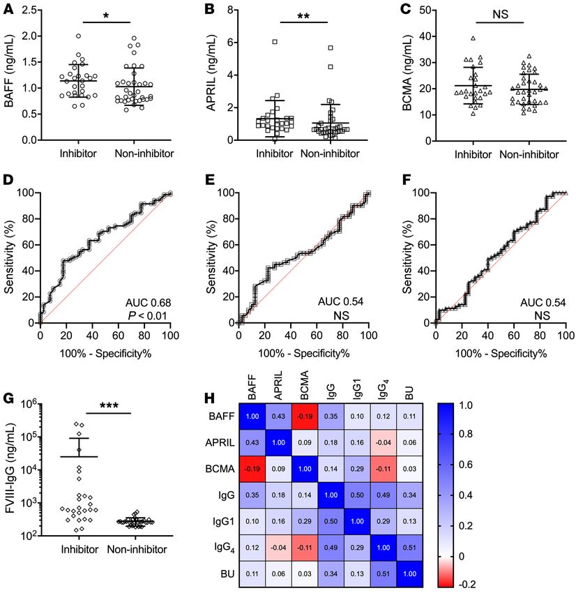
The Journal of Clinical Investigation                                                                                          RESEARCH ARTICLE

Figure 1. B cell cytokine levels in pediatric patients with hemophilia A. B cell cytokines in pediatric HA patients with FVIII inhibitors (n = 24) or without
FVIII inhibitors (n = 45). (A) BAFF levels. (B) APRIL levels. (C) BCMA levels via unpaired t test. Longitudinal analysis of (D) BAFF, (E) APRIL, and (F) BCMA
levels in pediatric patients with inhibitors who failed immune tolerance induction (n = 4) or succeeded (n = 6) via paired t test. (G) Peripheral T-helper
cytokine levels in pediatric HA patients with (red squares) and without (blue squares) inhibitors via unpaired t test. (H) Heatmap of Spearman’s correla-
tion of Bethesda titer, α-FVIII IgG subclasses, and cytokines. Box-and-whisker plots show median with 25%–75% IQR, whiskers delineate 10th and 90th
percentiles, with values outside these ranges shown as symbols. Other data plotted as mean ± SD. *P < 0.05. NS, not significant.

were not different between pediatric HA patients with and with-                   risk of autoimmune disease in patients of Italian, particularly
out inhibitors (Figure 1, B and C), or between those who achieved                 Sardinian, descent (46). Thus, we sought to determine if HA
and failed to achieve FVIII tolerance (Figure 1, E and F).                        inhibitor patients from Italy had increased B cell cytokine lev-
    Levels of the T-helper cytokines IFN-γ, TNF-α, IL-2, IL-4,                    els. Demographic data from 46 predominantly adult patients
and IL-10 did not differ between pediatric HA patients with and                   followed at the Careggi HTC are summarized in Table 1. Of
without inhibitors (Figure 1G). BAFF levels correlated with α-FVIII               the 46 patients, 22 (47.8%) had inhibitors and 24 (52.2%) did
IgG1 (Spearman’s correlation coefficient [ρ] = 0.16, P < 0.05) and                not have inhibitors. Of the 22 patients with inhibitors, 5 had
α-FVIII IgG4 (Spearman’s ρ = 0.18, P < 0.01) and most strongly with               achieved FVIII tolerance, 7 were on ITI, and 10 had failed ITI.
the α-FVIII Bethesda titer (Spearman’s ρ = 0.19, P < 0.005). Cor-                 Patient age did not differ between those with inhibitors (medi-
relation plots are shown in Supplemental Figure 1 (supplemental                   an 54, IQR 18–63 years) versus noninhibitor controls (median
material available online with this article; https://doi.org/10.1172/             44, IQR 28–57 years; P > 0.05 by Mann-Whitney U test). All
JCI142906DS1). In contrast, none of the other cytokines correlat-                 patients were of Caucasian descent and there was no difference
ed with Bethesda titer or IgG subclasses, with Spearman’s ρ rang-                 in disease severity or F8 mutations between the cohorts. Lev-
ing from –0.03 to 0.14 (P > 0.05; correlation heatmap, Figure 1H).                els of BAFF (1.14 ± 0.31 vs. 1.03 ± 0.36 ng/mL, P = 0.041) and
    BAFF and APRIL levels are associated with inhibitor presence                  APRIL (1.33 ± 1.11 vs. 1.06 ± 1.14 ng/mL, P = 0.008) were higher
in adult HA patients. Recent genomic studies have identified 2                    in patients with inhibitors versus noninhibitors (Figure 2, A and
variants that lead to elevated BAFF levels and confer increased                   B). Levels of BCMA were not different between patients with

                                           J Clin Invest. 2021;131(8):e142906 https://doi.org/10.1172/JCI142906                                           3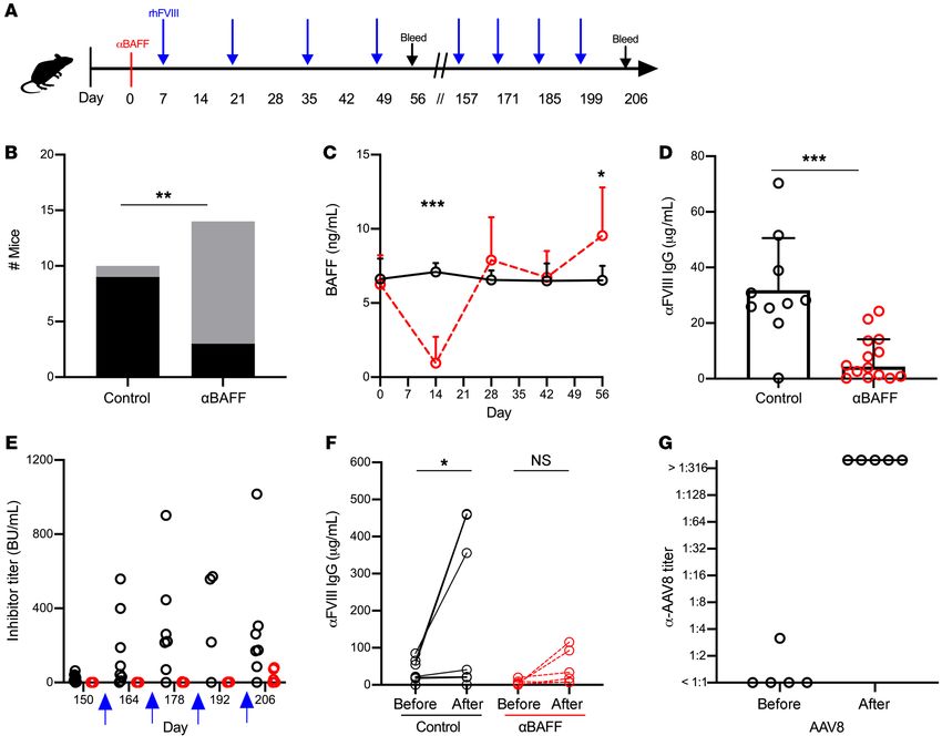
RESEARCH ARTICLE                                                                                              The Journal of Clinical Investigation

Figure 2. B cell cytokine levels in adult Italian hemophilia A patients. B cell cytokine and α-FVIII IgG levels in adult HA patients with (n = 22) or without
(n = 24) FVIII inhibitors. (A) BAFF levels. (B) APRIL levels. (C) BCMA levels. Receiver operating characteristics of (D) BAFF, (E) APRIL, and (F) BCMA for
pediatric and adult HA patients. (G) α-FVIII IgG in adult HA patients. (H) Spearman’s correlation heatmap of B cell cytokines and α-FVIII IgG in adult HA
patients. *P < 0.05, **P < 0.01, ***P < 0.001 by Mann-Whitney U test. NS, not significant.

and without inhibitors (Figure 2C). As expected, FVIII-specif-                       Finally, we combined the pediatric and adult HA patient
ic IgG was considerably higher in the inhibitor cohort (25,039                   cohorts to determine whether BAFF, APRIL, and BCMA lev-
± 65,555 vs. 275.5 ± 77.8 ng/mL, P < 0.001; Figure 2G) and                       els can be used to discern the presence of FVIII inhibitors by
BAFF levels correlated with total IgG by Spearman’s correla-                     receiver-operating characteristic (ROC) analysis. ROC curves
tion analysis (ρ = 0.35, P < 0.01; Figure 2H). The BAFF levels                   measure the probability of a test to distinguish a binary outcome
observed in the adult Italian HA inhibitor cohort were similar                   at various thresholds and the area under the curve (AUC) rep-
to those seen in the pediatric HA inhibitor cohort from the                      resents the degree of separation. Thus, the higher the AUC, the
United States (1.14 ± 0.31 vs. 1.30 ± 0.61 ng/mL, respectively;                  more likely the test performs well in discerning disease state.
P > 0.05) and higher than those in the noninhibitor pediat-                      In our analysis, the AUC was statistically significant for BAFF
ric cohort (1.14 ± 0.31 vs. 0.99 ± 0.47 ng/mL, respectively; P                   at 0.68 (95% CI 0.57–0.78, P < 0.01; Figure 2D) but not APRIL
< 0.05). Although APRIL and BCMA levels trended higher in                        (AUC 0.54, Figure 2E) or BCMA (AUC 0.54, Figure 2F). Total
the pediatric inhibitor cohort, they were not statistically differ-              operating characteristic curves of these cytokines are similar to
ent between the inhibitor-positive adult and pediatric cohorts                   the ROC curves (Supplemental Figure 2). BAFF levels greater
(P > 0.05). Noninhibitor adult Italian patients had lower APRIL                  than 1.03 ng/mL had 68.3% sensitivity, 63.8% specificity, and
levels (1.06 ± 1.14 vs. 2.97 ± 7.49 ng/mL, P < 0.05) and higher                  likelihood ratio of 1.89 for the presence of FVIII inhibitors, sug-
BCMA levels (20.66 ± 5.42 vs. 17.99 ± 4.08 ng/mL, P < 0.05)                      gesting that BAFF could be a potential harbinger of an ongoing
than the pediatric noninhibitor cohort.                                          α-FVIII humoral immune response.

   4                                      J Clin Invest. 2021;131(8):e142906 https://doi.org/10.1172/JCI142906
The Journal of Clinical Investigation RESEARCH ARTICLE
although levels did rise in this population as well (0.78 ± 0.42 to
20.60 ± 29.66 ng/mL, P > 0.05). In studies of other immune-me-
diated processes, the rise in BAFF following rituximab therapy has
been shown to preclude antigen tolerance (54–56). Given the rise
in BAFF in HA inhibitor patients treated with rituximab, we inves-
tigated this hypothesis in HA animal models.
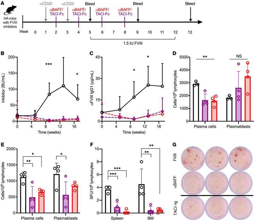
Inhibitor prevention and eradication studies in HA mouse models
We hypothesized that the elevated levels of BAFF in human HA
inhibitor patients could serve as a survival signal for FVIII-reactive
B cells and targeting BAFF may be of therapeutic value in FVIII
inhibitors. Here, we tested the hypothesis that blocking BAFF
could be effective in the prevention and/or eradication of FVIII
inhibitors in animal HA models. Doering et al. have shown that the
use of murine FVIII protein does not induce inhibitor formation
in HA mice (57); to overcome this limitation, recombinant human
FVIII (rhFVIII) protein concentrates are used. To avoid strain-spe-
cific results and limit the potential bias in the assessment of the
immune responses, we used distinct HA strains on a C57BL/6-129
background (colony at CHOP) or BALB/c background (colony at
Indiana University). The HA phenotype is similar between these
strains but immune responses to pathogens, proteins, and gene
therapy are known to differ (58–63). Further, we used 2 distinct
murine α-BAFF (α-mBAFF) mAbs: (a) clone 10F4, which is a ham-
ster IgG1 mAb with a half-life of approximately 2 weeks (64); and
Figure 3. BAFF levels in HA inhibitor patients treated with rituximab. (A) (b) clone Sandy-2, which is a mouse IgG1 mAb with a half-life of
Schema for rituximab therapy. Adult and pediatric HA patients with refrac- approximately 10 days (65). These antibodies are biologically
tory inhibitors were treated with rituximab (black diamonds) and plasma equivalent in their inhibition of TR, follicular, and MZ B cells, with
samples (red drops) were obtained before and after therapy. Patients (n = 8)
10F4 taking longer (~8 weeks) for immune reconstitution com-
received concurrent FVIII ITI or not (n = 9). (B) BAFF levels before and after
rituximab therapy in HA patients who did (black circles, n = 3) or did not pared with Sandy-2 (6 weeks) (64, 65).
(red circles, n = 14) achieve FVIII tolerance at the end of their ITI course. α-mBAFF therapy prevents FVIII inhibitor development in
**P < 0.01 by paired t test. NS, not significant. FVIII-naive HA C57BL/6-129 mice. As BAFF is necessary for the
survival of TR and MZ B cells, the latter of which have been impli-
cated in the initiation of the FVIII immune response in mice (30,
Increase in BAFF levels following rituximab therapy in adult and 31), we investigated whether prophylactic α-mBAFF mAb ther-
pediatric HA patients. Next, we investigated whether a rise in BAFF apy could prevent FVIII inhibitor formation in HA C57BL/6-129
after rituximab-based therapy precludes tolerance to FVIII, as mice, which mount a robust immune response to rhFVIII protein
seen in other allo- and autoimmune disease contexts. BAFF levels compared with BALB/c mice (63). FVIII-naive C57BL/6-129 HA
were measured from samples obtained from a total of 17 HA inhib- mice (n = 10–14/group) were given α-mBAFF mAb (Sandy-2) or
itor patients. Of these, 9 were enrolled in the only prospective trial isotype control prior to immunization with rhFVIII and followed
of rituximab alone for ITI-refractory FVIII inhibitors (RICH trial) for FVIII inhibitor development (Figure 4A). Only 3 of 14 mice
wherein rituximab was dosed at 375 mg/m2 weekly for 4 weeks. in the α-mBAFF group developed inhibitors, with Bethesda titers
The remaining patients were enrolled from HTCs at Emory Uni- ranging from 0–150 BU with a median titer of 0 BU (IQR 0–0.5)
versity (n = 6) and CHOP (n = 2) who received the same dose of compared with 9 of 10 mice in the control group with a range of
rituximab with concurrent FVIII protein replacement ITI. Plasma 0–254 BU and median titer of 21.1 BU (IQR 2.5–157.3), resulting
samples were obtained at baseline and following the last dose of in a significantly reduced relative risk of 0.23 (95% CI 0.08–0.57)
rituximab (Figure 3A) and all patients were followed longitudinal- with α-mBAFF therapy (Figure 4B).
ly for inhibitor titers. Of this cohort of 17 HA patients, 3 of 17 (17%) BAFF levels in α-mBAFF–treated mice were depleted at 14 days
achieved tolerance to FVIII (1 treated with rituximab alone and 2 after injection (0.94 ± 1.78 vs. 7.10 ± 0.60 ng/mL, P < 0.001) and
with rituximab and FVIII ITI), as defined by a negative Bethesda levels equalized by day 28 between groups (Figure 4C). α-FVIII IgG
titer, and 14 of 17 (82%) did not achieve FVIII tolerance. Within was lower in the α-mBAFF–treated group (7.52 ± 8.07 μg/mL) com-
the nonresponding cohort, 8 were treated with rituximab only pared with controls (31.83 ± 18.77 μg/mL, P < 0.001; Figure 4D). Of
and 6 were treated with rituximab and FVIII ITI. In the patients note, these experiments were also conducted in HA BALB/c mice,
who failed to achieve FVIII tolerance (Figure 3B), BAFF levels a model that requires weekly rhFVIII immunization to mount an
increased 3-fold from baseline (0.89 ± 0.39 to 2.66 ± 2.03 ng/ inhibitor response. The data showed decreased α-FVIII IgG, with a
mL, paired t test P = 0.007). The relatively low number of patients median of 0.19 (IQR 0.12–0.52) in treated versus 0.75 (IQR 0.42–
who achieved FVIII tolerance prevented statistical conclusions, 2.43) μg/mL in control mice (P < 0.05) (data not shown).
J Clin Invest. 2021;131(8):e142906 https://doi.org/10.1172/JCI142906 5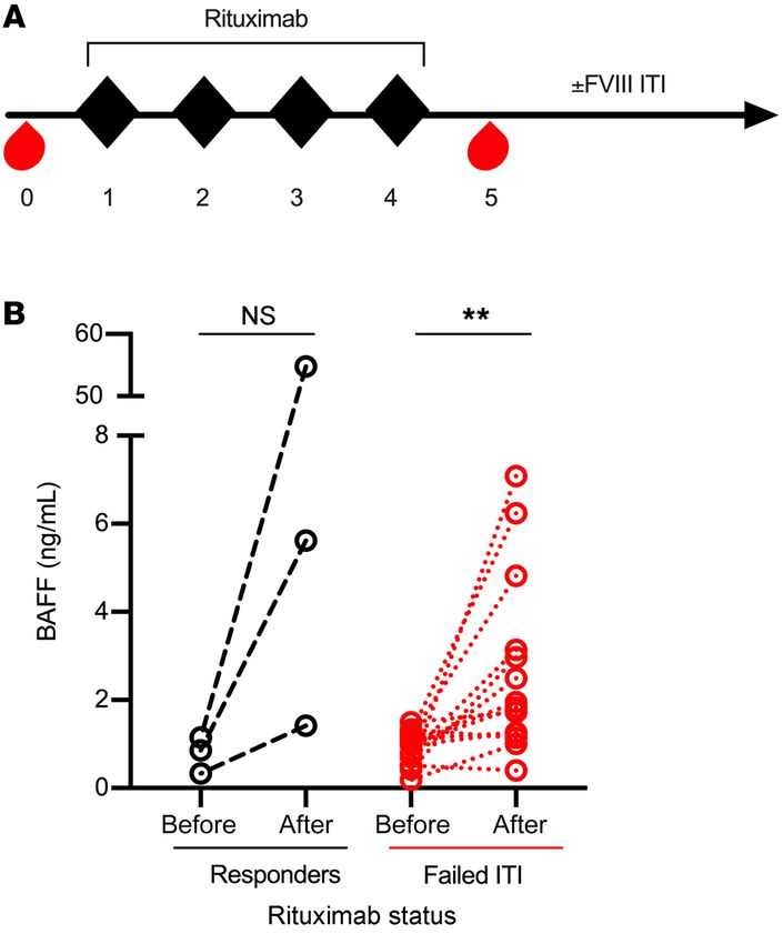
RESEARCH ARTICLE                                                                                                     The Journal of Clinical Investigation

Figure 4. α-mBAFF antibody therapy for prevention of FVIII inhibitors in HA mice. (A) C57BL/6-129 HA mice (n = 10–14/group) were injected with α-mBAFF
antibody prior to immunization with FVIII and followed longitudinally. (B) Number of inhibitor-positive (black bars) or -negative (gray bars) mice in controls
versus α-mBAFF–treated groups. (C) BAFF levels over time in the α-mBAFF (red circles) and control (black circles) groups. (D) α-FVIII IgG in the α-mBAFF group
(red circles) compared to controls (black circles) on day 56. FVIII inhibitor titers (E) and α-FVIII IgG (F) after remote FVIII challenge (blue arrow) in control (black
circles) and α-mBAFF–treated (red circles) mice. (G) Titers of neutralizing antibodies against AAV8. AAV8 was injected 17 weeks after mice were treated with
α-mBAFF antibody and α-AAV8 antibody titers were measured before and 4 weeks after AAV8 injection (n = 6). *P < 0.05; **P < 0.01; ***P < 0.001 by Fisher’s
exact (B), 2-way ANOVA (C), Mann-Whitney U (D and E), Wilcoxon’s matched signed rank (F), and paired t (G) tests. NS, not significant.

     In the HA C57BL/6-129 mice, 22 weeks after initial α-mBAFF                       BAFF levels (4 weeks). To ensure that mice were capable of mount-
mAb, long-term tolerance to FVIII was tested by rhFVIII injections                    ing an immune response to a T cell–dependent antigen, a second
in 6 mice from the α-mBAFF treatment group (Figure 4E). No mice                       cohort of α-mBAFF–treated mice were challenged with adeno-asso-
developed a high-titer (BU > 5) FVIII inhibitor response after the first              ciated virus type 8 (AAV-8) vector at 17 weeks and developed robust
challenge and, thus, immunizations were continued for a total of 4                    neutralizing antibody responses, with titers higher than 1:316 dilu-
challenges. After the fourth challenge, control mice had a median                     tion (Figure 4G). Thus, the lack of a robust neutralizing antibody
inhibitor titer of 174.6 BU (IQR 85.6–305.6) compared with 8.5 BU                     responses with FVIII challenge in these mice suggests that prophy-
(IQR 0.9–74.1) in α-mBAFF–treated mice (P < 0.05). Remarkably,                        lactic α-mBAFF mAb therapy during initial FVIII exposure may bias
only half of the mice from the α-mBAFF group developed high-ti-                       the immune system specifically toward FVIII-antigen tolerance.
ter inhibitors (15–80 BU), whereas the remaining mice had inhibi-                          Combination α-mCD20/α-mBAFF mAb therapy induces tolerance in
tor titers of less than 2 BU. Corresponding FVIII-specific IgG levels                 HA BALB/c mice with established FVIII inhibitors. The clinical burden of
before and after the 4 challenge rhFVIII doses are presented in Fig-                  disease in HA resides with patients with established inhibitors. Thus,
ure 4F. Thus, a single dose of α-mBAFF was sufficient to prevent the                  we sought to determine if α-mBAFF–based therapy could be effective
formation of high-titer inhibitors in HA mice, with a sustained effect                in eradicating FVIII inhibitors. HA BALB/c mice with inhibitors were
(>22 weeks) beyond the relative short initial period of reduction of                  treated with α-mCD20 alone, α-mBAFF alone (GlaxoSmithKline

   6                                         J Clin Invest. 2021;131(8):e142906 https://doi.org/10.1172/JCI142906
The Journal of Clinical Investigation RESEARCH ARTICLE
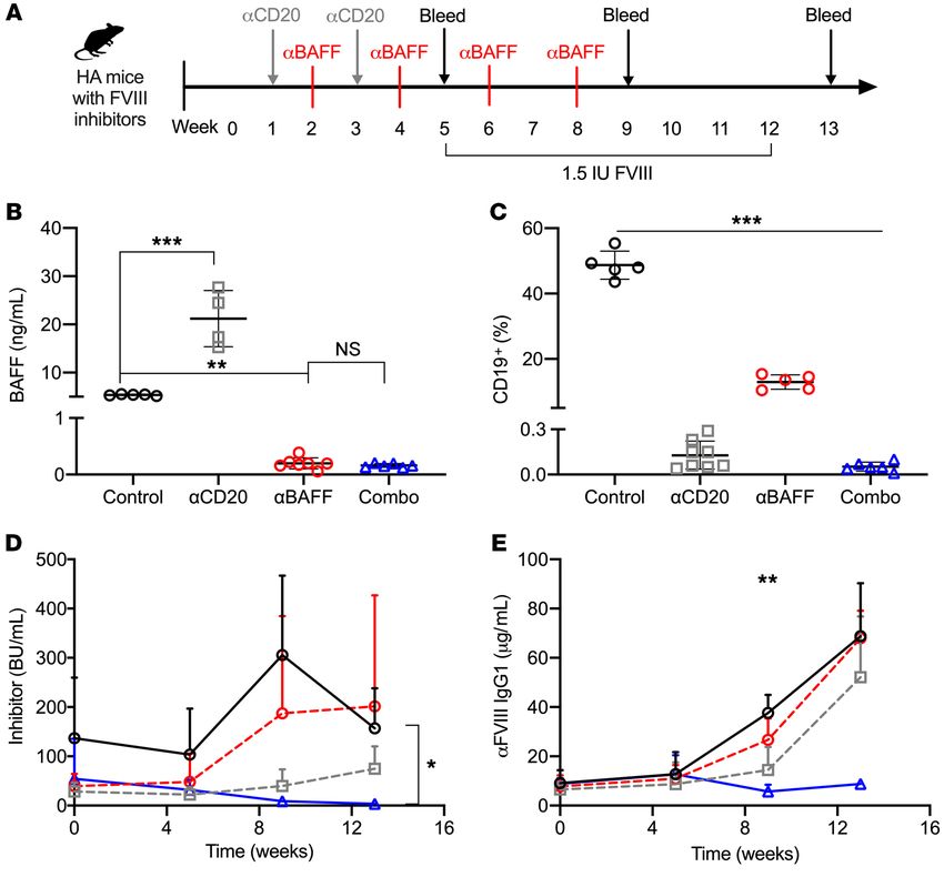
Figure 5. Combination of α-mBAFF and α-mCD20 therapy for FVIII tolerance induction. (A) Schema for combination α-mCD20 and α-mBAFF therapy.
HA-BALB/c mice with established inhibitors were treated with α-mCD20 (gray squares, n = 8), α-mBAFF (red circles, n = 6), combination therapy (blue tri-
angles, n = 6), or no treatment (black circles, n = 5) and followed for 13 weeks. (B) BAFF levels at week 5, (C) peripheral CD19+ B cells at week 5, (D) inhibitor
titer, and (E) α-FVIII IgG1. *P < 0.05; **P < 0.01; ***P < 0.001 by 1-way ANOVA (B and C) or mixed-effects ANOVA (D and E). NS, not significant.
clone 10F4), or combination α-mCD20/α-mBAFF mAb therapies, as tors and maintaining immune tolerance despite continued challenges
depicted in Figure 5A. Recapturing the human data following ritux- with the protein. In the more immunogenic HA C57BL/6-129 mice,
imab therapy, HA-BALB/c mice treated with an α-mCD20 antibody inhibitor titers decreased from 70 BU to less than 5 BU with 2 cycles of
had a 3-fold increase in mBAFF levels at week 5 (Figure 5B) compared this combination therapy regimen (data not shown).
with control mice (15.57 ± 0.47 vs. 5.37 ± 0.15 ng/mL, P < 0.001), and For preliminary quantification of immune cell subsets involved
this rise was ameliorated by addition of α-mBAFF mAb to α-mCD20 in the induction of tolerance to FVIII, HA BALB/c mice were treat-
(0.16 ± 0.34 ng/mL, P < 0.001 vs. α-mCD20 alone), which was simi- ed with the various mAb regimens to monitor B cell repopulation
lar to α-mBAFF mAb alone. APRIL levels were not elevated (data not at weeks 5 and 9 (Supplemental Figure 3, A–F). Compared with
shown). Compared with control mice, all treatment group mice had control mice, spleens of combination-treated mice had universally
lower peripheral CD19+ B cell percentages at week 5, with levels of 48.7 lower B cell subsets, including follicular, MZ, MEM, TR, and PCs
± 4.3 in controls, 0.1 ± 0.1 in α-mCD20, 12.9 ± 2.2 in α-mBAFF, and as well as plasmablasts at week 5 (P < 0.001), of which follicular
0.1 ± 0.0 in combination therapy (P < 0.001 by 1-way ANOVA, Figure (P < 0.001), MZ (P < 0.01), and PC (P < 0.05) depletion persist-
5C). Only the combination therapy resulted in a substantial decrease ed at week 9. Consistent with previous reports (67–69), BAFF-R
in inhibitor titer (3.7 ± 1.7 vs. 156.9 ± 81.3 BU, P < 0.05) and α-FVIII IgG1 expression was present in follicular and MZ B cell subsets, splenic
(8.28 ± 1.46 vs. 68.76 ± 21.51 μg/mL, P < 0.01) compared with con- plasmablasts, and PCs, with highest expression in GC B cells and
trols (Figure 5, D and E), even following repeated rhFVIII challenge. lower expression in BM plasmablasts and PCs (Supplemental Fig-
From prior experiments, B cell repopulation was seen by 4 weeks ure 3G). In contrast, TACI expression was highest in splenic and
after α-mCD20 mAb administration, so these challenges occurred BM plasmablasts and PCs (Supplemental Figure 3H). These initial
during or after B cell recovery (66). Thus, the combination therapy of data suggested that a PC-dependent mechanism was responsible
α-mBAFF mAb and α-mCD20 is effective in eradicating FVIII inhibi- for the ability of combination therapy to induce FVIII tolerance.
J Clin Invest. 2021;131(8):e142906 https://doi.org/10.1172/JCI142906 7RESEARCH ARTICLE The Journal of Clinical Investigation
Figure 6. Combination therapy with α-mCD20 with α-mBAFF or mTACI-Fc in FVIII inhibitor mice. (A) Schema for combination α-mCD20 and α-mBAFF
or mTACI-Fc therapy. HA inhibitor mice were treated with α-mCD20 with α-mBAFF (red circles, n = 10), α-mCD20 with mTACI-Fc (purple diamonds, n
= 8), or no treatment (black circles, n = 8) and followed for (B) Bethesda titer and (C) α-FVIII IgG1. At 16 weeks from start of regimen, spleens (D) and
bone marrow (E) were harvested for quantification of plasmablasts and plasma cells by flow cytometry (values normalized per million lymphocytes). (F)
FVIII-specific B cell ELISPOT from splenic and bone marrow plasma cells (conducted in triplicate from n = 4 mice per group), with representative images
of samples (G). *P < 0.05; **P < 0.01; ***P < 0.001 by mixed-effects ANOVA (B and C) or 1-way ANOVA (D–F). NS, not significant.
Either BAFF or APRIL can support PC survival in the BM, indicat- process. Littermate controlled HA BALB/c mice with preexisting
ing a redundant role for BAFF in LLPC maintenance (70). inhibitors were treated with α-mCD20 and α-mBAFF or mTACI-Fc
α-mBAFF performs similarly to mTACI-Fc in combination with mAb, as delineated in Figure 6A. Compared with control mice,
α-mCD20 for FVIII inhibitor eradication. To investigate the PC α-mCD20 plus mTACI-Fc or α-mBAFF mAb resulted in signifi-
compartment further, we compared performance of α-mCD20 cantly lower inhibitor titers starting at 2 months (84.1 ± 34.9, 3.5 ±
mAb with α-mBAFF or mTACI-Fc mAb. Consisting of the Fc 3.5, and 1.6 ± 1.8 BU, respectively; P < 0.001) but no difference was
region of IgG and the binding domain of the TACI receptor, mTA- seen between the mBAFF- and mTACI-targeted groups (Figure
CI-Fc (atacicept in humans, ref. 71) can bind and inactivate both 6B). A higher proportion of α-mBAFF mice had low-titer inhibitors
BAFF and APRIL in their soluble forms and thereby inhibit down- (BU < 5) at week 13 compared with mTACI-Fc (88.9% vs. 62.5%,
stream signaling (72). As TACI-Fc is known to target PC survival respectively) but this was not statistically significant. Similarly, the
(70), similar results between these 2 mAb therapies would confirm α-FVIII IgG1 titer (Figure 6C) was higher in control mice compared
that combination therapy exerts its effect through a PC-mediated with the α-mBAFF or mTACI-Fc mice at 2 months (16.9 ± 10.9 vs.
8 J Clin Invest. 2021;131(8):e142906 https://doi.org/10.1172/JCI142906The Journal of Clinical Investigation RESEARCH ARTICLE
1.5 ± 1.4 or 1.5 ± 0.7 μg/mL, respectively; P < 0.001). Tolerance was humans is characterized by both low- and high-affinity antibodies,
sustained for the duration of the experiment (4 months), even after which correspond to nonneutralizing and neutralizing antibodies,
weekly immunological challenges with rhFVIII protein (months 2 respectively (76, 77). Our pediatric HA patient data indicate that
and 3). These data support our findings that combination therapy BAFF levels correlate most strongly with the FVIII-inhibitor and
targeting mCD20 and mBAFF eradicates FVIII inhibitors through neutralizing-IgG4 titers but also with α-FVIII IgG1, whereas other
a PC-mediated process. T-helper cytokines tested here did not. Prior studies have shown
Combination therapy targeting mBAFF or mTACI suppresses correlation of polymorphisms in regulatory elements of certain
PCs. To determine the effect of combination therapies on PCs, cytokines with inhibitors but few have assessed cytokine levels;
spleens and BM were harvested from mice at the 4-month time the results from either method are inconsistent between popula-
point for flow cytometric quantification of PCs and plasmablasts tions of distinct geographic origins (78–82). As genetic polymor-
and an FVIII-specific B cell ELISPOT assay. Mice treated with phisms that predispose patients to elevated BAFF levels have
α-mBAFF or mTACI-Fc had a reduction in splenic PCs compared been characterized in people of Sardinian and continental Italian
with controls (1593 ± 379, 1646 ± 49, and 2927 ± 171 cells/106 descent compared with northern Europeans (46), we measured
lymphocytes, respectively; P < 0.01) but not splenic plasmablasts BAFF levels from an Italian adult HA cohort. BAFF and APRIL
(3501 ± 1026, 2611 ± 1273, and 1836 ± 150 cells/106 lymphocytes, levels were elevated in our cohort of adult Italian HA inhibitor
respectively), as seen in Figure 6D. There was a decrease in BM patients. Comparing the Italian adult to the US pediatric inhibi-
PCs (653 ± 77, 491 ± 332, and 1124 ± 152 cells/106 lymphocytes, tor-positive cohort, levels of BAFF, APRIL, and BCMA did not dif-
respectively, P < 0.01; Figure 6E) across all groups. BM plasmab- fer between the 2 sites. However, in noninhibitor patients, APRIL
last counts were higher in control (1216 ± 341) versus mTACI-Fc– levels were higher and BCMA levels were lower in the pediatric US
treated mice (565 ± 411, P < 0.05) but not α-mBAFF mice (837 ± cohort compared with the Italian adult cohort. Of note, APRIL and
107 cells/106 lymphocytes). These results were consistent with BAFF levels are known to decrease with age (83) and are thought
ELISPOT analysis, which showed fewer spot-forming units (SFU) to help maintain the peripheral B cell pool, which also decreases
in the experimental versus treated mice (Figure 6, F and G). Com- with age (84). The similarity of the BAFF levels between adult and
bined with data from earlier time points (Supplemental Figure 3), pediatric inhibitor patients, thus, suggests that ongoing elevated
these data suggest that combination mAb treatment could bias BAFF may contribute to circulating α-FVIII B cells. Naive B cells
the immune system toward FVIII immune tolerance via sustained and plasmablasts rely on APRIL for survival, which may explain
depletion of FVIII-specific PCs. the higher APRIL levels in pediatric patients (85). Nevertheless,
upon combination of these 2 groups (n = 115), BAFF had an AUC of
Discussion 0.68 in the ROC analysis for inhibitor presence and greater than
FVIII is one of the most immunogenic biologics (1) and inhibitors 60% sensitivity and specificity for inhibitors at a cutoff of 1.03
of FVIII pose a significant barrier to optimal care, thereby increas- ng/mL. Although statistically significant, the differences in the
ing patient morbidity and mortality. Understanding mechanisms adult and pediatric populations do need to be taken into account
that predispose and/or drive FVIII immune responses is thus of in using BAFF levels as a diagnostic marker of inhibitor presence.
clinical significance. ITI protocols are not always effective at erad- Rather, ongoing elevation or a rise in BAFF levels over time in a
icating inhibitors and combination immunosuppressive strategies patient with an inhibitor could potentially serve as a marker of
to date have had meager results, with toxicity-related limitations. incomplete tolerance induction (given its correlation with α-FVIII
The α-CD20 mAb rituximab held promise for FVIII inhibitor IgG) or harbinger of impending ITI failure.
eradication in early reports, but the modest efficacy observed in a The data suggest a role for BAFF as a modulator of FVIII inhib-
prospective study of monotherapy with rituximab (26) has damp- itors but the underlying mechanism and timing of BAFF elevation
ened this optimism. This suboptimal efficacy may partially be remains to be determined. Presentation of FVIII during times of
explained by the persistence of rituximab-resistant FVIII+ B cell immune activation is thought to increase the likelihood of inhibi-
subsets. In the context of autoimmune cytopenias, rituximab par- tor development (86). Correlation of BAFF with the proinflamma-
adoxically promotes the rapid repopulation of cells with an LLPC tory cytokines IFN-γ and IL-2 in the pediatric cohort may support
phenotype in extrafollicular foci in the spleen (54, 73, 74). Fur- the idea that BAFF participates in this cascade to elicit a strong
ther, increased availability of BAFF as a result of B cell depletion immune response to FVIII. An alternative hypothesis is that BAFF
is thought to possibly exacerbate autoimmune disease in patients levels are elevated prior to inhibitor development and serve as an
(55, 56) and increase alloantibody responses in graft-versus-host adjuvant to FVIII. Certainly, coadministration of BAFF protein
disease (75) or transplant rejection (51). Thus, we hypothesized with vaccines has been shown to increase antibody titers (87).
that BAFF plays a role in the initiation and/or maintenance of the Finally, BAFF levels could be a surrogate marker of unregulated
FVIII immune response, and so we used distinct HA patient sam- B cell activity. The continued high levels of BAFF seen in adult
ples and mouse models to address this hypothesis. Our data show inhibitor patients and in pediatric patients who fail ITI would sup-
a potential role for BAFF inhibition in both prevention and eradi- port this possibility.
cation of FVIII inhibitors. Additionally, utilizing rare plasma specimens from refractory
Using a rare cohort of longitudinal samples from pediatric HA HA inhibitor patients who received rituximab-based ITI, we show
patients, we show that levels of BAFF are elevated in pediatric HA that those who fail to establish tolerance to FVIII have BAFF lev-
inhibitor patients and a decrease in BAFF correlates with success- els that rise 3-fold from baseline, a finding that is mirrored in HA
ful FVIII tolerance induction with ITI. The α-FVIII IgG response in mice that receive α-mCD20 mAb therapy. Together, our data sug-
J Clin Invest. 2021;131(8):e142906 https://doi.org/10.1172/JCI142906 9RESEARCH ARTICLE The Journal of Clinical Investigation
gest that BAFF may be associated with the α-FVIII B cell response. combination therapy is likely the combination of initial MEM B
Further, as seen in the 6 successfully tolerized patients in the pedi- cell depletion with α-CD20 therapy followed by prevention of new
atric cohort, BAFF levels in conjunction with α-FVIII IgG could FVIII+ PCs by α-BAFF therapy.
potentially be used as a surrogate for likelihood of successful ITI. The dramatic reduction in inhibitor levels with preemptive
Identification of BAFF modifiers could provide additional insight α-mBAFF mAb is explained by the reliance of MZ B cells on BAFF
into the reason behind this elevation. for survival and differentiation (94). We postulate that these mice
Next, we establish that a single dose of α-mBAFF mAb in naive have lower rates of GC B cell reactions and consequently fewer
HA mice prevents inhibitor development (even after immune PCs and MEM B cells, allowing for tolerance to FVIII. This is sup-
reconstitution) and that, in mice with preexisting inhibitors, the ported by the fact that nearly half of these mice did not generate
combination of α-mBAFF and α-mCD20 dramatically reduces a high-titer immune response despite 4 remote FVIII challeng-
and/or eliminates FVIII inhibitors, with sustained suppression es after immune reconstitution, suggesting that FVIII exposure
of FVIII-specific PCs. Recent evidence suggests that the initial during B cell reconstitution after initial mAb therapy may have
FVIII immune response is mediated by MZ B cells (30, 31), which shifted the immune balance toward tolerance. Notably, these
can rapidly differentiate into short-lived antibody-secreting cells mice are able to mount a robust immune response against an
(ASCs) (88). However, high-affinity antibodies typically result unrelated antigen, supporting the safety and specificity of this
from GC reactions that result in BCR rearrangement, leading to strategy. Our data, combined with studies in enzyme replace-
differentiation into PCs or MEM B cells. Within GCs, Tfh cells ment therapy (95), indicate that BAFF plays a role in the immu-
secrete BAFF in order to promote the selection of high-affinity GC nogenicity of biotherapeutics. As some HA mice still developed a
B cell clones (69, 89). In HA inhibitor mice, Reipert’s group has high-titer α-FVIII antibody response, future studies are needed to
shown that MEM B cells can drive ASC generation (90). Although determine whether modification of dose and/or treatment dura-
rituximab is thought to work primarily by suppressing MEM B tion would help prevent inhibitors completely. Certainly, before
cells, the poor response to rituximab ITI in HA patients suggests translational studies, additional data regarding the safety of this
that MEM B cells are not solely responsible for the FVIII immune regimen are necessary, especially as patients with inhibitors are
response. This finding is supported by additional murine studies typically less than 2 years of age. Using a specific immune modu-
in which depletion of PCs was necessary for long-term tolerance latory strategy in these young patients may be safer than general
induction (91). Additional studies are needed to further confirm immunosuppressive regimens tested in hemophilia (21, 24, 96)
these findings. and other genetic diseases (97–100).
We hypothesize that combination therapy with α-CD20 and Our study does have some limitations. First, there are likely
α-BAFF prevents the selection of high-affinity B cell clones in the differences in the established, longer immune response seen in
GC and depletes FVIII-specific PCs. In our study, neutralization adults versus pediatric inhibitor patients whose immune responses
of downstream signaling from BAFF and APRIL via combina- may still be evolving, as noted in the HIPS study (34). However, the
tion therapy with α-mCD20 and mTACI-Fc did not additionally continued elevation of BAFF in the adult HA inhibitor population
improve inhibitor eradication over combination with α-mBAFF. (which otherwise should fall) supports the hypothesis that BAFF
As TACI-Fc is known to target PC survival and spare MEM B cells, modulates the FVIII immune response. Second, mice are geneti-
consistent with the PC depletion seen in our experiments, this cally more homogeneous in comparison with humans; we attempt-
supports the role of PCs in the FVIII immune response. Although ed to ameliorate this by using a variety of antibody reagents and
inhibitor titers were still dramatically low in the combination 2 different HA strains. Finally, although common to all small-an-
therapy groups at 16 weeks, there was a small increase in the titer imal HA models, the rhFVIII immune response studied in mice is
between weeks 12 and 16 (when further FVIII was not given). This a xenoprotein response and thus may not be directly applicable to
may point to a resurgence of MEM B cells that are driving new PC the human experience. However, as both are alloantibodies and
generation, as suggested by the data from Reipert et al. (90). Nota- given that most inhibitor patients do not make endogenous FVIII,
bly, however, low BU titers (≤5) were sustained at the 16-week we think characterizing the response to rhFVIII in a mouse model
time point, which would allow inhibitor patients to resume FVIII provides valuable insight into both inhibitor formation and poten-
replacement therapy, which is the main goal of successful ITI. tial therapeutics targeting human α-FVIII antibodies.
In a trial of kidney transplant recipients who received the In summary, our data establish the potential to use α-BAFF
α-BAFF mAb belimumab in an effort to decrease de novo IgG pro- therapy in conjunction with α-CD20 therapy for eradication of
duction and limit allograft rejection, MEM B cell numbers were FVIII inhibitors in patients with HA. Belimumab is FDA approved
not different between the belimumab-treated and control groups in pediatric and adult patients and trials of combination thera-
(92). However, de novo IgG production still dropped 3-fold in the py with rituximab are ongoing in autoimmune disease contexts
belimumab group even after discontinuation of therapy and was in adults (NCT02631538, NCT02260934, NCT03967925,
associated with a skewing of cytokine production favoring IL-10 NCT03747159, etc.). These data, along with pediatric trial data,
over IL-6 in TR and MEM B cells, thus supporting a tolerogenic could provide important safety information for use in young
immune profile. Finally, the lack of tolerance induction in pre- inhibitor patients. Future studies aimed at understanding the
existing-inhibitor mice with α-BAFF therapy alone mimics the longevity and exact mechanism of this response are needed.
human experience in HLA sensitization (93). BAFF is not known to Determining whether genetic variants in BAFF (46) or other
affect preexisting MEM B cells in isolation without α-CD20 ther- BAFF modifiers are present in HA inhibitor patients could help
apy. Thus, the mechanism of success with α-CD20 and α-BAFF identify those at high risk of inhibitor development and/or ITI
10 J Clin Invest. 2021;131(8):e142906 https://doi.org/10.1172/JCI142906The Journal of Clinical Investigation RESEARCH ARTICLE
failure. Finally, as HA patients enrolled here were treated both For inhibitor prevention experiments, 8- to 12-week-old HA
with recombinant and plasma-derived FVIII concentrates, BAFF C57BL/6-129 mice (n = 10–14/group) were treated with either
levels seem to be associated with inhibitor development in gen- α-mBAFF mAb (65) or IgG1 isotype control (Adipogen) at 2 mg/
eral. Proving this concept with the highly immunogenic FVIII kg once and subsequently immunized every 2 weeks with rhFVIII
protein could allow for expansion of this strategy for other dis- (Takeda Pharmaceuticals) i.v. at 2 IU for 4 injections. Four additional
eases complicated by an immune response to biotherapeutics: rhFVIII challenges were conducted at 22 to 30 weeks to test longevity
for instance, hemophilia B and/or enzyme replacement therapy of tolerance induction. Retro-orbital blood was collected longitudinal-
in other genetic diseases. ly to monitor BAFF and α-FVIII antibody titers.
FVIII antibody ELISA. Murine IgG antibodies against FVIII were
Methods detected using an ELISA as previously described (66, 102, 103).
HA patients. Pediatric HA patients (n = 69) were recruited consecu- Human α-FVIII IgG1 and IgG4 ELISAs were performed as described
tively from CHOP and predominantly adult HA patients from Careg- by Whelan et al. (77), with minor modifications as detailed in Supple-
gi University Hospital (n = 46) HTCs. In addition, adult and pediatric mental Methods.
HA patients treated with rituximab for ITI-refractory inhibitors were Bethesda assay. For human samples, Bethesda titers from citrated
recruited from the phase II trial “Rituximab for the Treatment of plasma specimens were quantified after heat inactivation at 56°C for
Inhibitors in Congenital Hemophilia A” (RICH trial, NCT00331006, 30 minutes to remove residual FVIII (104). Murine samples were used
n = 9; ref. 26), CHOP (n = 2), and Emory University (n = 6). Patients directly without heat inactivation. Bethesda assays were conducted as
received 4 doses of 375 mg/m2 rituximab alone (RICH) or rituximab previously described (66, 103). Bethesda titer was calculated as per-
with FVIII ITI (CHOP and Emory). Each patient’s baseline pre-ritux- centage residual activity against a known noninhibitor control (5); lev-
imab sample served as their internal control. Patients were enrolled in els greater than 0.6 BU were considered positive.
the RICH trial if they had severe congenital HA, were over 18 months Cytokine levels. BAFF levels from mouse plasma samples were
of age, and had a historical Bethesda titer of greater than 5 BU and measured by ELISA (R&D Systems) per the manufacturer’s instruc-
excluded if they had received immunomodulatory therapy within tions (105). Peripheral IFN-γ, TNF-α, IL-2, IL-4, and IL-10 levels were
30 days. Patients opted to enroll in a separate biorepository and only measured by a customized Luminex bead array (MilliporeSigma)
these patients who had samples from before and after rituximab ther- following the manufacturer’s instructions (106). BAFF, APRIL, and
apy were included in the present study. BCMA levels from the CHOP and Careggi patient samples were mea-
Specimens were processed within 1 hour of blood collection. Citrat- sured by a multiplex ELISA at the University of Pennsylvania Transla-
ed plasma was aliquoted and frozen at –80°C until ready for analysis. tional and Correlative Studies Laboratory (see Supplemental Methods
Animal studies. F8 exon 16–knockout hemophilic mice were on for details). Because of limitations on sample transport, BAFF levels
a BALB/c background (BALB/c F8e16 –/Y) bred at Indiana University from rituximab-exposed patients were measured by ELISA (R&D Sys-
(gift from David Lillicrap, Queen’s University, Kingston, Ontario, tems) per the manufacturer’s instructions (107) with controls to nor-
Canada) or on a C57BL/6-129 background bred at CHOP (gift from malize data between laboratories.
Haig Kazazian, University of Pennsylvania). Animal studies were ELISPOT assays. The frequency of FVIII-specific immunoglob-
done using littermate controls. ulin-secreting B cells was quantified by a B cell ELISPOT assay as
For inhibitor eradication experiments, 8- to 10-week-old HA BAL- described previously (108). Briefly, RBCs from splenocyte or BM
B/c mice (n = 5–8/group) were immunized i.v. with 1.5 IU B domain– single-cell suspensions were lysed (eBioscience) and double filtered
deleted rhFVIII (BDD-rhFVIII) (Pfizer) weekly to establish inhib- through 70-μm cell strainers. Cells (1 × 10⁶) were seeded in triplicate
itors (28–136 BU). Mice were subsequently treated with (a) 250 μg in RPMI 1640 plus 10% FBS onto B cell ELISPOT–specific plates (Mil-
α-mCD20 mAb at 21-day intervals for 2 doses, (b) α-mBAFF mAb at lipore) precoated with BDD-rFVIII (2 g/mL). After overnight incuba-
2.8 mg/kg at 14-day intervals for 2 doses followed by 1.6 mg/kg every tion at 37°C in 5% CO2, cells were removed by washing in PBS plus
14 days for 2 doses, (c) α-mCD20 followed by α-mBAFF, or (d) no treat- 0.5% Tween 20. Rat α-mIgG1–HRP (AbD Serotec) was used for detec-
ment (Figure 5A). α-mCD20 IgG2a (clone 18B12) was purified from tion followed by addition of AEC substrate (BD Biosciences) for spot
transfected HEK293 cells (ATUM) (101) and α-mBAFF mAb (clone development. Plates were analyzed using the ImmunoSpot system
10F4) was from GlaxoSmithKline (64). All animals received weekly 1.5 (Cellular Technology Limited).
IU BDD-rhFVIII i.v. from weeks 5 to 12. BM and spleens were harvest- Statistics. Data were analyzed using GraphPad Prism version 8.
ed from n = 4 mice at weeks 5 and 9 for lymphocyte subset analysis. Patient demographic data were analyzed by χ2 analysis (for categorical
Data were analyzed by FlowJo version 10 (FlowJo LLC) or FCS Express variables) or Mann-Whitney U test (for continuous variables). Spear-
7 (De Novo Software). Flow cytometric antibody panels, lymphocyte man’s correlation was used to analyze cytokine levels with FVIII inhibitor
subsets, and details can be found in Supplemental Table 1, with the titer and α-FVIII IgG. Comparison of 2 groups was done by t tests (with
gating strategy shown in Supplemental Figure 4. Each panel includ- paired t test for before/after intervention experiments) or Mann-Whitney
ed fluorescence minus one, single-stained, and negative controls. In U test. Parametric versus nonparametric tests were used after normality
a parallel set of experiments, HA BALB/c mice with established inhib- was tested using the Shapiro-Wilk test. For multiple group comparisons,
itors were treated with mTACI-Fc antibody (Biolegend) at 2.8 mg/kg 1-way ANOVA was used for single-time-point studies and repeated-mea-
every 2 weeks for 4 doses starting 1 week after α-mCD20 mAb. Ani- sures mixed-effects ANOVA was used for longitudinal studies, both with
mals were followed longitudinally for α-FVIII IgG1 and Bethesda titer Tukey’s correction for multiple comparisons. Relative risks were calcu-
and sacrificed at 4 months for quantification of lymphocyte subsets lated by Fisher’s exact test. Data are presented as mean ± SD unless oth-
and enzyme-linked immunospot (ELISPOT) analysis. erwise stated, with P less than 0.05 considered significant.
J Clin Invest. 2021;131(8):e142906 https://doi.org/10.1172/JCI142906 11RESEARCH ARTICLE The Journal of Clinical Investigation
Study approval. Human subject investigation was done according (to SLM), and National Heart, Lung, and Blood Institute grants U54
to Declaration of Helsinki principles and was approved by the IRBs of HL112309 (to SLM) and U54-HL142012 (to VRA). BAFF antibody for
CHOP (IRB08-7008) and Emory (IRB00006290). At Careggi Uni- animal studies at Indiana University was a gift from GlaxoSmithKline.
versity Hospital, written consent was obtained from patients to use The authors would like to thank Jan Voorberg at Sanquin Research
stored samples for research purposes. Data use and material trans- for α-FVIII human IgG1 and IgG4 antibodies. We thank the UPENN
fer agreements were approved by CHOP for use of the RICH trial Human Immunology Core for assistance with multiplex bead array
samples. All animal studies were conducted in accordance with the assays, Simon Lacey at the UPENN Translational and Correlative
GlaxoSmithKline policy on the care, welfare, and treatment of laborato- Studies Laboratory for assistance with multiplex B cell cytokine ELI-
ry animals (https://www.gsk.com/media/2936/care-welfare-and-treat- SAs, and the flow cytometry core at Indiana University Simon Cancer
ment-of-animals-policy.pdf) and were approved by respective IACUCs Center. Additionally, we thank Rodney Camire at UPENN for review
(CHOP IAC20-001269, Indiana University 18037). of the manuscript. Finally, we would like to thank the investigators
of the RICH trial, the patients who contributed to the study across all
Author contributions institutions, and the Emory, CHOP, and Careggi HTCs who assisted
BSD, JR, MAS, RK, and JSSB conducted the experiments. BSD and in patient recruitment and sample collection.
MB designed the experiments and analyzed data. GC, SLM, and
CL provided samples. BSD, MB, and VRA wrote the manuscript. Address correspondence to: Moanaro Biswas, Indiana Univer-
MB and VRA directed the study. sity School of Medicine, Ped Research, 1044 W, Walnut Street,
Indianapolis, Indiana 46202, USA. Phone: 317.278.4303; Email:
Acknowledgments nbiswas@iu.edu. Or to: Valder R. Arruda, The Children’s Hospi-
This work was supported by grants from the Bayer Hemophilia tal of Philadelphia, 5056 Colket Translational Research Building,
Awards Program (to BSD), American Society of Hematology (to BSD), 3501 Civic Center Blvd, Philadelphia, Pennsylvania, USA. Phone:
National Hemophilia Foundation (to MB), Hemophilia of Georgia 215.590.4907; Email: arruda@email.chop.edu.
1. Baker MP, et al. Immunogenicity of protein comes and surgical procedures. Haemophilia. 2015;13(11):1980–1988.
therapeutics: The key causes, consequences and 2020;26(4):631–636. 21. Berntorp E, et al. Immune tolerance induction
challenges. Self Nonself. 2010;1(4):314–322. 12. National Hemophilia Foundation. Recommenda- and the treatment of hemophilia. Malmö protocol
2. Iorio A, et al. Establishing the prevalence and tion on the use and management of emicizumab- update. Haematologica. 2000;85(10 suppl):48–50;
prevalence at birth of hemophilia in males: a Kxwh (Hemlibra) for hemophilia a with and with- discussion 50–51.
meta-analytic approach using national registries. out inhibitors. hemophilia.org/sites/default/files/ 22. Gruppo RA, et al. Induction of immune tolerance
Ann Intern Med. 2019;171(8):540–546. document/files/258_emicizumab.pdf. Updated in patients with hemophilia A and inhibitors. Am
3. Manco-Johnson MJ, et al. Prophylaxis versus March 16, 2020. Accessed November 3, 2020. J Pediatr Hematol Oncol. 1992;14(1):82–87.
episodic treatment to prevent joint disease 13. Pipe SW, et al. Executive summary of the NHLBI 23. Doshi BS, et al. Combined anti-CD20 and mTOR
in boys with severe hemophilia. N Engl J Med. State of the Science (SOS) workshop: overview inhibition with factor VIII for immune tolerance
2007;357(6):535–544. and next steps in generating a national blueprint induction in hemophilia A patients with refractory
4. Peyvandi F, et al. A randomized trial of factor VIII for future research on factor VIII inhibitors. Hae- inhibitors. J Thromb Haemost. 2020;18(4):848–852.
and neutralizing antibodies in hemophilia A. mophilia. 2019;25(4):610–615. 24. Wermes C, et al. Immune tolerance in an inhib-
N Engl J Med. 2016;374(21):2054–2064. 14. DiMichele DM. Inhibitors in childhood hemo- itor patient with severe hemophilia A — com-
5. Lossing TS, et al. Detection of factor VIII inhibi- philia A: genetic and treatment-related risk parison of two different treatment schedules
tors with the partial thromboplastin time. Blood. factors for development and eradication. Pediatr including rituximab. In: Scharrer I, Schramm
1977;49(5):793–797. Blood Cancer. 2013;60 Suppl 1:S30–S33. W eds. 34th Hemophilia Symposium. Springer;
6. Walsh CE, et al. Impact of inhibitors on hemo- 15. Gouw SC, et al. F8 gene mutation type and inhib- 2005:253–256.
philia A mortality in the United States. Am J itor development in patients with severe hemo- 25. Collins PW, et al. Rituximab and immune
Hematol. 2015;90(5):400–405. philia A: systematic review and meta-analysis. tolerance in severe hemophilia A: a consec-
7. Eckhardt CL, et al. Inhibitor development and Blood. 2012;119(12):2922–2934. utive national cohort. J Thromb Haemost.
mortality in non-severe hemophilia A. J Thromb 16. Valentino LA, et al. US guidelines for immune tol- 2009;7(5):787–794.
Haemost. 2015;13(7):1217–1225. erance induction in patients with haemophilia a 26. Leissinger C, et al. Rituximab for treatment of
8. Darby SC, et al. Mortality rates, life expectancy, and inhibitors. Haemophilia. 2015;21(5):559–567. inhibitors in haemophilia A. A phase II study.
and causes of death in people with hemophilia A 17. Van Dijk K, et al. Use of implantable venous Thromb Haemost. 2014;112(3):445–458.
or B in the United Kingdom who were not infect- access devices in children with severe hemo- 27. Kaveri SV, et al. Factor VIII inhibitors: role of von
ed with HIV. Blood. 2007;110(3):815–825. philia: benefits and burden. Haematologica. Willebrand factor on the uptake of factor VIII by
9. Leissinger C, et al. Assessing the impact of age, 2004;89(2):189–194. dendritic cells. Haemophilia. 2007;13 suppl 5:61–64.
race, ethnicity and inhibitor status on func- 18. Hay CR, et al. The principal results of the Inter- 28. Qadura M, et al. Reduction of the immune response
tional limitations of patients with severe and national Immune Tolerance Study: a randomized to factor VIII mediated through tolerogenic factor
moderately severe haemophilia A. Haemophilia. dose comparison. Blood. 2012;119(6):1335–1344. VIII presentation by immature dendritic cells.
2011;17(6):884–889. 19. DiMichele D, et al. The maintenance of tolerance J Thromb Haemost. 2008;6(12):2095–2104.
10. Castaman G, et al. Emergency management in after successful immune tolerance induction in 29. Herczenik E, et al. Uptake of blood coagula-
patients with haemophilia A and inhibitors on hemophilia A and B: the North American Regis- tion factor VIII by dendritic cells is mediated
prophylaxis with emicizumab: AICE practical try. Factor VIII/IX Subcommittee of the Interna- via its C1 domain. J Allergy Clin Immunol.
guidance in collaboration with SIBioC, SIMEU, tional Society for Thrombosis and Hemostasis. 2012;129(2):501–509.
SIMEUP, SIPMeL, and SISET. Blood Transfus. Haematologica. 2000;85(10 suppl):40–42. 30. Navarrete A, et al. Splenic marginal zone
2020;18(2):143–151. 20. Antun A, et al. Inhibitor recurrence after antigen-presenting cells are critical for the
11. McCary I, et al. Real-world use of emicizumab immune tolerance induction: a multicenter primary allo-immune response to therapeutic
in patients with haemophilia A: Bleeding out- retrospective cohort study. J Thromb Haemost. factor VIII in hemophilia A. J Thromb Haemost.
12 J Clin Invest. 2021;131(8):e142906 https://doi.org/10.1172/JCI142906You can also read