Atypical Legionnaires' Disease in the Setting of Suspected Recurrent Lung Cancer - Cureus
←
→
Page content transcription
If your browser does not render page correctly, please read the page content below
Open Access Case
                               Report                                                 DOI: 10.7759/cureus.24760

                                                Atypical Legionnaires' Disease in the Setting of
                                                Suspected Recurrent Lung Cancer
Review began 04/29/2022
                                                Merna Haridi 1 , Alana Hutcheson 1 , Beatriz De Faria 2 , Mohamed Saleh 3
Review ended 05/04/2022
Published 05/05/2022                            1. Family Medicine, St. Martinus University, Willemstad, CUW 2. Family Medicine, Nova Southeastern University Dr.
© Copyright 2022                                Kiran C. Patel College of Osteopathic Medicine, Fort Lauderdale, USA 3. Family Medicine, Pontiac General Hospital,
Haridi et al. This is an open access article    Pontiac, USA
distributed under the terms of the Creative
Commons Attribution License CC-BY 4.0.,
                                                Corresponding author: Merna Haridi, m.haridi93@gmail.com
which permits unrestricted use, distribution,
and reproduction in any medium, provided
the original author and source are credited.

                                                Abstract
                                                Legionnaires’ disease is a type of pneumonia caused by Legionella bacteria. This type of bacteria can be
                                                found anywhere across the world, mostly in moist environments (e.g., ground soil, rivers, lakes). More
                                                importantly, Legionella can multiply in water systems such as air conditioners, which is a common source of
                                                outbreaks nationwide, particularly during the summer months. We present a unique clinical course of
                                                Legionnaires’ disease with suspected underlying recurrent lung cancer in a 77-year-old man during an
                                                outbreak that originated in a small city near our hospital. The patient presented to Urgent Care and after
                                                initial assessment, was admitted to the Internal Medicine Unit. He underwent supportive treatment with
                                                antibiotic therapy and oxygen, and was discharged one week after admission with improvement. The patient
                                                returned to Urgent Care a few weeks later with worsening dyspnea, where he was then transferred to another
                                                hospital for admission to the Intensive Care Unit (ICU), and later died. We report this special case to bring
                                                awareness to physicians of the possibility and importance of early detection and prompt management of
                                                Legionnaires’ disease in lung cancer and critically ill patients with possible environmental risk factors.
                                                Prompt detection and management of Legionella pneumophila allows for a greater chance of a favorable
                                                prognosis, particularly in the immunocompromised.

                                                Categories: Family/General Practice, Medical Education, Epidemiology/Public Health
                                                Keywords: immunocompromised patients, infection prevention and control, lung cancer, legionnaires disease,
                                                legionella infection

                                                Introduction
                                                The incidence of Legionnaires’ disease is about 1.4-1.8 cases in 100,000 people [1]. Most cases are due to
                                                Legionella pneumophila, an aerobic, gram-negative, intracellular bacteria. Legionella is typically transmitted
                                                via inhalation of aerosols from contaminated water sources or soil [2]. Typically, it presents in the summer
                                                months between July and September and can present as an outbreak or as sporadic cases [3]. Per the
                                                Michigan Department of Health and Human Services, there was an increase in cases of Legionnaires’ disease
                                                in July 2021 in multiple counties across Michigan [4]. Legionnaires’ disease typically presents as pneumonia
                                                with fever, cough, and shortness of breath along with diarrhea and vomiting. Rust-colored sputum or
                                                confusion may also be present [3]. We bring forward a sporadic case that was presented in early fall with
                                                suspected underlying lung cancer to highlight the connection between critically ill patients, especially those
                                                with lung cancer, and the possibility of developing Legionnaires’ disease.

                                                Case Presentation
                                                A 77-year-old man who was an active smoker with a 30 pack-year smoking history and past medical history
                                                of hypertension, chronic obstructive pulmonary disease (COPD), and lung cancer with lobectomy presented
                                                to the urgent care with a one-day history of productive cough and dyspnea. He had noted progressively
                                                worsening cough, shortness of breath on exertion, and diarrhea. He denied any nausea, vomiting, chest
                                                pain, abdominal pain, or weight changes. He denied any sick contacts and did not travel anywhere recently.

                                                Upon arrival at the hospital, he was noted to have a temperature of 101.7℉, heart rate of 81 bpm, blood
                                                pressure 146/56, respiratory rate of 16, and O2 saturation was 93. Further physical examination revealed a
                                                moderately distressed male due to dyspnea. A pulmonary examination revealed bilateral crackles. He was
                                                noted to have normal heart sounds without any murmurs, rubs, or gallops. The abdominal examination was
                                                normal. He did not have rashes or peripheral edema. His respiratory distress was initially treated with non-
                                                invasive ventilatory support. His laboratory test results on admission are shown in Table 1.

                                How to cite this article
                                Haridi M, Hutcheson A, De Faria B, et al. (May 05, 2022) Atypical Legionnaires' Disease in the Setting of Suspected Recurrent Lung Cancer.
                                Cureus 14(5): e24760. DOI 10.7759/cureus.24760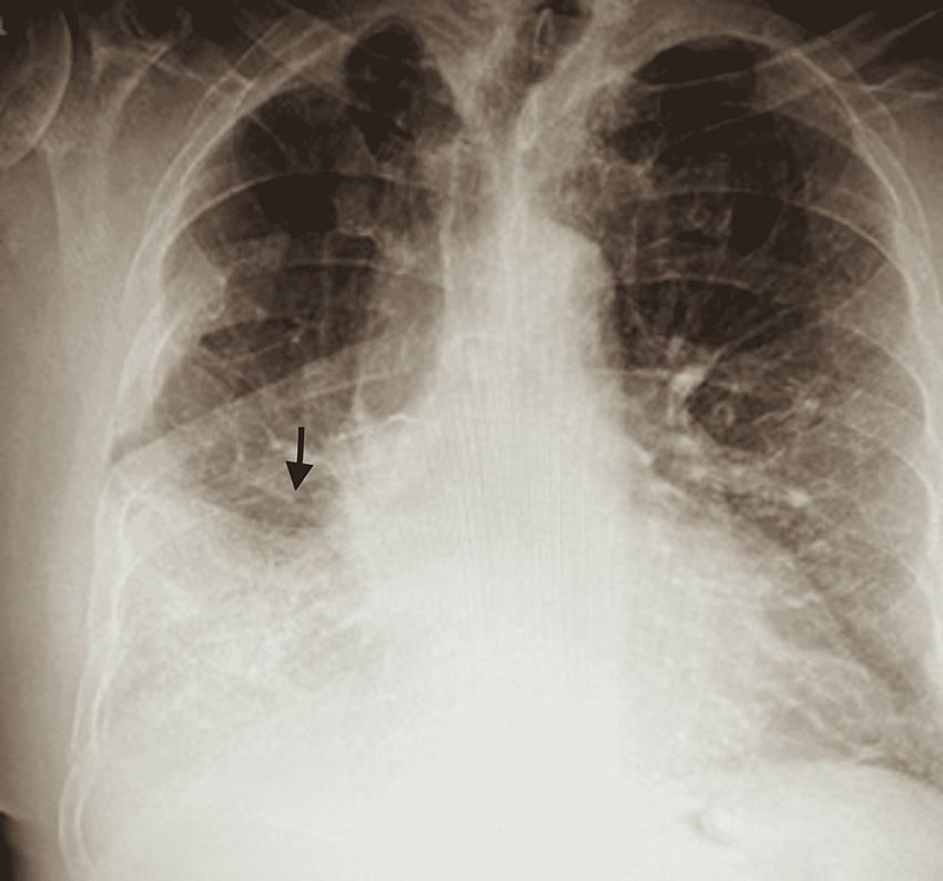
Tests                                                             Results (reference range)

               Sodium                                                            129 Lmmol/L (ref, 136-145)

               Potassium                                                         4.1 mmol/L (ref, 3.5-5.1)

               Creatinine                                                        1.5 Hmg/dL (ref, .8-1.3)

               Glomerular filtration rate                                        48 mL/min

               WBC                                                               10.5 K/uL (ref, 3.4-10.5)

               Neutrophils                                                       3.0%

               Lymphocytes                                                       12.0%

               Hemoglobin                                                        11.6 Lg/dL (ref, 11.7-16.0)

               Platelets                                                         230 K/uL (ref, 120-400)

              TABLE 1: Laboratory test results
              ref: reference range

                                                  He was treated with intravenous Lasix 20 milligrams, ceftriaxone 2 grams, and fluids for suspected
                                                  pneumococcal community-acquired pneumonia (CAP) in urgent care, and was admitted to the hospital.
                                                  Chest radiograph (CXR) done on the same day showed interval development of consolidation involving the
                                                  right lower lobe since prior examination (Figure 1). Upon admission, treatment was continued with 2 grams
                                                  of intravenous ceftriaxone every 24 hours and 100 milligrams of doxycycline every 12 hours. His oxygen
                                                  requirement remained high without any improvement. Due to concerns for CAP, blood cultures via
                                                  peripheral blood draw and urinary antigens for Mycoplasma pneumoniae and Streptococcus pneumoniae were
                                                  obtained.

2022 Haridi et al. Cureus 14(5): e24760. DOI 10.7759/cureus.24760                                                                                             2 of 5
FIGURE 1: Chest x-ray demonstrating right lung opacity
Arrow indicates the infiltrate
ECG revealed sinus arrhythmia with incomplete right bundle branch block (RBBB) and T-wave inversions.
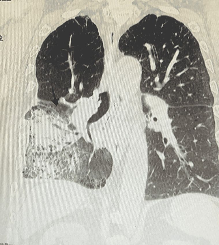
After day one of admission, a computed tomography (CT) scan showed prominent infiltrate in the right mid
and lower lung suggesting a large mass (Figure 2). On day two, he spiked a fever of 103.6℉ with increased
production of orange-like sputum. Legionella urine antigen was positive for L. pneumophila and the
antibiotics were discontinued. The infectious diseases team was consulted.
2022 Haridi et al. Cureus 14(5): e24760. DOI 10.7759/cureus.24760 3 of 5FIGURE 2: Chest CT demonstrating right lung opacity and infiltrating
mass
Arrow indicates the infiltrate
He was started on intravenous azithromycin 500 milligrams every 24 hours, levofloxacin 500 milligrams for
five days, and ceftriaxone 1 gram. ECG showed prolonged QT interval. On day three, his condition improved
dramatically; GI symptoms ceased and the fever subsided. ECG was repeated and showed sinus rhythm with
prolonged QT interval and T-wave inversions. On day six, his serum sodium level increased to 135 mEq/L,
and his chloride normalized to 98 mEq/L. Electrolytes and renal function improved. The patient’s clinical
status improved, and he was discharged home on 3L of oxygen and oral levofloxacin 500 milligrams after a
six-day hospital stay.
Three weeks following the discharge of the patient, he returned to Urgent Care with dyspnea and elevated
cardiac troponins. He was immediately transferred to the Cardiology Unit in another hospital and was
intubated. He died a few days after hospitalization. No autopsy was performed.
Discussion
Legionella is an intracellular, gram-negative bacteria that is commonly known to cause nosocomial
pneumonia. Legionella infection can present as a febrile illness, pneumonia, GI symptoms (nausea,
diarrhea), transaminitis, and hyponatremia. We report a case of L. pneumophila pneumonia in a patient with
suspected nodular lung cancer. This case demonstrates the prompt need to test for and treat Legionella in
immunosuppressed patients once it is suspected, despite the presence or absence of environmental factors.
CAP is defined as the presence of an infiltrate on the CXR, in addition to at least one of the following signs
and symptoms: dyspnea, cough, sputum production, and abnormal breath sounds [5]. On the other hand,
Legionella CAP includes the mentioned signs and symptoms, in addition to at least one positive
2022 Haridi et al. Cureus 14(5): e24760. DOI 10.7759/cureus.24760 4 of 5microbiological test for the organism. Identifying Legionella in patients presenting with CAP has proven to
be challenging over time [5]. However, several studies have proposed a clinical rule that can be used to
quickly identify it [6]. This rule is known as the CAP incidence study (CBPIS), which has a system of 17
points based on the evaluation of serum creatinine, sodium and lactate dehydrogenase (LDH), temperature,
headache, smoking, and vomiting [6]. Although this proposal has proven to have low sensitivity and/or
specificity, a scale with the different criteria in Legionella pneumonia is needed for future practice.
Legionella species cause a severe form of CAP as well as a high mortality rate of about 10% [7]. More
importantly, the mortality rate in patients with Legionella may be as high as 27% in patients without
adequate antibiotics management on admission [7]. Early identification of Legionella is of utmost
importance in patients presenting to the Emergency Department or Urgent Care with respiratory symptoms.
Research has shown that a delay in the appropriate therapy for L. pneumophila pneumonia is associated with
an increased rate of mortality [8]. A urinary antigen test has shown 100% specificity in detecting L.
pneumophila and, thus, should be considered in suspected cases. Physicians should consider testing for this
organism and administer the appropriate anti-Legionella antibiotics promptly to the patients with risk
factors, more importantly, the critically ill.
This patient presented to our hospital with a one-day history of productive cough and dyspnea. He has a
medical history significant for multiple lung pathologies, which are risk factors for developing Legionnaires'
disease [8]. He also had a suspected recurrence of his lung cancer, making him immunocompromised. We
hypothesized that our patient’s underlying lung pathologies made him more susceptible to developing
severe disease and put him at risk of a higher mortality rate. No autopsy was performed on the case under
review. However, prompt diagnosis and treatment may improve the prognosis of Legionnaires' disease in
immunocompromised patients.
Conclusions
Legionnaires’ disease is caused by a gram-negative intracellular bacteria known as Legionella. It has been
shown to rarely cause CAP but can be associated with mortality, especially in immunosuppressed
individuals. We report a case of a 77-year-old man with Legionnaires’ disease and underlying suspected lung
malignancy. The main goal of this case report is to facilitate early recognition and treatment of Legionella in
immunosuppressed patients with lung cancer who present with pulmonary symptoms. This approach will
lower the chances of mortality earlier in the disease course.
Additional Information
Disclosures
Human subjects: Consent was obtained or waived by all participants in this study. Conflicts of interest: In
compliance with the ICMJE uniform disclosure form, all authors declare the following: Payment/services
info: All authors have declared that no financial support was received from any organization for the
submitted work. Financial relationships: All authors have declared that they have no financial
relationships at present or within the previous three years with any organizations that might have an
interest in the submitted work. Other relationships: All authors have declared that there are no other
relationships or activities that could appear to have influenced the submitted work.
References
1. Priest PC, Slow S, Chambers ST, et al.: The burden of Legionnaires' disease in New Zealand (LegiNZ): a
national surveillance study. Lancet Infect Dis. 2019, 19:770-7. 10.1016/S1473-3099(19)30113-6
2. Increased Cases of Legionnaires Disease Investigated in Genesee County . (2016). Accessed: April 11, 2022:
https://www.michigan.gov/mdhhs/inside-mdhhs/newsroom/2016/01/13/increased-cases-of-legionnaires-
disease-investigated-....
3. Chahin A, Opal SM: Severe pneumonia caused by Legionella pneumophila: differential diagnosis and
therapeutic considerations. Infect Dis Clin North Am. 2017, 31:111-21. 10.1016/j.idc.2016.10.009
4. Newton HJ, Ang DK, van Driel IR, Hartland EL: Molecular pathogenesis of infections caused by Legionella
pneumophila. Clin Microbiol Rev. 2010, 23:274-98. 10.1128/CMR.00052-09
5. Fiumefreddo R, Zaborsky R, Haeuptle J, et al.: Clinical predictors for Legionella in patients presenting with
community-acquired pneumonia to the emergency department. BMC Pulm Med. 2009, 9:4. 10.1186/1471-
2466-9-4
6. Fernández-Sabé N, Rosón B, Carratalà J, Dorca J, Manresa F, Gudiol F: Clinical diagnosis of Legionella
pneumonia revisited: evaluation of the Community-Based Pneumonia Incidence Study Group scoring
system. Clin Infect Dis. 2003, 37:483-9. 10.1086/376627
7. Falcó V, Fernández de Sevilla T, Alegre J, Ferrer A, Martínez Vázquez JM: Legionella pneumophila. A cause
of severe community-acquired pneumonia. Chest. 1991, 100:1007-11. 10.1378/chest.100.4.1007
8. Heath CH, Grove DI, Looke DF: Delay in appropriate therapy of Legionella pneumonia associated with
increased mortality. Eur J Clin Microbiol Infect Dis. 1996, 15:286-90. 10.1007/BF01695659
2022 Haridi et al. Cureus 14(5): e24760. DOI 10.7759/cureus.24760 5 of 5You can also read