ANENCEPHALIC FETUS WITH CRANIOSPINAL RACHISCHISIS - CASE REPORT
←
→
Page content transcription
If your browser does not render page correctly, please read the page content below
Case Study                                               International Journal of Research - GRANTHAALAYAH
              ISSN (Online): 2350-0530                                                                 May 2021 9(5), 24–29
              ISSN (Print): 2394-3629

ANENCEPHALIC FETUS WITH CRANIOSPINAL RACHISCHISIS –
CASE REPORT
Ayse KONAC1
1
    Gelisim University Health Sciences, Istanbul, Turkey

                                            ABSTRACT
                                            Anencephaly, in which a substantial part of the brain, skull, or scalp is miss-
                                            ing, is a lethal neural tube defect (NTD) that occurs during the fourth week of
                                            pregnancy after failed cranial neuropore closure. One in every 1,000 births is
                                            anencephalic, and newborns with this NTD are not viable or treatable. Associ-
                                            ated with anencephaly is rachischisis, or severe incomplete neural tube closure
                                            and exposure of the spinal cord. Ultrasonography can quickly diagnose anen-
                                            cephaly. Like other NTDs, nutritional and environmental factors both play a
                                            role in the development of anencephaly. Here, we report and discuss an unusual
                                            case of a 12-week gestation anencephalic fetus with craniospinal rachischisis
                                            and its embryological roots.
                                                In our case, except from the low socio-economic life of the patient, the
Received 18 April 2021                      absence of a predisposing factor that could cause such an anomaly, the abor-
Accepted 4 May 2021                         tion being in the irst trimester and the occurrence in the irst pregnancy of the
Published 31 May 2021                       patient as a result of 5-year infertility made us think that pathology examination
Corresponding Author                        of the abortus material is important in complet or incomplete abortions.
Ayse KONAC, ayse.konac1@gmail.
com
DOI 10.29121/                            Keywords: Anencephaly, Neural Tube Defect, Rachischisis, İncomplet Abortion,
granthaalayah.v9.i5.2021.3899
                                         First Trimester
Funding: This research received
no speci ic grant from any funding
agency in the public, commercial,
or not-for-pro it sectors.               1. INTRODUCTİON
Copyright: © 2021 The                    Anencephaly is a lethal congenital neural tube defect (NTD) that occurs in 1 in every
Author(s). This is an open access        1,000 births. It is the most prevalent anomaly that affects the central nervous sys-
article distributed under the
terms of the Creative Commons            tem like other NTDs, anencephaly affects neural and non-neural tissues, including
Attribution License, which               vertebrae, muscles, and skin Gupta (2004); Salder and Lagman’s (2012).
permits unrestricted use,
distribution, and reproduction in            The Genesis of Anencephaly is directly related to neur like other NTDs (e.g., Spina
any medium, provided the original        Bi ida, Encephalocele, Cranioracalvaria), Anencephaly affects neural and non-neural
author and source are credited.
                                         tissues, including vertebrae, muscles, and skinulation, or the process in which the
                                         and neurulation is the process in which the neural plate, a neuroectoderm derivative,
                                         forms a neural tube that produces the primitive CNS. The lateral edges of the neural

How to cite this article (APA): KONAC, A. (2021). Anencephalic fetus with craniospinal rachischisis – case report. International   24
Journal of Research - GRANTHAALAYAH, 9(5), 24-29. doi: 10.7821/granthaalayah.v9.i5.2021.3899
KONAC Ayse

                                        plate elevate and form neural folds at the end of the third week of pregnancy. In the
                                        midline, later neural folds begin to fuse, spreading cranially and caudally to create a
                                        neural tunnel. The cranial neuropore closes at or near day 25 of gestation, resulting
                                        in a closed tubular structure of the primordial CNS and caudal neuropore by day 28.
                                        The neural tube’s cranial neuropore then goes on to form the hippocampus, and the
                                        caudal neuropore grows into the spinal cord. NTDs in the third and fourth weeks of
                                        development typically arise from abnormal closures of the neural folds Moore and
                                        Persud (2009); Salder and Lagman’s (2012).
                                            Anencephaly speci ically is caused during the fourth week of pregnancy after the
                                        failed closure of the cranial neuropore (1). As a consequence, the main part of the
                                        brain is abnormal, and the development of the calvaria is faulty. Most of the ner-
                                        vous tissue is then subject to degeneration or atrophy by exposure or extrusion from
                                        the skull. While this NTD is commonly referred to as anencephaly (“without brain
                                        tissue”), meroanencephaly (“partial lack of brain tissue”) might be a better word for
                                        some iterations of this phenomenon, as rudimentary neural tissue can still be present
                                        (2). Anencephaly is also associated with acrania (absence of the calvaria or skull-
                                        cap) and rachischisis, which is the full exposure or extensive de icient closure of the
                                        spinal cord. As a consequence of defective notochosis induction, rachischisis affects
                                        the axial structures as well Moore and Persud (2009); Salder and Lagman’s (2012).
                                        NTDs are strongly suspected in utero when maternal serum and amniotic luid show
                                        a high level of the alpha-fetoprotein (AFP) (4). This report outlines one such case of
                                        anencephaly with rachischisis.

                                        2. CASE REPORT
                                        A 26-year-old woman at gravida 1 was admitted to the Istanbul Training and
                                        Research Hospital for surgical pregnancy termination after the fetus was diagnosed
                                        with craniospinal rachischisis anencephaly. The patient had been married 5 years
                                        in a non-consanguineous marriage and smoked 3–4 cigarettes a day, but did not
                                        drink alcohol. The patient had endured 1 year of male factor infertility complaints
                                        and became pregnant as a result of infertility treatment. She also fell into a low
                                        socioeconomic status.
                                           The patient was at 12 weeks gestation according to the date of her last menstrual
                                        cycle. With natural discharge, her menstrual cycle was typical prior to pregnancy.
                                        Hypertension, gestational diabetes mellitus, diabetes mellitus, and cardiac and renal
                                        disease had not emerged in her brief pregnancy history. Since the beginning of her
                                        pregnancy, the patient had been taking iron and folic acid pills. She had no family
                                        history of NTDs, and she did not report any drug use. Further, the patient was mod-
                                        erately developed upon inspection, measured 161 cm tall, and weighed 56 kg. Her
                                        heartbeat, pulse, and blood pressure were natural, with a result of 109 mg/dl for
                                        spontaneous blood glucose. Tests for urinaryenfection, HIV, and HbSAg were nega-
                                        tive.

International Journal of Research - GRANTHAALAYAH
                                                                                                                            25
Anencephalic fetus with craniospinal rachischisis – case report
Figure 1 Acrania fetus irst trimester
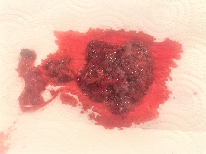
Figure 2 Material of Abortion
The patient came to the clinic with complaints of excessive bleeding and abdomi-
nal pain. Upon examination, abortus materials in the vagina and residual pregnancy
materials in the endometrium were found by Ultrasonography and the patient was
diagnosed with incomplete abortion. She was hospitalized on the same day and
under general anesthesia revision curettage made on the same day. After curettage,
both the vaginal and endometrium material were sent for pathological testing.
The pathology results reported a fetus with anomaly compatible with 12 weeks
of gestation. The indings signaled anencephaly, rachischisis (craniospinal), as well
International Journal of Research - GRANTHAALAYAH
26KONAC Ayse
as pulmonary extra-lobar sequestration (localized in the abdomen) and gastroschis.
There were no anomalies in the other internal organs.
3. DİSCUSSİON
The prevalence of anencephaly varies according to region, race, gender, and environ-
mental factors. Per a population-based study from India, every 2.5 in 1,000 births
had anencephaly, with NTDs in general ranging from every 6.57 to 8.21 in 1,000
births Cherian et al. (2005). In another prospective study of 3,500 consecutive births
from southern India, 11.4/1,000 births with NTDs occurred, with 5.1/1,000 births
attributable to craniospinal rachischisis anencephaly. In cases where there was a
history of NTDs or consanguineous marriage, there was an increased probability of
NTD occurrence Kulkarni et al. (1989). Overall, China had the highest incidence of
live births with NTDs (11.39/1,000), while the lowest incidence of live births with
NTDs was recorded in the U.S.A. (1/1,000) Detrait et al. (2005); Li et al. (2006). Large
numbers of congenital malformations and genetic abnormalities are also one of the
factors of child mortality and morbidity in developing countries. For instance, one
survey carried out on 94,640 newborns to clarify the incidence of malformations
noted a 2.03% rate of malformed babies and NTDs, with musculoskeletal disabilities
the most prevalent Verma (2000).
It is thought that NTDs arise from complex interactions between environmen-
tal and genetic variables. Such environmental variables include age, periconceptual
infection, recreational drug use, caffeine, smoking, and alcohol consumption Detrait
et al. (2005); Moore and Persud (2009); Verma (2000). Extensive clinical and epi-
demiological research has also shown that the incidence of NTDs increases with inad-
equate maternal nutrition, primarily low folic acid levels, and low socioeconomic sta-
tus.
For the neural tube to properly develop, other micronutrients such as vitamins
B6 and B12 and minerals such as zinc are also necessary, though holotranscobal-
amin helps monitor vitamin B12 speci ically as a sensitive predictor Ray et al. (2007).
Further, NTDs can form by taking certain medications, such as valproic acid and anti-
convulsants, and being exposed to high levels of vitamin A Berry et al. (1999); Blanc-
quaert et al. (2010); Ray et al. (2007).
As for genetic variables, research implies that genes involved especially in
the metabolism of folate are signi icant in generating NTDs. For instance, the
methylenetetrahydrofolate reductase mutation is responsible for folate-related
NTDs Put et al. (1998). However, during the periconceptual period, administering
food with folic acid (400 µg) and synthetic vitamin B12 decreased the risk of NTDs
in the U.S.A. by 50–70% Houk et al. (1992); Put et al. (1998).
In conclusion, assessing the Alpha Feto Protein level and early USG in at-risk preg-
nancies can assist in the early detection of NTDs. Genetic counseling is also useful
for planning future pregnancies; as there are no viable curative modalities, though,
International Journal of Research - GRANTHAALAYAH
27Anencephalic fetus with craniospinal rachischisis – case report
therapeutic emphasis largely falls on preventive step Houk et al. (1992). Studies have
shown that by taking folic acid supplements daily during pregnancy, the frequency of
NTDs can be decreased. Because most pregnancies are unplanned, even the adminis-
tration of multivitamins containing 400 µg of folic acid to all women of childbearing
age should be suggested to decrease the chance of NTD genesis Tuncbilek (2004).
Neural tube defects are among the most severe congenital anomalies in Turkey
and epidemiological indings show that the prevalence varies according to regional
and demographical characteristics. According to the studies conducted in various
cities of Turkey, NTD incidence varies between 3 and 5.8 per thousand Tuncbilek
(2004).
Although there is no predisposing factor for fetal anomaly especially in infer-
tile cases, if there is abortion, even if the gestational week is small, talking to the
patient and informing her and sending the curettage material to pathology exami-
nation would be helpful in detecting unexpected anomalies. In our case, there was
no history of giving birth with other anomalies in the family of the patient and her
husband.
REFERENCES
Berry, R. J., Li, Z., Erickson, J. D., Li, S., Moore, C. A., Wang, H., & Mulinare, J. (1999). Prevention
of neural-tube defects with folic acid in China. Engl J Med.
Blancquaert, D., Storozhenko, S., Loizeau, K., Steur, H. D., Brouwer, V. D., Viaene, J., Ravanel, S.,
Rébeillé, F., Lambert, W., & Straeten, D. V. D. (2010). Folates and Folic Acid: From Fun-
damental Research Toward Sustainable Health. Critical Reviews in Plant Sciences, 29(1),
14–35. Retrieved from https://dx.doi.org/10.1080/07352680903436283 10.1080/
07352680903436283
Cherian, A., Seena, S., Bullock, R. K., & Antony, A. C. (2005). Incidence of neural tube defects in
the least-developed area of India: a population-based study. The Lancet, 366(9489),
930–931. Retrieved from https://dx.doi.org/10.1016/s0140-6736(05)67319-9 10
.1016/s0140-6736(05)67319-9
Detrait, E. R., George, T. M., Etchevers, H. C., Gilbert, J. R., Vekemans, M., & Speer, M. C. (2005).
Human neural tube defects: Developmental biology, epidemiology, and genetics. Neu-
rotoxicology and Teratology, 27(3), 515–524. Retrieved from https://dx.doi.org/10
.1016/j.ntt.2004.12.007 10.1016/j.ntt.2004.12.007
Gupta, H. (2004). Neural tube defects and folic acid. Indian Pediatr, 41577586–41577586.
Houk, V. N., Oakley, G. P., Erickson, J. D., Mulinare, J., & James, L. M. (1992). Recommendations
for the Use of Folic Acid to Reduce the Number of Cases of Spina Bi ida and Other Neural
Tube Defects. Atlanta: Centers for Disease Control, 26, 23–34.
Kulkarni, M. L., Mathew, M. A., & Reddy, V. (1989). The range of neural tube defects in southern
India. Archives of Disease in Childhood, 64(2), 201–204. Retrieved from https://dx.doi
.org/10.1136/adc.64.2.201 10.1136/adc.64.2.201
Li, Z., Ren, A., Zhang, L., Ye, R., Li, S., & Zheng, J. (2006). Extremely high prevalence of neural
tube defects in a 4-county area in Shanxi Province China. BDR: A Clinical and Molecular
Teratology, 4237240–4237240.
Martinelli, M., Scapoli, L., Pezzetti, F., Carinci, F., Carinci, P., Stabellini, G., Bisceglia, L., Gom-
International Journal of Research - GRANTHAALAYAH
28KONAC Ayse
bos, F., & Tognon, M. (2001). C677T variant form at the MTHFR gene and CL/P: A risk
factor for mothers? American Journal of Medical Genetics, 98(4), 357–360. Retrieved
from https://dx.doi.org/10.1002/1096-8628(20010201)98:43
.0.co;2-f 10.1002/1096-8628(20010201)98:43.0.co;2-f
Moore, K. L., & Persud, T. (2009). The Developing Human - Clinically Oriented Embriology,
8th edn. Sauders, 62–75.
Put, N. V. D., Gabrels, F., Stevens, E., & Frans, J. M. (1998). A second common mutation in the
methylenetetrahydrofolate reductase gene: an additional risk factor for neural-tube
defects? Am J Human Genet, 510441051–510441051.
Ray, J. G., Wyatt, P. R., Thompson, M. D., Vermeulen, M. J., Meier, C., Wong, P.-Y., Farrell, S. A.,
& Cole, D. E. C. (2007). Vitamin B12 and the Risk of Neural Tube Defects in a Folic-
Acid-Forti ied Population. Epidemiology, 18(3), 362–366. Retrieved from https://dx
.doi.org/10.1097/01.ede.0000257063.77411.e9 10.1097/01.ede.0000257063.77411
.e9
Salder, T. W., & Lagman’s. (2012). Medical Embryology, 63(70), 296–297.
Tuncbilek, E. (2004). neural tube defect prevalence and high in Turkey what can be done to
prevent. Child Health and Diseases Journal, 47, 79–84.
Verma, I. C. (2000). Burden of genetic disorders in india. The Indian Journal of Pediatrics,
67(12), 893–898. Retrieved from https://dx.doi.org/10.1007/bf02723953 10.1007/
bf02723953
International Journal of Research - GRANTHAALAYAH
29You can also read