A genetically encoded tool for reconstituting synthetic modulatory neurotransmission and reconnect neural circuits in vivo - Nature
←
→
Page content transcription
If your browser does not render page correctly, please read the page content below

ARTICLE
                  https://doi.org/10.1038/s41467-021-24690-9                OPEN

                  A genetically encoded tool for reconstituting
                  synthetic modulatory neurotransmission and
                  reconnect neural circuits in vivo
                  Josh D. Hawk          1,2 ✉,   Elias M. Wisdom            2,   Titas Sengupta2, Zane D. Kashlan2 & Daniel A. Colón-Ramos                         2,3 ✉
1234567890():,;

                  Chemogenetic and optogenetic tools have transformed the field of neuroscience by facil-
                  itating the examination and manipulation of existing circuits. Yet, the field lacks tools that
                  enable rational rewiring of circuits via the creation or modification of synaptic relationships.
                  Here we report the development of HySyn, a system designed to reconnect neural circuits
                  in vivo by reconstituting synthetic modulatory neurotransmission. We demonstrate that
                  genetically targeted expression of the two HySyn components, a Hydra-derived neuropeptide
                  and its receptor, creates de novo neuromodulatory transmission in a mammalian neuronal
                  tissue culture model and functionally rewires a behavioral circuit in vivo in the nematode
                  Caenorhabditis elegans. HySyn can interface with existing optogenetic, chemogenetic and
                  pharmacological approaches to functionally probe synaptic transmission, dissect neuropep-
                  tide signaling, or achieve targeted modulation of specific neural circuits and behaviors.

                  1 Grass Laboratory, Marine Biological Laboratory, Woods Hole, MA, USA. 2 Department of Neuroscience and Department of Cell Biology, Program in Cellular

                  Neuroscience, Neurodegeneration and Repair, Yale University School of Medicine, New Haven, CT, USA. 3 Instituto de Neurobiología, Recinto de Ciencias
                  Médicas, Universidad de Puerto Rico, San Juan, Puerto Rico. ✉email: jsh.hawk@gmail.com; daniel.colon-ramos@yale.edu

                  NATURE COMMUNICATIONS | (2021)12:4795 | https://doi.org/10.1038/s41467-021-24690-9 | www.nature.com/naturecommunications                             1
ARTICLE                                                                NATURE COMMUNICATIONS | https://doi.org/10.1038/s41467-021-24690-9

R
        ecent advances in optogenetic and chemogenetic tools have           Results
        provided unprecedented in vivo access to neurons, enabling          In Hydra, the loosely connected nerve net uses neuropeptides to
        manipulation of neuronal activity patterns at will1,2 and           produce volumetric neurotransmission11. We reasoned that this
facilitating examination of their roles in behaviors. Moreover,             property of Hydra peptidergic synapses, if reconstituted in other
synapse-specific labeling methods, such as Trans-TANGO3 and                  systems, would enable examination of adjacent chemical synaptic
synaptic GRASP4, have enabled visualization of individual                   relationships, as well as the reconstitution of new functional
synaptic connections in the context of the intact circuits, and in          connections between neurons that are not adjacent to each other.
the case of Trans-TANGO, genetic access to transsynaptically                It would also allow orthogonal neuromodulation of targeted
labeled neurons. Tools like these have transformed the field of              endogenous circuits at a distance, similar to how neuromodula-
neuroscience by facilitating the examination and manipulation of            tory systems work in vivo. We, therefore, built HySyn using a
existing circuits. Yet, the field lacks tools that enable rational           Hydra-derived RFamide-related peptide (HyRFamide I/II), and
rewiring of circuits via the creation or modification of synaptic            its postsynaptic cognate receptor (HyNaC 2/7/9) that fluxes
relationships. Engineering de novo circuit relationships could              calcium12,13. Importantly, the primary sequence of this Hydra-
reveal important components of the underlying circuit structures,           derived peptide is distinct from RFamide-related peptides found
and rational rewiring of circuits could prove a powerful strategy           in other organisms14,15 and specific to its receptor12. Because
towards understanding how neuronal relationships generate                   presynaptic neuropeptide processing, transport and release
behaviors.                                                                  mechanisms14 and postsynaptic calcium signaling are conserved
   Ideally, de novo synapses, like other orthogonal genetic tools           throughout evolution16–18, expression of this neuropeptide
used for neuronal manipulation, should be generated from spe-               ligand-receptor pair, while orthogonal, would still harness fun-
cific, controllable modules that enable modification of circuits,             damental and conserved signaling mechanisms in the desired
but avoid undesirable or uncontrollable interactions with endo-             cells. The harnessing of conserved biological pathways with an
genous components of the system. Cross-species transplantation              orthogonal and specific neuropeptide–receptor pair achieved two
of channels (like Channelrhodopsin) and gap junctions (like the             goals: (1) it allowed reconstitution of neuronal connections with
use of Connexins in invertebrates5–7) have been powerful in                 minimal components and (2) it resulted in a versatile tool for use
providing the desired modularity and specificity to the engineered           in different biological contexts. The divergent evolution of this
systems2,5,6. For example, heterologous expression of the Droso-            neuropeptide–receptor pair, in the context of conserved cell
phila allostatin receptor in vertebrates can be used to silence             biology, could be exploited to produce a bipartite synthetic con-
neurons upon the exogenous application of the allostatin                    nection whose components would be (individually) inert when
peptide4. Similarly, expression in C. elegans neurons of the Dro-           expressed in systems other than Hydra. Yet when targeted, spe-
sophila HisCl channel enables chemogenetic inhibition of neu-               cific bipartite reconstitution of HySyn could be used to reconfi-
ronal activity8. In invertebrates, heterologous expression of               gure modulatory and functional relationships. To emphasize how
vertebrate gap junction proteins, called connexins, within specific          the HySyn system compares to existing tools: canonical neu-
neurons of the C. elegans nerve ring result in the predictable              roscience approaches, like optogenetics, control specific neurons,
creation of artificial electrical connections between adjacent               whereas the synthetic synaptic approach of HySyn was designed
neurons5,6, the rewiring of the circuit, and the recoding of a              to control relationships between neurons.
learned behavioral preference7. Yet, no existing technology                    To drive heterologous expression, processing, and transport of
enables the purposeful creation of new, synthetic and manip-                the cnidarian neuropeptide, we designed a genetically encoded
ulatable chemical synaptic connections for re-configuring neural             pre-pro-peptide carrier, “HyPep”, that harnesses the universality
networks in diverse organisms.                                              of the neuropeptide processing pathway (Supplementary Fig. 1
   For these reasons, we developed a system that allows the                 and Fig. 1a, “Hydra RFamide”). Previous approaches to label
engineering of synthetic relationships between neurons through              neuropeptides concatenated a reporter onto an existing full-
the targeted reconstitution of modulatory neurotransmission                 length natural neuropeptide precursor19,20. We built upon the
between selected partners, eliciting orthogonal circuit control             knowledge of neuropeptide synthesis from these and other
over neuromodulatory connectivity. Because neuromodulation is               studies15,19–21 to create HyPep as a synthetic pre-pro-peptide
a powerful way of re-configuring neural circuits to produce dis-             carrier that would enable targeting and processing of hetero-
tinct behavioral outcomes, because neuromodulation is not con-              logous neuropeptides using the endogenous neuropeptide pro-
strained by the architecture of the nervous system, and because             cessing pathway (Supplementary Fig. 1a). HyPep consists of a
the role of neuromodulators and their long-range effects remains            signal peptide that directs trafficking, acidic spacers with enzy-
largely unexplored9,10, we focused our efforts on a system that             matic recognition sites for cleavage of the neuropeptide, and the
enables reconstitution of synthetic modulatory chemical synaptic            sequence encoding the heterologous neuropeptide itself. We
connectivity. We note that we use the term “chemical synaptic               based our signal peptide on neuropeptide Y (Supplementary
connectivity” to refer to peptidergic synaptic relationships                Fig. 1b, “Signal Peptide”), a ubiquitously expressed neuropeptide
between neurons, be it junctional relationships between abutting            in vertebrates14. We designed artificial neuropeptide spacers
partners, or partners that communicate via volumetric neuro-                containing consensus cleavage sites (Supplementary Fig. 1b, red
transmission and at a distance. We prioritized the design of a              lines) for pre-pro-convertase (PC2), a conserved neuropeptide
system that would be (1) versatile to function in a wide range of           endopeptidase15. Because the cross-species alignment of neuro-
cell types and organisms, (2) modular to allow independent                  peptides revealed a strong bias for acidic residues between the
genetic targeting of pre- and postsynaptic components, (3) spe-             dibasic cleavage sites, we created acidic linkers between these
cific to modulate only the intended target cells while being inert           cleavage sites, which in turn flanked the heterologous cnidarian
to endogenous neurotransmission, (4) robust by targeting con-               neuropeptide we sought to express with the HyPep system
served intracellular signaling cascades, and (5) synergistic to             (Supplementary Fig. 1b, “Hydra RFamide”).
interface with existing optogenetic and chemogenetic technolo-                 Expression of the HyPep synthetic pre-pro-peptide carrier in
gies. Informed by these goals, we engineered “HySyn”, a Hydra-              mammalian Neuro2a cells resulted in localization and transport
derived, two-component system that creates synthetic neuromo-               of a fluorescent reporter to the expected intracellular compart-
dulatory connections to manipulate intracellular calcium within             ments and vesicular release sites (Supplementary Fig. 1c, see also
in vivo neural circuits (Fig. 1a, left).                                    Fig. 3). This observation is consistent with our hypothesis that the

2               NATURE COMMUNICATIONS | (2021)12:4795 | https://doi.org/10.1038/s41467-021-24690-9 | www.nature.com/naturecommunications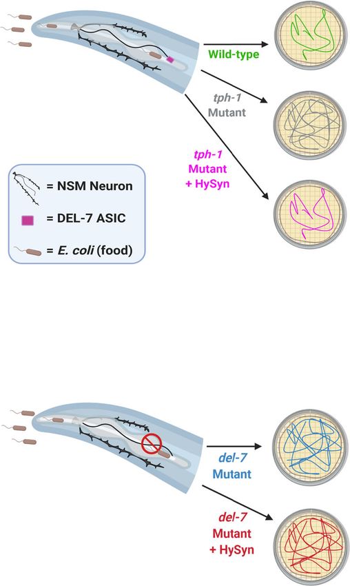
NATURE COMMUNICATIONS | https://doi.org/10.1038/s41467-021-24690-9                                                                                                                              ARTICLE

                                                                                                                Optogenetic
    a                                                                                                           Stimulation

                                                                                                                              Optogenetics
        ‘Presynaptic’ Cell                                ‘Postsynaptic’ Cell
                                                                                                                                         Na+
                                                                                                                                                            Ca2+
                                                Ca   2+                                                                                                                        HyCal Receptor
                                                                      2+                                                        Na   +                                  Ca2+    Expressing
          Hydra
                                                                 Ca        Ca2+                                                                                                     Cell
         RFamide                                          Ca2+                                                                Hydra
                                                                                                                             RFamide
                                                                 Ca2+ Ca2+                                                                                                         Control
                                                                                                                                                                                    Cell

                                  HyPep           HyCal
                                Transmitter      Receptor

     b                                                                                             c                                     d                                        e
                                                                                                      Optogenetics                             Untransfected                               GFP
                                                                                                   + HyPep Transmitter                            Control                             + HyCal Receptor

                                                                                  Current Traces

                                                                                                               20 pA

                                                                                                                                                        20 pA

                                                                                                                                                                                                  20 pA
                                                                                                                  10s                                      10s                                       10s
                                                                             Lights: On                   On            On                On       On              On               On       On            On

Fig. 1 Hydra-derived synthetic synapse (HySyn) engineered through heterologous expression of Hydra neuropeptide (HyPep) and receptor (HyCal). a
(left) Schematic describing “HySyn”: Hydra-derived neuropeptide (blue) is released from the “presynaptic” cell and interacts with the HyCal receptor
(yellow) to flux calcium in the “postsynaptic” cell. a (right) Schematic illustrating the experimental paradigm to assay HySyn function by whole-cell patch-
clamp electrophysiology. b Micrograph showing co-culture of Neuro2a neuroblastoma cells expressing either “presynaptic” HyPep with ChRoME
(magenta), “postsynaptic” HyCal with GFP (green), or untransfected (grayscale). Boxed regions correspond to the micrographs in c–e, with the color of the
box matched to the color of the title of the micrograph. c–e Identification of cell populations based on fluorescent markers (top, micrograph, 20μm scale
bar) enabling whole-cell current recording (bottom, “current traces”) from three distinct populations during optogenetic stimulation (480 nm, blue-shaded
windows in “current traces”, “On”): c “presynaptic” cells expressing HyPep with ChRoME (pseudocolored magenta) consistently produced step-wise
optogenetic currents, d unlabeled cells remain unaltered by optogenetic activation of neighboring HyPep+ cells, and e green “postsynaptic” cells
expressing the HyCal receptor show an increasing inward current during light stimulation. The downward deflection of traces during optogenetic
stimulation in blue windows indicates an inward current, suggesting a depolarizing cation current. The persistence of HyCal current between stimulation
(e) is consistent with the lack of desensitization of HyCal channel and suggests an accumulation of HyPep neuropeptide in the bath solution. Transfection
and reporter expression (micrographs in b–e, see also Fig. 2a) was reproducibly observed, including in the 22 cases where successful electrophysiological
recordings were made (Supplementary Fig. 2), and in 3 independent populations with GCaMP expression (Fig. 2).

engineered HyPep carrier harnesses the universality of the neu-                                        currents (Fig. 1d). But when we optogenetically stimulated the
ropeptide processing pathway to target the Hydra neuropeptide                                          “presynaptic” Neuro2a cells, we observed distinct “postsynaptic”
processing, transport, and release. Next, we used whole-cell                                           currents in co-cultured cells expressing the “postsynaptic” HyCal
patch-clamp electrophysiology to test whether the “presynaptic”                                        receptor (Fig. 1e). These results indicate the existence of synthetic
HyPep, when paired with the “postsynaptic” HyCal receptor, is                                          peptidergic connections between the “presynaptic” HyPep- and
capable of producing a functional neuromodulatory connection                                           “postsynaptic” HyCal-expressing cells. Considering the absence of
(Fig. 1a). We created an optogenetically excitable population of                                       currents without the receptor (Fig. 1d, see also Supplementary
“presynaptic” cells by co-expressing HyPep with mRuby-labeled                                          Fig. 2), we conclude that these results show the creation of de
ChRoME22, an optogenetic tool for depolarizing neurons (Fig. 1b,                                       novo peptidergic synaptic relationships through the specific
magenta). We then co-cultured these “presynaptic” cells with                                           reconstitution of the HySyn system.
other cells expressing the “postsynaptic” receptor HyCal (Fig. 1b,                                        We observed that repeated optogenetic stimulation of the
green), as well as untransfected control cells (Fig. 1b, unlabeled).                                   “presynaptic” HyPep-expressing cells produced an integrating
As expected22, optogenetic activation (480 nm) of ChRoME-                                              current in the “postsynaptic” HyCal receptor-expressing cells
expressing “presynaptic” cells produced a sustained step-like                                          (Fig. 1e, bottom). With each stimulation, the inward current
current consistent with direct channel opening by light (Fig. 1c).                                     (downward trace deflections) increased to a new plateau level.
Neighboring cells expressing neither the ChRoME optogenetic                                            This phenomenon is consistent with the known neuromodulatory
tool nor the HyCal receptor did not exhibit light-activated                                            properties of the peptide–receptor pair. Specifically, the HyCal

NATURE COMMUNICATIONS | (2021)12:4795 | https://doi.org/10.1038/s41467-021-24690-9 | www.nature.com/naturecommunications                                                                                        3
ARTICLE                                                                 NATURE COMMUNICATIONS | https://doi.org/10.1038/s41467-021-24690-9

receptor does not desensitize13, and neuropeptides act at low                cells, the majority (8/14) responded during both of the HyPep+
concentrations across large volumes15. Thus, this integrating                solution applications, and with a calcium rise greater than 3 stan-
current suggests that an increasing fraction of HyCal channels               dard deviations beyond any changes observed before the applica-
open as neuropeptide release and stimulation persist. The                    tion. We note, however, that cells responded differentially to the
selective presence of these currents in HyCal-expressing “post-              two applications, and that some cells responded to only a single
synaptic” cells aligns with the expected specificity of this system.          HyPep+ solution application. These results are consistent with the
These data show that combining “presynaptic” HyPep and                       expected neuromodulatory nature of the HySyn system, and suggest
“postsynaptic” HyCal creates an artificial coupling of activity in a          that while HyCal modulates the responsiveness of Neuro2a cells, it
vertebrate neuronally-derived cell culture model, likely by utiliz-          is not the sole determinant of activity. Importantly, these results
ing volume neurotransmission.                                                provide further support that HySyn functions through volume
   Next, we used calcium imaging to examine the extent and                   transmission and that the HyCal receptor may be used with exo-
reliability of HySyn neuromodulation in a population of cells. We            genously applied neuropeptide for pharmacological or chemoge-
used our established approach to activate “presynaptic” HyPep-               netic manipulations of intracellular calcium.
expressing cells through optogenetics, but used the red-shifted                 We then sought to determine whether HySyn could be used
variant Chrimson23 (Fig. 2a, magenta cells) that is compatible               in vivo to modulate organismal behavior by expressing HySyn in
with GCaMP imaging. In parallel, we monitored activation of                  the nematode C. elegans. As a first step, we harnessed the well-
“postsynaptic” cells with the calcium-sensitive fluorophore                   characterized and stereotyped distribution of synaptic speciali-
GCaMP6f24 (Fig. 2a, green cells). In GCaMP-labeled cells lacking             zations to examine the subcellular localization of HySyn com-
the HyCal receptor, we did not observe light-evoked calcium                  ponents in vivo. We focused our characterization of HySyn on the
activity (Fig. 2b), which was consistent with our electro-                   AIB interneurons—a pair of symmetric interneurons with a
physiological data (Fig. 1d). These results support that HyPep is            polarized and distinguishable distribution of pre- and post-
inert in mouse Neuro2a cells. In contrast, when “presynaptic”                synaptic sites in the distal and proximal neurite, respectively25–27
HyPep-positive cells were optogenetically stimulated, co-cultured            (Fig. 3a). Expression of the transmembrane dense-core vesicle
cells expressing the “postsynaptic” HyCal receptor showed rising             marker, IDA-1/phogrin::mCherry, resulted in the localization of
GCaMP signals (Fig. 2c, responses are organized based on the                 the transmembrane receptor to presynaptic sites in the distal
response magnitude to highlight the frequency of responses). We              neurite (Fig. 3b). Co-expression of IDA-1::mCherry and GFP-
classified ~34% (14/44) of quantified cells as clear “responders”,             tagged HyPep resulted in colocalization of both components to
based on a change in GCaMP signaling 3× standard deviations                  presynaptic regions of the neurite (Fig. 3c–e), consistent with
(or greater) than signal observed prior to light stimulation.                HyPep being targeted to presynaptic dense core vesicle-rich
Quantification of this increase in calcium over the course of the 4-          regions, as expected (and consistent with our observations in
min stimulation (Fig. 2d and Supplementary Figure 2) highlights              Neuro2a cells, Supplementary Fig. 1c). Moreover, expression of
an overall doubling of the calcium signal compared to the initial            the postsynaptic receptor GLR-1::GFP in AIB resulted in its
signal, but some cells experienced changes in the GCaMP signal               localization to the postsynaptic region of AIB (Fig. 3g–i). Simi-
as high as 6-fold. We did not find a clear spatial pattern of                 larly, expression of the postsynaptic HySyn component, the
activation with respect to the location of the “presynaptic” cells.          HyCal receptor was enriched in the postsynaptic proximal neurite
This observation is consistent with our electrophysiological data            (Fig. 3h). Together, our data indicate that expression of the
and our expectations for a neuropeptide that functions through               HySyn components in vivo results in their expected subcellular
volume transmission. “Postsynaptic” calcium responses selec-                 localization and trafficking.
tively in HyCal-expressing cells provide further support for the                To determine whether HySyn could modulate organismal
reconstitution of the HySyn peptidergic functional connection,               behavior, we then expressed GFP-tagged HyPep throughout the
and the specificity of HySyn. Our finding that robust calcium                  nervous system and the HyCal receptor either in muscle tissues,
responses occur in a third of Neuro2a cells is consistent with a             pan-neuronally, or in GABAergic inhibitory locomotory inter-
neuromodulatory function (as compared to the more determi-                   neurons (Fig. 3j–l and Supplementary Fig. 3a). We predicted that
nistic relationship often evoked by a classical neurotransmitter).           activating the HyCal receptor in muscles, all neurons, or in
Together, these observations support the idea that HySyn creates             inhibitory neurons, would result in paralysis that would prevent
neuromodulatory connectivity in a vertebrate neuronally-derived              proper worm locomotion. Indeed, the reconstitution of HySyn,
cell culture model. Our methods also demonstrate that HySyn is               through the expression of the HyPep and the HyCal in the spe-
compatible with existing optogenetic and calcium imaging                     cific tissues resulted in severely uncoordinated animals (Supple-
approaches.                                                                  mentary Fig. 3a and Supplementary Movies 1, 2). Importantly,
   We reasoned that if HySyn acts through volume transmission,               expression of the pan-neuronal HyPep alone, or of HyCal alone
optogenetic stimulation of a culture of “presynaptic” HyPep-                 in the indicated tissues, did not produce any observable pheno-
expressing Neuro2a cells would generate a HyPep+ solution that               types or locomotory defects (Fig. 3l and Supplementary Fig. 3a),
could act as a chemogenetic stimulator when added to a separate              consistent with these elements being inert in vivo in C. elegans
culture of “postsynaptic” HyCal-expressing cells (Fig. 2e). To test          when expressed on their own. Transgenic animals with pan-
this idea, we collected the solution from optogenetically stimulated         neuronal HyPep and muscle-targeted HyCal (i.e., neuromuscular
HyPep-expressing Neuro2a cells (HyPep+ solution) and added it to             HySyn) performed very little crawling behavior, as illustrated by
a separate HyCal-expressing cell culture. Prior to adding media,             the trajectory of worms observed for 30 min on an agar pad
GCaMP signals were stable in all HyCal-expressing “postsynaptic”             (Fig. 3k, right) and by the reduced velocity of travel (Fig. 3l, blue).
Neuro2a cells (Fig. 2f), including the 14 cells (~45% of 31 exam-            To better quantify the phenotypes, we used DeepLabCut to train a
ined) that ultimately showed responses to at least one application of        neural network to identify worm postures and quantify worm
HyPep+ solution. Application of HyPep+ solution produced an                  locomotion28,29 (see “Methods” section). We observed that in
increase in the calcium signal, while subsequent washout and                 swimming assays, animals with reconstituted HySyn displayed a
application of untreated solution led to a decline in the calcium            reduced head radial velocity (Supplementary Fig. 3a, b), con-
signal (quantified in Fig. 2g). When the HyPep+ solution was                  sistent with the uncoordinated phenotypes that we detected
repeatedly added after washout cycles, and individual responding             during locomotion on solid agar surfaces (Fig. 3k, l). We note that
cells were tracked, we observed that of the responding Neuro2a               HyCal expression in muscle, with HyPep expression in the

4                NATURE COMMUNICATIONS | (2021)12:4795 | https://doi.org/10.1038/s41467-021-24690-9 | www.nature.com/naturecommunications
NATURE COMMUNICATIONS | https://doi.org/10.1038/s41467-021-24690-9 ARTICLE
a HyPep HyCal b c d
Presynaptic Cell Postsynaptic Cell No Receptor + HyCal Receptor 6 *
Optogenetics
Lights Off Lights On Lights Off Lights On
GCaMP Signal
GCaMP Signal
GCaMP 5
Na+ Imaging
Average
Average
20% 20%
GCaMP signal change
Ca 2+ ∆F/F ∆F/F
Na+ Ca2+ 50 sec 50 sec 4
Ca2+
Hydra Ca2+
Fold ∆F/F
Ca2+
RFamide
4 4
3
HyCal Receptor 5 5
HyPep Transmitter
10
3
10
3
2
Individual Cells
Individual Cells
Optogenetics GCaMP 15 15
MERGE
Fold ∆F/F
Fold ∆F/F
+ HyPep Transmitter + HyCal Receptor
20 20 1
2 2
25 25 n.s.
30 30
1 1
0
35 35
40 40 Light: off ON off ON
No Receptor + HyCal
e f Baseline HyPep+ Washout 1 HyPep+ Washout 2
g 6
Solution Solution *
Optogenetic
*
Stimulation
*
GCaMP Signal
4
*
Average
GCaMP signal change
50%
∆F/F
HyPep 50 sec
2
Fold ∆F/F
Cell
4
2
HyPep+ Solution
0
4 3
GCaMP
Individual Cells
Fold ∆F/F
Imaging 6
2
-2
8
Ca2+
HyCal 10
Cell 1 -4
12
14
-6 HyPep HyPep
Fig. 2 HySyn produces volumetric neuromodulation of postsynaptic calcium. a Schematic illustrating the experimental paradigm used to characterize
HyPep to HyCal signaling by using calcium imaging, top. Micrograph illustrating transfected Neuro2a cells used in these experiments, bottom (20 μm scale
bar). Chrimson was used for optogenetic stimulation (591 nm, 500 ms at 1 Hz) of “presynaptic” cells expressing the HyPep carrier of the neuropeptide
(“Hydra RFamide”). GCaMP was expressed in distinct cells either alone (b) or with the cognate receptor HyCal (c), and both of these two groups of cells
were co-cultivated with HyPep “presynaptic” cells. b Average GCaMP signals, top bold line with shaded 95% confidence intervals, and individual cell
heatmap profiles, bottom, for Neuro2a cells expressing GCaMP alone. Without receptor expression, but even in the presence of HyPep-secreting cells co-
expressing the optogenetic tool (Chrimson), GCaMP signals remained stable in these cells both without (left, “Lights Off”) and with (right, “Lights On”,
yellow shading) optogenetic stimulation of the HyPep cells. c As in b, but in cells also expressing the HyCal receptor in the presence of HyPep-secreting
cells co-expressing the optogenetic tool (Chrimson). We observe calcium signal rise over the course of light stimulation. Out of 44 cells, ~34% (14)
showed changes in fluorescence over 3 standard deviations beyond the mean change observed prior to light stimulation. d Quantification of the change in
GCaMP signal (mean ΔF/F in final 30 s minus mean ΔF/F in first 30 s) revealed that optogenetic stimulation of co-cultured HyPep-expressing cells co-
expressing the optogenetic tool (Chrimson) did not result in stimulation of potential “postsynaptic” cells lacking the HyCal receptor (but expressing
GCaMP). In contrast, they resulted in a doubling of intracellular calcium signal in cells expressing the HyCal receptor (p = 1.0 for “No Receptor” (n = 43)
Lights off vs ON, p = 0.00002 for “+HyCal Receptor” (n = 44) Lights off vs ON). e Schematic illustration of solution transfer experiments. After
optogenetic stimulation (as in a for 5 min), the bathing solution from HyPep-expressing cells (“HyPep+ Solution”) was transferred to naive “postsynaptic”
cells in another culture dish expressing GCaMP and the HyCal receptor. f Following a stable baseline, applying the HyPep+ solution increased the GCaMP
signal, and this rise was reversed after washout and applying the fresh bathing solution (Washout). Repeated cycles of washout and application of the
HyPep+ solution reproducibly increased the GCaMP signal in the same responding cells (n = 14, p = 0.00004 1st addition, p = 0.0001 Washout 1, p =
0.0003 2nd addition, p = 0.0003 Washout 2). g To highlight the kinetics of individual responding cells (from panel f), we quantified (as in d) GCaMP
changes for cells that displayed responses. Error bars (and shaded regions in b, c, f) indicate 95% confidence intervals and * indicates p < 0.05 using two-
tailed Mann–Whitney–Wilcoxon test with Bonferroni correction for multiple comparisons. Transfection and reporter expression (micrographs in a) were
reproducibly observed, including in the 22 cases where successful electrophysiological recordings were made (Supplementary Fig. 2), and in 3 independent
populations with GCaMP expression (Fig. 2). Source data are provided as a Source Data file.
intestines, was insufficient to drive paralysis or uncoordinated observation provides an opportunity for a forward genetic screen
phenotypes (Fig. 3l, “Intestinal HyPep + Muscle HyCal”, to identify novel genes required for neuropeptide processing and
magenta), consistent with these phenotypes emerging from release. To test this idea, we examined whether a mutation known
neuron-to-neuron or neuron-to-muscle synthetic interactions to impair neuropeptide function, in the endogenous C. elegans
introduced by HySyn reconstitution. pre-pro-convertase enzyme egl-3, suppresses the neuromuscular
The neuromuscular HySyn establishes a robust and easily HySyn-induced paralysis phenotype (Supplementary Movie 1 and
observed behavioral phenotype, paralysis. This ease of Fig. 3l). Although the HySyn system components were both
NATURE COMMUNICATIONS | (2021)12:4795 | https://doi.org/10.1038/s41467-021-24690-9 | www.nature.com/naturecommunications 5ARTICLE NATURE COMMUNICATIONS | https://doi.org/10.1038/s41467-021-24690-9
b presynaptic c IDA-1 d HyPep e merge
a
postsynaptic
presynaptic postsynaptic
worm brain
AIB neuron
AIB
D
f presynaptic g GLR-1 h HyCal i merge
A P
V postsynaptic
AIB
j k Neuronal HyPep l
Wild-type n.s.
Control + Muscle HyCal n.s.
n.s.
*
Migration speed (mm/sec)
Initial Position
0.2
0 min
0.15
Neuronal HyPep
0.1
15 min 0.05
0 No
Ne usc
Int usc
Ne
No
Ne sc
+M
+M
+M
es
uro le
uro
uro le
Tra
Tra
tin le Hy
u
na HyC
na
na HyC
ns
ns
al
lH
lH
lH
ge
ge
Hy Cal
30 min
yP al
yP
yP al
ne
ne
Pe
ep
ep
ep
p
Nerve Ring
50mm
Wild-type Background egl-3(n150)
Fig. 3 HySyn components localize in vivo to appropriate synaptic compartments, and modify animal behavior. a Schematic of the worm head (black
outline) illustrating the polarized distribution of synaptic specializations in the neuron AIB (brown) as determined by previous cell biological and electron
microscopy studies25,26. Presynaptic sites are restricted to the region of the neurite most distal to the soma (indicated in red, “presynaptic”). Postsynaptic
specializations are enriched in the posterior soma-proximal region of the neurite (indicated in green, “postsynaptic”). b–e Schematic illustrating enrichment of
presynaptic specializations in the neuron AIB (b red shading) and representative confocal images of the presynaptic dense-core vesicle marker IDA-1/phogrin::
mCherry46, (c red), and the presynaptic component of HySyn, HyPep-eGFP (d cyan). Overlay (e “merge”) showing colocalization of these markers in the
presynaptic compartment of the neurite (red arrow marks the position of the distal neurite enriched for presynaptic specializations, as shown in schematic in b).
The scale bar in c is 10 µm. f–i Schematic illustrating enrichment of postsynaptic specializations in the neuron AIB (f green shading) and representative confocal
images of the postsynaptic receptor GLR-1::GFP26 (g green), and the postsynaptic component of HySyn, HyCal-mCherry (h magenta). Overlay (i merge)
showing that within the AIB neurite these two elements colocalize predominantly to the soma-proximal postsynaptic region (green arrow marks the position of
the proximal neurite enriched for postsynaptic specializations, as shown in the schematic in f). Bright-field (j top) and fluorescence (j bottom) micrographs
show the pattern of expression for HyPep-GFP under the control of a pan-neuronal promoter in the nematode C. elegans. The fluorescence pattern (green,
bottom panel) shows a dense collection of puncta in the nerve ring (inset), a synaptic-rich neuropil. k Illustration of trajectories for 24 worms over a 30 min
observation period. Each line represents a single worm track from a commonly aligned initial position (red dot) after either a 0 min (top), 15 min (middle), or 30
min (bottom) monitoring period. Compared to the dispersion of wild-type control (left), transgenic animals expressing the synthetic HySyn connection between
neurons and muscles (right, “Neuronal HyPep + Muscle HyCal”) showed substantially reduced migration over time. l During infrequent bouts of detectable
migration in this 30 min interval, those animals expressing the full HySyn system (“Neuronal HyPep + Muscle HyCal”, blue) move at a slower speed than
control animals without HySyn (“No Transgene”, black) (p = 0.000001 for wild-type (n = 43) vs “Neuronal HyPep + Muscle HyCal” (n = 24)). Neither the
neuropeptide itself (“Neuronal HyPep”, green) nor the receptor in the presence of intestine-produced neuropeptide (“Intestinal HyPep + Muscle HyCal”,
magenta) altered migration speeds (p = 1.0 for wild-type (n = 43) vs “Neuronal HyPep” (n = 68), p = 1.0 for wild-type (n = 43) vs “Intestinal HyPep + Muscle
HyCal” (n = 45)). Migration in animals carrying a mutation of egl-3(n150), a gene required for neuropeptide maturation. The egl-3(n150) mutation suppressed
the function of the reconstituted HyPep-HyCal connection (right-most bar, blue) based on comparison of egl-3(n150) mutants with “Neuronal HyPep + Muscle
HyCal” (n = 37) to wild-type (n = 43, p = 1.0) or egl-3(n150) mutant animals without transgene expression (n = 52, p = 0.1243). Error bars indicate 95%
confidence intervals and * indicates p < 0.05 using two-tailed Mann–Whitney–Wilcoxon test with Bonferroni correction for multiple planned comparisons.
Source data are provided as a Source Data file.
6 NATURE COMMUNICATIONS | (2021)12:4795 | https://doi.org/10.1038/s41467-021-24690-9 | www.nature.com/naturecommunicationsNATURE COMMUNICATIONS | https://doi.org/10.1038/s41467-021-24690-9 ARTICLE present, the ability of HySyn to produce paralysis was suppressed relationships towards understanding circuit functions in varying in the egl-3(n150) mutants, and worm locomotion reverted to systems. wild-type levels (Fig. 3l, right-most bar). These results support the Because NSM is selectively active in the presence of food, and specificity of this system by demonstrating that functional pre- because cell-specific expression of HyPep in NSM results in its synaptic HyPep is required to produce the observed paralysis specific release upon NSM activation, we could examine the phenotype with postsynaptic HyCal. This dependence on neu- extinguishing effects of HySyn in animals in which its release was ropeptide processing also highlights the potential of this system reduced upon removal from food (and NSM inactivation). We for forward genetic screening to identify novel components of characterized the persistent effects of NSM-released HyPep by neuropeptide processing and/or release, which can rescue this monitoring the mean speed in HySyn expressing animals upon paralysis. their removal from food (Supplementary Fig. 3d, e). We observed To then examine whether HySyn could be used to reconnect that the effect of HySyn (decreased movement, or “dwelling”), in specific circuits, we used it to repair a broken neuromodulatory pNSM::HySyn animals was slowly extinguished over a period of connection. In C. elegans, the serotonergic neuron called NSM roughly 70 min off food, consistent with its role as a long-lived regulates a behavioral switch between two states: a high-speed neuromodulator. The half-maximal behavioral effect of HySyn, roaming state that occurs in the absence of food, and a low-speed which was ~40 min after removing animals from food, is con- dwelling state that occurs in the presence of food30,31. NSM is an sistent with in vivo half-lives of neuropeptides such as enteric neuron in the pharynx that senses the presence of bacteria vasopressin35 and neuropeptide Y36,37. The duration of neuro- (C. elegans food) via the use of an acid-sensing ion channel (an modulatory effect of neuropeptides is often limited by non- ASICs channel) called DEL-732. In the presence of food, NSM specific extracellular proteases38,39, leading us to speculate that releases serotonin to modulate dwelling and, in the absence of this time-dependent reduction in HySyn activity is similarly food, reduced levels of serotonin result in roaming30. Mutants regulated in vivo. that affect this process, including mutants for DEL-7 (incapable of sensing food), ablations of NSM, or mutants that affect the serotonergic biosynthetic pathway, such as the tph-1(mg280) Discussion allele33, all result in animals that abnormally roam even in the The versatility of our engineered HySyn system is illustrated by its presence of food (Fig. 4a, c). The genetics of the serotonergic functioning in both tissue culture Neuro2a cells and in vivo in C. system in C. elegans are well-established, with five distinct ser- elegans. The system is also modular, which facilitates modifying otonin receptors, including two (ser-4 and mod-1) that are and integrating its components with other approaches. For expressed in both neurons and muscle to inhibit locomotion34. example, the GFP-labeled version of HyPep can be used on its Thus, the modulation of locomotion is genetically and anatomi- own to track neuropeptide processing, trafficking or release. Prior cally distributed30, with parallel pathways that converge from work has shown that the HyCal channel, when expressed het- high-level sensory neurons, onto muscle, to mediate slowing. We erologously, creates a non-inactivating calcium current with reasoned that HySyn could be used to bypass interneurons and direct peptide addition and with dose-dependent currents ran- reconfigure synaptic relationships in vivo to suppress the ging, from minimal current in the sub-nanomolar range, to abnormal roaming behaviors, “repairing” food-induced dwelling maximal current in the micromolar range12. Future experiments in serotonergic mutants. For example, we would expect that could examine the ability of synthetic HyPep to induce cellular HySyn-based functional rewiring between NSM and muscles responses at different concentrations in varying tissues. The would suppress the tph-1 mutants, as HySyn would “repair” the HyCal receptor could then be utilized in chemogenetic approa- neuromodulatory connection lost in serotonin mutants. But we ches to pharmacologically enhance calcium in genetically targeted would not expect HySyn to suppress the del-7 mutant roaming cells. Calcium, in turn, can alter synaptic plasticity, gap junction phenotype, as these animals would be incapable of sensing food, function, and gene expression at different timescales and based on and thus, of releasing HyPep from NSM in the presence of food. the persistence of the signal18. HyCal activity can be blocked with Indeed, when we expressed HyPep in NSM, and expressed HyCal small-molecule pharmacological agents12, providing another in the body wall muscles (pNSM::HySyn, Fig. 4b) of tph-1 mutant potential approach for temporal gating of HySyn, an enabling animals, we observed that pNSM::HySyn effectively suppressed characteristic that should be experimentally validated in varying roaming, restoring instead a “dwelling-like” state in tph-1 mutant tissues. Because these components are genetically controlled, they animals (Fig. 4b, pink). Importantly, pNSM::HySyn animals failed could be linked to conditional approaches based on heterologous to suppress the del-7 mutant phenotype, as we predicted, due to inducible promoters40 or recombination-based activation41, a their inability to sense food, activate NSM, and release neuro- form of temporal gating that may be useful in some systems. peptides (including HyPep). These findings are consistent with The power of the HySyn system lies in the ability to create a HySyn expression reconstituting an NSM-activity-dependent, new functional connection to bias the relationship between a neuromodulatory connection. Also consistent with HySyn particular synaptic input and postsynaptic intracellular calcium. resulting in the reconstitution of a neuromodulatory connection, We acknowledge, however, that this neuromodulatory aspect also we observed we could eliminate the behavioral effects of HySyn in represents a limitation of the system that needs to be further the neuropeptide processing mutant, egl-3(n150) (Supplementary explored in its specific applications. For example, while HySyn Fig. 3c, white). These experiments demonstrate that the HySyn- provides tissue-level specificity via the controlled expression of its reconstituted NSM-to-muscle communication is sufficient to components, we note the persistence of HySyn-mediated effects reconstitute food-mediated animal dwelling. upon release, both in tissue culture cells and in vivo. The kinetics Together, these results show that the HySyn system can we observe are consistent with in vivo kinetics observed for other reconfigure neural circuits in vivo to alter organismal behavior neuropeptides35–37, yet we anticipate that distinct kinetics of and could be used to probe genetic requirements of neuropeptide HySyn effects might be achieved in the context of distinct signaling and neuromodulation. We note that these types of behavioral circuits, and recommend controls, like the ones per- experiments, which use orthogonal synthetic connections to formed in this study, to characterize the extinction of the HySyn recover neuromodulatory signaling, are not possible with other circuit modulation. In this study, we did not examine propagation existing tools in neuroscience. The approach presented here could of action potentials in the context of the neuromodulatory effects be extended to repair connections or create new neuronal of HySyn, another factor that would be useful to characterize in NATURE COMMUNICATIONS | (2021)12:4795 | https://doi.org/10.1038/s41467-021-24690-9 | www.nature.com/naturecommunications 7
ARTICLE NATURE COMMUNICATIONS | https://doi.org/10.1038/s41467-021-24690-9
ns
a b
✱✱✱✱
✱✱✱✱
Number of squares entered
Dwelling 150
100
Roaming 50
0
pe
M t
t
-1 tan
-ty
an
Dwelling
ut
ild
ph u
;t 1M
W
yn h-
yS tp
H
::
M
S
pN
c d ns ns
Number of squares entered
150
100
Roaming
50
) )
gk
)
k
gk
)
ok
Roaming ( (o
t(
t( nt nt
an
a
an
a
ut ut
ut
ut
M
M
M
M
-7 l-7
l-7
el
l-7
de
de
;d
de
;
S yn yn
Hy yS
:: ::H
SM S M
pN pN
Fig. 4 Suppression of abnormal behavioral states via targeted reconstitution of HySyn neuromodulatory connections. a A schematic of a nematode
head, and the pharyngeal, food-sensing enteric neuron NSM (black). NSM senses the presence of bacteria (food) via the DEL-7 acid-sensing ion channel
(ASIC) receptor (purple box), and releases serotonin, resulting in a “dwelling” behavioral state in wild-type animals (green), which can be scored by
quantifying animal displacement in the food-covered Petri dishes, as depicted in the schematic and described30,32. Mutant animals for serotonin biosynthesis,
such as tph-1(mg280)33, are incapable of releasing serotonin and remain in a state of “roaming” even in the presence of food (gray). Reconstitution of circuit
connectivity by using HySyn to connect serotonin-releasing NSM neuron and muscles. Note this configuration of HySyn can suppress the abnormal “roaming”
state in tph-1(mg280) mutants. b Quantification of the roaming and dwelling phenotypes, as described30,32, for the indicated genotypes. Each dot in the graph
represents an individual animal, and a total of ten animals were blindly scored per genotype (p < 0.0001 for tph-1 mutant vs. wild-type, p < 0.0001 for tph-1
mutant vs. pNSM::HySyn; tph-1 mutant, n = 10 independent biological replicates for all groups). c As a, but for mutant animals lacking the del-7 receptor,
which makes them incapable of sensing food. Note that these animals are wild-type for the serotonin biosynthetic pathway, but phenocopy serotonin mutant
animals because the NSM enteric neuron is not activated in the presence of food30,32. d As b, but for two comparable alleles of del-7 (n = 10 independent
biological replicates for all groups). del-7(ok1187) (light blue) contains a deletion for the intercellular region of DEL-7, and del-7(gk688559) (dark blue) contains
an early stop codon. Note that del-7 mutants display roaming behaviors, but that the reconstituted HySyn (dark and light red), as expected, is incapable of
suppressing del-7, as del-7 mutant animals are incapable of sensing food, incapable of activating NSM and therefore incapable of inducing the release of HyPep
in the presence of food. This experiment demonstrates that the HySyn suppression of the tph-1 mutants results from the activity-dependent release of HyPep
from NSM upon encountering food. One-way ANOVA followed by a Tukey’s multiple comparison post hoc test was used to compare the means of each
group. * indicates p < 0.05, ns indicates that no statistically significant difference was observed. Error bars (black) represent the mean of each group and 95%
confidence intervals. Schematics were made with BioRender43. Source data are provided as a Source Data file.
context-specific applications. In future studies, it will be useful to The HySyn system is fully compatible with existing tools to
characterize HySyn dynamics in the context of tissue culture, slice optogenetically or chemogenetically control neural circuits but
preparation, and intact circuits using both mass spectrometry- provides an innovative and complementary avenue to control the
based analyses of HyPep output and synthetic peptide-based wiring of these circuits. As a volume-transmission neuromodu-
analyses of HyCal activation dynamics. latory connection, the synthetic connections created by HySyn
8 NATURE COMMUNICATIONS | (2021)12:4795 | https://doi.org/10.1038/s41467-021-24690-9 | www.nature.com/naturecommunicationsNATURE COMMUNICATIONS | https://doi.org/10.1038/s41467-021-24690-9 ARTICLE
could be applied in a wide range of neuroanatomical configura- Worm transgenesis and behavior. Transgenic lines were created by micro-
tions, including in the absence of direct ultrastructural synaptic injection into the distal gonad syncytium as previously described10 and selected
based on the expression of co-injection markers, Punc-122::GFP or Punc-122::
contact. Our engineered HySyn system, therefore, enables biasing dsRed (Supplementary Data 1). Confocal images of transgene expression were
or re-configuring neural circuits into discrete states for in vivo acquired using Volocity (Perkin Elmer) on the UltraView VoX spinning disc
dissection of the role of neuromodulation to establish neural confocal microscope with a NikonTi-E stand and a ×60 CFI Plan Apo VC,
circuit logic and connectivity. NA1.4, oil objective. Figures were prepared with FIJI42, Adobe Illustrator (2020
24.3.0), and BioRender43. Animal migration on an agar pad was monitored for
30 min at 2 fps using a MightEx camera (BCE-B050-U). Trajectories were ana-
Methods lyzed using an adaptation of the MagatAnalyzer software package as previously
Molecular biology. Optogenetic and calcium imaging plasmids were obtained described44. Track analyses and planned comparisons using non-parametric
from Addgene (ChRoME22, 108902; Chrimson22, 105447; GCaMP6f24, 40755). All Mann–Whitney–Wilcoxon test were implemented in MATLAB. A list of
HySyn synaptic components were synthesized (Gene Universal, Newark DE USA) nematode strains and the corresponding genotypes used in this study can be
with flanking attB1/B2 sites for subsequent Invitrogen BP Gateway recombina- found in Supplementary Data 2.
tional cloning into pDONR221 entry vector (Thermofisher, Waltham MA USA).
To produce mammalian expression constructs, Gateway LR recombination was Off-food exploration assays. Synchronized young adult populations were washed
performed into the pEZY3 expression construct (Addgene 18672). For C. elegans in M9 buffer then transferred by pipette to the 20 °C behavioral test plate (22-cm ×
expression, we used LR recombination subcloning with a Multisite Gateway 22-cm agar plates). Worms were obliquely illuminated using an array of 624 nm
system7 to generate expression constructs. Core HySyn components are available LEDs and migration was monitored for 120 min at 2fps using a MightEx camera
from Addgene, and all plasmid information, including sequences, are listed in (BCE-B050-U). Animal speed was analyzed using LabView (2011, v11.0) and an
Supplementary Data 1. The primers used to genotype the mutant C. elegans strains adaptation of the MagatAnalyzer (v1.0) software package44,45 and custom
are listed in Supplementary Data 3. MATLAB scripts. Briefly, MagatAnalyzer uses a published and well-characterized
machine vision approach to extract animal centroid position over time7,44,45. To
estimate speed over the entire trajectory, as in Fig. 3I, we calculated the slope of a
Cell culture and transfection. Neuro2a neuroblastoma cells (gift from Zhao-Wen linear fit of the displacement for this centroid over time.
Wang, UConn, Neuro-2a (ATCC CCL-131)) were cultured in Opti-MEM (Ther-
moFisher) supplemented with 5% fetal bovine serum (FBS, ThermoFisher) and
On-food exploration assays. Using methods adapted from prior work30,32, 10
penicillin/streptomycin. Cell transfection was performed with Lipofectamine 2000
young adult animals (for each of the tested genotypes) were cultivated at 20 °C
(ThermoFisher) in Opti-MEM media according to the manufacturer’s protocol.
and picked to individual 60 mm NGM plates uniformly seeded with E. coli
For electrophysiology experiments, ChRoME-mRuby2 fusion construct (Addgene
strain OP50. After 16 h, individual animals were removed from plates, and
108902) was used to label cells co-transfected with HyPep and the optogenetic tool,
plates were superimposed on a 3.5 mm square grid. Using a dissecting scope, the
whereas HyCal-expressing cells were labeled with pIRES2-GFP (ClonTech,
number of squares containing a worm track (out of a maximum of 178) were
PT3267-5). For calcium imaging experiments, a Chrimson-mRuby2 fusion con-
counted. All genotypes were tested in parallel within the same 24 h period to
struct (Addgene 105447) was used to label cells co-transfected with HyPep and the
account for any day-to-day variation in behaviors, and the scorer was blinded to
optogenetic tool, whereas HyCal-expressing cells were labeled with pCMV-
the genotypes.
GCaMP6f (Addgene 40755). Transfections were performed in separate dishes for
the pre- and postsynaptic components. After ~24 h, transfected cells were dis-
sociated with trypsin-EDTA (ThermoFisher) followed by mixing to co-culture the Head thrashing assays. L1-staged animals, with co-injection markers for both
separately transfected populations on poly-L lysine-coated coverslips. Electro- HyPep and HyCal, as well as appropriate tissue expression (pan-neuronal, body
physiology and calcium imaging were performed 24–48 h after co-culture. For wall muscle, GABAergic neurons), were picked from an NGM plate uniformly
electrophysiology experiments, Chrimson-mRuby fusion construct were used to seeded with E. coli strain OP50 and submerged into an NGM plate containing M9
label cells transfected with the optogenetic tool, whereas HyCal-expressing cells buffer. Animals were allowed to swim for 30 s to remove any residual OP50 before
were labeled with pIRES2-GFP (ClonTech, PT3267-5). being filmed for ~10 s using IC Capture Easy Image Acquisition software (Version
2.5.1525.3931, 64 bit) set to 30 fps. Swimming videos were filmed using a USB 3.0
Industrial Color Camera (Model# DFK 23UX236, The Imaging Source, LLC)
Electrophysiology and calcium imaging. Coverslips with Neuro2a cells were attached to a Leica 165 at a 10.0 zoom. Animals were age-matched and scored at
mounted in the QE-1 chamber (Warner Instruments, Hamden CT USA) on an the L1 stage to eliminate possible effects from developmental arrest resulting
MT1000 stage (Sutter Instruments, Novato CA, USA) at room temperature under from expression of the pan-neuronal reconstitution of HySyn (in rab-3::HySyn
an Axio Vert.A1 microscope (Zeiss, Jena, Germany) equipped with filter set 63HE animals).
for mRuby and 38HE for GFP and GCaMP. Visualization and imaging of cells were
performed with Obj. EC Plan-Neofluar ×5/0.16 M27 (420330-9901-000) or Obj. W
Head thrashing analysis. Worm behavior was tracked using DeepLabCut (version
Plan-Apochromat ×40/1.0 DIC M27 (421462-9900-000) on poly-L lysine-coated
2.2b8), a deep convolutional network that utilizes pretrained residual networks to
coverslips. Cells were bathed in an external solution containing (in mM): 140 NaCl,
robustly track animal behavior28. We labeled 10 videos using DeepLabCut’s GUI
1.3 KCl, 4 CsCl, 2 TEACl, 1 NaH2PO4, 1.8 CaCl, 0.8 MgSO4, 5.5 D-glucose, 10
toolbox with the following changes to default config file parameters: 8 evenly-
HEPES. Illumination for imaging and optogenetics was achieved using the
spaced body parts from head to tail were labeled across 15 frames (set as “num-
Lambda-421 optical beam combiner (Sutter Instruments) using maximal output
frames2pick” value); label size was set to 2, alpha value to 0.7, p-cutoff to 0.9.
from the following LEDs: OBC-440, OBC-480, OBC-506, OBC-561, and OBC-590.
Frames to be labeled were extracted using the k-means clustering function. Other
Patch pipettes were pulled with a P1000 micropipette puller (Sutter Instruments) to
default parameters for extracting frames were maintained. Following this step, the
3-5 MΩ, then filled with an internal solution containing (in mM): 140 KCl, 1
frames were labeled manually, a training set was created using the resnet_50
MgCl2, 5 K4BAPTA, 3 CaCl2, 25 HEPES. Electrophysiological data were acquired
network and default augmentation method, and the network was trained with the
with the Double Integrated Patch Amplifier and Data Acquisition unit and Sut-
default parameters outlined in prior work29. The resulting network was used to
terPatch software (Sutter Instruments). Cells were held at −70 mV for optogenetic
label all frames across each video and body part locations in each frame were saved
experiments and all recordings were performed sequentially. Individual recordings
for downstream analyses. To estimate thrashing, the head angle vector, described
of optogenetically evoked responses are shown in Supplementary Fig. 2. For cal-
by the first two points along the body axis in the head and the neck, was quantified
cium imaging, imaging and stimulation were achieved by alternate 500 ms exposures
across all frames. After aligning to a common origin, the change in angle of this
with the OBC-480 + 38HE and the OBC-590 + 63HE LED/filter combinations. In the
vector was calculated between successive frames captured at 30 fps. The median
“Light Off” condition (Fig. 2b, c, left panels), the OBC590 was turned off at the
head angle change per second value was used to quantify individual animal
hardware switch. Solution transfer and washout experiments for calcium imaging
thrashing. Illustration of angle quantification is available in Supplementary
were achieved manually with the aid of a micropipette in a total 1 ml bath solution.
Movies 2 (full speed) and 3 (10× slower speed clip).
Calcium imaging, also on the Axio Vert.A1, was captured with an ORCA-Flash4.0 LT
(Hamamatsu, Hamamatsu City Japan) controlled by μManager9. ROIs were manually
generated around individual cells (and a reference background sample) in a max- Reporting summary. Further information on research design is available in the Nature
imum intensity projection using ImageJ (1.52)42. Mean ROI intensities were quan- Research Reporting Summary linked to this article.
tified for each frame and exported to MATLAB for image quantification and planned
comparisons using the non-parametric Mann–Whitney–Wilcoxon test. Briefly, the
background sample was first subtracted from each ROI, then a ΔF/F value was Data availability
calculated for each frame, F, using the minimal fluorescence over the sample window The source data generated in this study (plasmids and vectors) are available at Addgene
as F0 in the equation (F − F0)/F0. Because HySyn led to a gradual rise in GCaMP (Deposit #78628). All relevant sequences are available in Supplementary Data 1. The data
signal over time (Fig. 2c, f), we compared mean ΔF/F signals in the initial 2 min with for the head thrashing assays (Supplementary Fig. 3a) is available on GitHub (https://doi.
those in the final 2 min of the recording interval to calculate a change in GCaMP org/10.5281/zenodo.4782623). Source data are provided with this paper.
signal (ΔF/Ffinal − ΔF/Finitial) in Fig. 2d, g.
NATURE COMMUNICATIONS | (2021)12:4795 | https://doi.org/10.1038/s41467-021-24690-9 | www.nature.com/naturecommunications 9You can also read